What is joint dysplasia
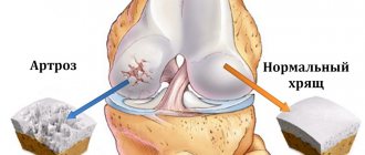
Each joint is formed by the articular surface of the rounded ends of the bones, surrounded by an articular capsule. Inside there is an articular cavity filled with synovial fluid. Normally, all components of the joint correspond to each other and are firmly held by ligaments.
With dysplasia, due to developmental deficiency, the mobility of bones inside the joint increases, a pathological gap is formed between them, as a result of which the joint cannot fully function.
Congenital and acquired dysplasia
Cats have congenital and acquired joint dysplasia. The largest percentage of cases is a congenital anomaly that occurs during embryonic development. But at present, scientists cannot identify a specific gene responsible for the development of dysplasia. They believe that this defect is formed under the influence of a specific set of chromosomes or a certain sequence of their connection.
According to another hypothesis, dysplasia is provoked by the synthesis of an insufficient amount of hyaluronic acid, which is part of the synovial fluid. This indicator is laid down at the genetic level, and it is impossible to influence it by external factors.
Acquired dysplasia can be caused by:
- intensive growth of kittens;
- an unbalanced diet, when the body receives an excess amount of phosphorus with a lack of calcium;
- birth injuries.
Note! The most intensive growth in the first months of life is observed in cats of the Asher breed. And although the debate about the recognition of this breed still does not subside, when choosing a pet, owners need to approach the assessment of its constitutional characteristics “with passion”, and also very carefully select the diet.
Second and third types
In general, the second type is almost completely similar to the first. The only difference is the slower rate of development of the disease. As a rule, the course of the pathology is acute, but it develops gradually, more insidiously. In such cats, you can see a reluctance to run, jump, they may have a stooped, “hunched” back. This disease is more common in older animals whose age has already passed the mark of five to eight years.
The third type of discopathy is most uncharacteristic for cats . In this case, the disease is characterized by a sudden onset. As a rule, it develops in the case of severe injuries: due to the extreme load of an impact or fall, the normal core of the intervertebral disc can simply “explode”, and its contents immediately compress the spinal cord, penetrating deeply into the cavity of the spinal canal. In most cases, a hernia forms.
Causes of dysplasia in cats
Owners should understand that genetic predisposition to the disease is not a death sentence. Thanks to proper feeding and maintenance, the degree of development of pathology can be minimized. Therefore, you need to know the reasons for the development of dysplasia in cats in order to avoid mistakes in care.
- Obesity. Excess weight significantly increases the load on the animal's osteoarticular apparatus, leading to its deformation. In addition, an obese pet leads a sedentary lifestyle, as a result of which the muscles and ligaments weaken and no longer fully support the skeleton. Therefore, any sudden movement or physical activity can provoke pathological changes in the joint, leading to dysplasia.
- Injuries. After bone fractures, joint dislocations, sprains (ruptures) of ligaments, muscles or tendons, a natural redistribution of weight occurs, during which the load on healthy limbs increases, and, as a result, the risk of developing joint dysplasia increases. If during an injury the joint capsule was damaged and synovial fluid leaked out, then irreversible changes will certainly occur in the joint, leading to dysplasia.
- Low level of physical activity. Even pets with normal weight should have a well-developed muscular corset that holds the articular surfaces of the bones at a certain distance from each other. With low muscle tone, compression of articular cartilage and menisci occurs, protrusions, hernias and dysplasia occur.
- Early sterilization. The optimal age for sterilization of cats is considered to be from seven months to two years. Removing the gonads too early leads to changes in overall hormonal levels, slower bone growth and the deposition of subcutaneous fat.
- Rickets. Insufficient intake of calcium and vitamin D during the period of intensive growth and intrauterine development of kittens leads to the development of rickets. It manifests itself as curvature of the spine, deformation of the limbs and chest.
Note! Vitamin D is not synthesized in the skin of cats under the influence of sunlight, as it happens in humans. Therefore, it makes no sense to “replenish” your pet’s reserves of this vitamin by walking in the fresh air.
- Hormonal disorders. Impaired bone mineralization occurs when the thyroid gland malfunctions, since it is responsible for the rate of bone tissue regeneration. Dysplasia can also form due to insufficient production of growth hormone synthesized by the pituitary gland.
- Poor nutrition. Even though cats are carnivores, their diet should include grains, vegetables and fruits. Feeding only meat and meat products does not provide the pet with all the necessary microelements and vitamins, and the lack of fiber negatively affects the functioning of the gastrointestinal tract, the absorption of nutrients and the development of beneficial microflora. It is also worth noting that ready-made economy and premium food do not provide animals with the necessary substances for growth and development, which also leads to osteoporosis and dysplasia.
Treatment of discospondylitis in dogs
Discospondylitis is a therapeutic pathology with neurological disorders and early treatment is usually therapeutic.
Therapeutic treatment includes the prescription of specific antibiotics, for example for brucellosis.
If discospondylosis of unknown etiology occurs, broad-spectrum antibiotics are prescribed - fluoroquinolones, cephalosporins, penicillin antibiotics - for at least 6 weeks. In some cases, antibiotics are used for up to six months.
The course of the disease is monitored using MRI and special examinations.
Painkillers and anti-inflammatory drugs (gabapentin, Previcox, Trocoxil, meloxicam and others) are used in combination with antibiotics. The duration of medication administration is determined by the attending veterinarian.
If fungal infections are present, systemic antifungal drugs such as ketaconazole or fluconazole are prescribed. The duration of medication administration is also determined by the veterinarian.
Glucocorticosteroids are not used in the treatment of this disease.
Surgical treatment of discospondylitis is applicable to eliminate compression of the spinal cord and nerve roots using various techniques: dorsal laminectomy, hemilaminectomy, corpectomy, as well as in the presence of localized spinal abscesses and proliferation of granulation tissue. Sometimes, when spinal instability develops, various stabilizing structures are used (transpedicular fixation, etc.).
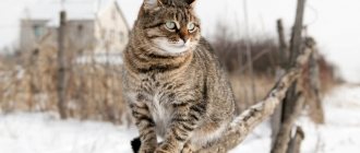
Symptoms and first signs of dysplasia in cats
Characteristic symptoms and the first signs of dysplasia in cats begin to appear during a period of intensive growth. Abnormalities of the elbow joints become noticeable only from the age of six months.
The degree of their manifestation depends on the stage of development of the pathology. The first warning signs are:
- tension and unnatural gait;
- lameness after sleep, and in severe cases - constantly;
- curvature of limbs;
- muscle atrophy and enlargement of limb joints;
- inactivity;
- a crunch made by joint surfaces rubbing against each other during movement.
Young kittens may meow frequently in pain while moving. They try not to make sudden movements and jumps. Adult animals cannot bend their hind legs under themselves when sitting and hide them under their stomach while lying on a horizontal surface.
Symptoms
Age-related changes appear in growing pets. For example, elbow deformity develops within six months and is immediately accompanied by symptoms.
Signs of the disease depend on the severity of the pathology:
- the appearance of an unsteady gait;
- the cat stops jumping on high surfaces;
- kittens develop lameness (at first - when the pet lies in one position for a long time, as the pathology progresses - the symptom becomes permanent);
- the paws become bent and become X-shaped;
- a frisky cat stops moving quickly;
- muscles begin to atrophy, accompanied by weakness of the paws and weight loss of the pet;
- thickening at the bends of the limbs;
- the cat cannot sit in the “Egyptian” pose;
- when the pet lies on its stomach, spreads its paws to the sides;
- A crunching sound can be heard when the cat moves.
If the initial stage of the disease is not treated and osteoarthritis begins, the pet begins to move with great difficulty or completely loses the ability to walk independently.
Breed predispositions
It is worth noting, first of all, such large cats as the Maine Coon. 18% of this breed suffer from dysplasia.
The following are:
- British;
- Scottish fold;
- Siamese;
- Persian;
- Norwegian forest
Tests and methods for diagnosing dysplasia
It is impossible to make an accurate diagnosis and begin proper treatment using clinical signs alone at home. Therefore, you should take the animal to the clinic, where tests will be prescribed.
- Collection of anamnestic data. During the survey, the specialist examines the pedigree (if any), finds out the presence of the disease in the parents and other kittens from the litter, the characteristics of pregnancy and childbirth, the presence of birth injuries, and the time of the first signs.
- Clinical examination. It includes examination of the limbs and spinal column, palpation, determination of the presence and degree of bone deformation.
- Laboratory analysis of blood and urine. An increased number of leukocytes indicates the development of inflammation in the body and the presence of associated pathologies.
- Study of the functioning of the cardiovascular system. Since general anesthesia is required for radiography, before administering it the doctor must make sure that the animal does not suffer from heart disease.
- Arthroscopy. During this procedure, a specialist can assess the condition of the inner surface of the joint using an arthroscope inserted into it through a pinhole puncture.
Note! To conduct this examination, you need expensive equipment, appropriate qualifications and experience of specialists. Therefore, not every clinic can afford such a “luxury”. In addition, the high cost of the study does not make it possible to make it widespread and publicly available.
The final diagnosis is made based on radiography of the joint.
What kind of disease is this?
Scottish fold cats often develop pathologies such as osteochondrodystrophy and osteochondrodysplasia. A feature of the first and second diseases is that, due to a genetic defect in the animal, cartilage and bone structures develop unevenly in the body. As a result of such a defect, the osteochondral system suffers, which is accompanied by systemic damage to the skeleton, joints, and spine.
Chondrodysplasia is often diagnosed in Scottish Fold cats - Scottish Fold and Highland Fold. The disease can also appear in British cats, provided that they have the lop-eared gene. One of the forms of osteochondrodystrophy is achondroplasia. With this pathology, the bones do not grow to the required size, as a result of which the animal develops dwarfism. In achondroplasia, as in the case of osteochondrodystrophy and osteochonrhodysplasia, the main factors giving rise to the pathology are gene mutations.
Test for dysplasia in cats
In 1983, scientists proposed a test for dysplasia called PennHIP. After completion in 1993, it became widespread, as it allows, with certain equipment, to recognize pathology at the earliest stages of development.
After general anesthesia and muscle relaxants that relax the skeletal muscles are administered, the cat is fixed on a special device in a stretched state. This makes it possible to move the femoral head away from the acetabulum to the maximum distance.
The doctor then takes several x-rays and calculates the DI displacement index by comparing the data obtained with the breed standard.
- A DI value of “0” indicates a severe degree of dysplasia and the presence of degenerative processes.
- DI=1 indicates low joint mobility.
- With an average degree of pathology, this coefficient is 0.5-0.6.
X-ray of the joint
The main method for diagnosing dysplasia today remains x-ray of the joint. It allows you to accurately assess the degree of tissue damage and bone condition.
The disadvantages of this survey are:
- age restrictions (the pet must be 2 years old at the time of the procedure);
- the need to administer general anesthesia to ensure complete immobility.
Note! If a cat suffers from heart pathologies and at the same time has a calm disposition, then radiography can be performed without anesthesia, but with careful fixation.
Diagnosis of discospondylitis in dogs
Discospondylitis is a common disease in veterinary practice, but it is quite difficult to diagnose at the initial stage of disease development. When diagnosing, veterinarians often confuse it with spondylosis, although it has its own distinctive features in the clinical picture and diagnosis. Diagnosis of discospondylitis should include, in addition to a general and neurological examination, special research methods such as X-ray examination, MRI and CT.
In addition to spondylosis, differential diagnosis should take into account oncological diseases of the spine - tumors of the vertebral bodies. A distinctive feature of vertebral neoplasms is the intactness of the disc boundaries and lesions within the same vertebra.
Rice. 1. Discospondylitis in a dog with CT diagnosis.
On radiographic examination, characteristic signs of discospondylitis will be narrowing of the intervertebral spaces, demineralization and erosion of the subchondral plates, unevenness of the endplates, proliferation of bone tissue with the formation of spondylophytes, even fusion of the vertebrae. Expansion of the intervertebral space and erosion of the vertebral bodies may be observed.
Rice. 2. Discospondylitis in a dog Th9-L4.
Computed tomography is a more detailed examination of bone tissue; the signs will be the same as in radiographic diagnostics.
Magnetic resonance imaging for discospondylitis allows you to see the disease at an early stage of development. With MRI, you can determine the involvement of the spinal cord in the process of inflammation, see compression problems and, accordingly, evaluate the pathological process in more detail. Characteristic signs on MRI in T-1 mode are a decrease in the MR signal from the disc and the part of the vertebral body adjacent to the disc. In T-2 mode, the MRI signal from these structures increases.
When studying discospondylitis, it is necessary to determine the root cause of this pathology, therefore dogs are necessarily examined for brucellosis (ELISA diagnostics), and bacteriological cultures are done during CT-assisted biopsy of the intervertebral disc.
In case of pain and the presence of discospondylitis on X-ray diagnostics, MRI diagnostics is mandatory.
Consequences of the disease
Conservative methods cannot completely restore the integrity of the joint and ensure its full functioning. Therefore, the consequences of the disease are:
- joint deformation;
- degeneration of bone and cartilage tissue;
- secondary osteoarthritis;
- hip subluxation;
- hip dislocation;
- pelvic deformity;
- arthritis;
- complete loss of joint mobility.
As a result of these complications, cats become very difficult to move and experience increased pain.
What treatment is prescribed?
There is no cure for osteodystrophy in cats. However, the use of analgesics and non-steroidal anti-inflammatory drugs will help reduce inflammation, reduce pain, and improve the general condition of the pet. To prevent bone tissue destruction, chondroprotectors are prescribed, such as Chondrotin sulfate and Glucosamine. In addition, veterinarians recommend giving the cat a special massage, which can improve blood circulation in the affected limbs, so that degenerative processes stop progressing. If conservative treatment does not bring results, surgery is prescribed - arthrodesis or osteotomy.
Effective methods of combating the disease are not available in clinics in our country.
In England, osteochondrodysplasia is treated with radiation. This is a new, effective conservative treatment that relieves pain and prevents further progression of the pathology. However, the problem is that radiation therapy is not common in domestic clinics, since the use of this technique requires expensive equipment.
Studies were conducted at the Veterinary Medicine Teaching Hospital of Seoul National University (Korea) that showed the effectiveness of radiation therapy. After 6 irradiation sessions, the cat, which had massive bilateral exostoses and severe cartilage deformation, was completely cured. All diagnostic indicators returned to normal after a month of therapy, the animal was actively moving and could even climb a tree. On X-ray photographs, there were no new inflammatory foci of bone proliferation.
Prevention measures
The main measures to prevent dysplasia are:
- culling and sterilization of kittens with a genetic predisposition to the disease;
- feeding pets with high-quality food, balanced in the content of nutrients, vitamins, calcium, phosphorus and other minerals;
- preventing obesity;
- regular examination by an orthopedist.
The cat should have a soft bed. It should be placed in a warm place where there are no drafts, which can provoke inflammatory processes in the joints.
What symptoms indicate pathology?
The owner can suspect problems with the cat's spine by reducing the activity of the pet. Then symptoms such as:
- stiffness of movements;
- lameness.
Constant meowing due to pain and lameness are signs of the pathology.
A cat can limp on two or one paw at once, which is directly related to the segment of damage to the spinal column. The pet begins to meow for no reason and becomes restless. Such symptoms are caused by severe pain that is localized in the back area. If the owner tries to touch it, the cat may begin to hiss and scratch. As discospondylosis progresses, the animal becomes less oriented in space and its gait changes. The possibility of a convulsive syndrome cannot be excluded. If the lumbosacral spine is affected, the emptying process is disrupted.