- Causes of thromboembolism in cats
- Symptoms of thromboembolism in cats
- Diagnosis of thromboembolism in cats
- Treatment of thromboembolism in cats
- Forecast
- Prevention
Arterial thromboembolism (ATE) develops due to the formation of a blood clot and blockage of arterial vessels. This is an acute condition that is accompanied by persistent tissue ischemia below the formation of the embolus. Thromboembolism is a complication of congestive heart failure.
Causes
Thrombosis in cats is often a consequence of the progression of chronic heart diseases in the pet’s body, such as cardiomyopathy, arterial hypertension, etc. A blood clot often forms in the left atrium, from where it migrates with the blood flow to the farthest parts of the aorta. Thromboembolism can be arterial and venous, and a clot forms much less frequently in the lumen of the arteries. The reasons why a cat may develop thrombosis are:
- infections;
- sepsis;
- severe poisoning;
- cardiovascular disorders;
- diseases of oncological nature;
- presence of enzymes in the blood;
- mechanical damage affecting vascular structures;
- previous surgical treatment.
Treatment and prognosis
Prevention and treatment of animals with suspected thromboembolism involves the use of anticoagulant drugs (heparin, dalteparin, warfarin, aspirin, etc.) and the use of painkillers to reduce pain that occurs due to neuromyopathy. Surgical treatment is usually ineffective.
The prognosis for cats with thrombosis of the vessels of the extremities is cautious, often unfavorable. Most cats die early after thrombosis, or owners are forced to use euthanasia in order to relieve the animal from excruciating pain.
It is worth remembering that early diagnosis of hypertrophic cardiomyopathy (HCM), renal failure or thyroid pathologies allows for timely prevention of further thrombus formation.
What symptoms are you worried about?
Depending on the location, the disease is accompanied by different symptoms, one of which is cold pads.
Signs of illness in a cat are determined by the location of the blood clot. Typical symptoms of thrombosis are:
- Loss of coordination of movements, severe lameness.
- If the clot is located in the femoral artery, the animal's hind legs are paralyzed. The muscles of the limbs become very tense and hard.
- The pads become pale and cold.
- When the clot is localized in the arteries of the kidneys, the animal suffers from pain in the lumbar region. Due to impaired kidney function, the body becomes intoxicated, and the cat suffers from nausea and vomiting.
- Blockage of the abdominal aorta is accompanied by severe pain when palpating the abdomen. In addition, the cat suffers from gastrointestinal disorders; blood particles can be seen in the stool.
- If a blood clot clogs the blood vessels of the brain, the pet suffers from epileptic seizures, and in severe cases, coma occurs.
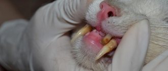
- The affected vessel in the lungs causes the development of symptoms such as a dry, painful cough, shortness of breath, decreased pulse, and pale mucous membranes.
If a cat's aorta is blocked by a blood clot and the blood clot comes off, it is impossible to save the animal. Therefore, if the owner notices that the pet’s condition has deteriorated and characteristic symptoms of the disease are present, it is dangerous to delay a visit to the veterinarian.
Diagnosis of thromboembolism in cats
If the symptoms described above are present, the veterinarian will evaluate the first 5 signs during a physical examination - pain, paresis/paralysis, a cold, pale limb and absence of pulse in the femoral or brachial artery. Thermometry is carried out, as a rule, hypothermia is observed, which leads to an unfavorable prognosis.
Next, auscultation is performed - shortness of breath and wheezing in the lungs are observed. Heart murmurs, gallop rhythm, arrhythmia.
Laboratory diagnosis - hyperglycemia, azotemia and hyperphosphatemia are observed. A sharp decrease in platelets and red blood cells due to coagulation.
X-ray – cardiomegaly, congestion, interstitial or alveolar pulmonary edema.
X-ray. Alveolar pulmonary edema.
Echocardiography - determines dilatation of the left atrium, the presence of a thrombus in the left atrium appendage or its cavity. Left ventricular hypertrophy/dilatation. Left ventricular systolic dysfunction. It is possible to identify a thrombus in the aorta using ultrasound.
Diagnostic measures
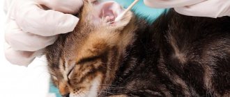
If there are signs of the disease, it is necessary to conduct a diagnosis, which includes electrocardiography.
If a cat suddenly loses its hind limbs, or there are other signs of thromboembolism, it is necessary to urgently take the pet to the hospital. After an initial examination and history taking, the doctor will give a referral to undergo a number of such diagnostic procedures:
- blood chemistry;
- Ultrasound of the heart;
- angiography;
- electrocardiography.
Diagnostic methods
With pronounced clinical symptoms, identifying thromboembolism is not particularly difficult for a doctor. If the signs are not so characteristic, then a number of procedures will help determine an accurate diagnosis. These include:
- Biochemical analysis of the animal’s blood, as well as an additional study of its clotting time.
- Ultrasound of the heart is aimed at assessing the speed at which myocardial contractions occur, as well as how much the atria have increased or decreased compared to normal.
- Angiography is a procedure through which it is possible to identify pathologies in the functioning of an animal’s blood vessels.
What treatment is prescribed?
Conservative
The earlier a cat is diagnosed with thrombophlebitis, the more favorable the prognosis for recovery. Drugs that prevent increased blood clotting will help restore blood flow in the femoral, abdominal, renal or pulmonary arteries. Heparin injections have proven themselves well, thanks to which they can successfully combat blood clots. However, the drug "Heparin" has many side effects, so the veterinarian must prescribe the dosage and treatment regimen. In addition to the use of thrombolytics, vitamins and agents that increase the animal’s immunity are additionally prescribed.
Surgical
Surgical intervention involves crushing the clot using a catheter inserted into the vessel.
Surgery to remove a blood clot is rarely prescribed. This is due to the fact that the procedure is very complex, is carried out only in specialized clinics, and even successful implementation does not guarantee the absence of relapses. Rheolytic thrombectomy is often prescribed to treat thrombophilia. The surgery is performed using general anesthesia. During the operation, a catheter is inserted into the affected vessel, with the help of which it is possible to break up the clot. In especially severe cases, when necrotic changes progress in the tissues surrounding the blocked vessel, the veterinarian will advise euthanizing the cat so that he does not die in agony.
Thromboembolism in cats - causes, symptoms, treatment
As a result of blockage of the arteries by a blood clot, a syndrome of disturbance of the general blood flow in the body occurs.
This condition is called thromboembolism and is often diagnosed in cats with serious extensive injuries.
Veterinary specialists also identify among the factors of thromboembolism - chronic myocardial insufficiency, as well as a violation of the functional characteristics of the renal structures.
The location of the embolus (clot), as a rule, is in the area where the aorta divides into two parts. But thromboembolism is also diagnosed in cats in the area of the renal arteries or pulmonary arteries.
The greatest danger of thrombus formation is that as the embolus moves through the choroid plexuses, there may be a complete cessation of blood supply to the hind or forelimbs. As a result of the pathological condition, the cat develops paresis and paralysis of the paws, and often a necrotic process occurs in the parts of the body where the blood clot that has blocked the vessel is located.
Blockage of an important artery by a blood clot leads to problems with the digestive system, kidneys, and also negatively affects the central nervous system, causing brain damage.
Every pet owner should clearly know what factors can lead to thromboembolism, the main symptoms of the pathology, which will allow them to contact a veterinary clinic for help as early as possible and save the life of their pet.
Causes of thromboembolism in cats
Veterinary medicine specialists distinguish two types of thromboembolism - arterial and venous. This is due to the location of the embolus formed from a blood clot.
The venous type of thromboembolism is diagnosed in veterinary clinical practice an order of magnitude more often than the arterial type. This is caused by a slower flow of blood through the veins.
In addition, arterial vessels have a smooth intima, preventing clots from forming in them.
But venous thromboembolism is not considered particularly dangerous, since it does not directly affect vital functions and does not lead to immediate death.
Thromboembolism in cats of the arterial type entails serious and dangerous consequences. The onset of arterial blockage of a vessel by a thrombus occurs in the left atrium (this occurs especially often with cardiomyopathy).
The thrombus formed at the site of bifurcation (the branching of the aorta into two branches) begins its movement. When the blood clot is small, it easily penetrates downwards, settling in the extremities, clogging one of the vessels. In venous vessels, blood clots form according to the same pattern, but only many times faster due to the lack of rapid blood flow in them.
With chronic diseases of the cardiovascular system, intoxication of the body with poisons, parasitic diseases, the risk of developing thromboembolism increases many times. The main causes of thromboembolism in domestic cats are:
- infectious diseases (blood poisoning);
- acute intoxication of the body with toxic chemicals;
- diseases of the heart and blood vessels that have a chronic course;
- malignant neoplasms;
- the presence of specific enzymatic substances in the systemic circulation;
- damage to vascular walls as a result of mechanical injuries;
- previous surgical interventions.
Symptoms of thromboembolism in cats
Symptoms of thromboembolism in cats directly depend on the vessel in which the blood clot forms, as well as on the fact how much the clot has blocked the blood flow. When aortic blockage by a thrombus and blockage of one vessel at the branch site are diagnosed, problems with the functionality of the hind legs are diagnosed. The cat will show signs of lameness and paralysis.
As a result of degenerative processes and disruption of tissue trophism, dangerous conditions such as gangrene and total necrosis of tissue structures develop. When the bifurcation is blocked by a blood clot, both hind limbs are taken away. Quite often, with thromboembolism, signs of myocardial failure develop.
When auscultating the heart, the specialist clearly hears murmurs in the heart area, but palpating the pulse in the affected limb does not produce results. The pulse wave is so disrupted that it is almost impossible to diagnose it.
Symptoms of thromboembolism in a cat also include a decrease in local temperature at the site of the thrombus, as well as hardening of muscle fibers. These signs are especially often observed when the area of a large artery in the thigh is blocked by a blood clot.
It is worth noting that with the development of the pathological process caused by blockage by a blood clot, the animal’s tail does not lose its mobility. The sick animal refuses to step on its hind legs and may also refuse to stand. As the pathology develops, the affected area becomes cyanotic in the skin, and the possibility of a shock in the cat cannot be ruled out.
The formation of a blood clot in a pet is characterized by the following symptoms:
- impaired coordination of the cat's movements;
- sudden appearance of lameness;
- paresis of the hind legs with hardening of the muscles on them;
- anemic (bloodless) paw pads;
- painful sensations in the cat in the lumbar region (with thromboembolism of the renal vessels);
- the occurrence of vomiting against the background of the accumulation of nitrogenous substances in the systemic bloodstream when blood functions are impaired;
- blockage of the abdominal aorta by a thrombus leads to severe disturbances of the digestive process (stool upset, eruption of gastric contents, stool mixed with blood, pain on palpation of the peritoneal area);
- vascular thrombosis in the brain is characterized by seizures resembling epilepsy, disturbances in the functioning of the vestibular apparatus, and comatose states;
- a blood clot in the area of the pulmonary artery provokes severe coughing and difficulty breathing, the visible mucous membranes acquire an initially pale appearance and then a cyanotic color, and the jugular veins in the animal’s neck swell.
It is important to remember that, according to statistics, animals with thromboembolism extremely rarely survive, since the presence of a blood clot in the artery provokes the penetration of ischemic toxins that spread throughout the body. Against this background, many different dystrophic processes develop in parallel in the body of a sick animal.
The development of thromboembolism begins suddenly and the main task of the owner is to promptly recognize the characteristic symptoms. Early diagnosis and adequate treatment significantly increase a cat's chances of survival.
Diagnosis and treatment of thromboembolism in cats
When contacting a veterinary clinic, the animal is immediately subjected to diagnostic measures, while simultaneously collecting an anamnesis. If thromboembolism is suspected in a cat, the doctor prescribes a number of tests, the main ones being thrombocoagulometry, blood clotting rate and biochemical blood test.
In addition, it is necessary to conduct neurological diagnostic studies, a general blood test, a cardiac examination and radiography with the use of contrast agents (angiography). In addition, ultrasound diagnostics are performed aimed at studying the patency of blood vessels and their tone (Doppler ultrasound).
The obtained research results make it possible to determine the group according to which further treatment tactics will be developed.
So, in veterinary medicine there are 3 groups:
- First group.
It includes animals with severe disorders of the nervous system with circulatory disorders and a slight degree of ischemia. Adequately prescribed treatment allows you to achieve the highest possible result by preserving the vital functions of the body (including the functioning of the musculoskeletal system). It has been proven that mild first-degree thromboembolism can self-limit without treatment, but in the future, without therapeutic measures, the risk of relapse increases significantly. The survival rate is 100%. - Second group
. This includes patients with neurological disorders, but much stronger than in the first case. Blood circulation is characterized by subcompensation, and the degree of ischemia is average. Animals classified in the second group survive in almost 85% of all clinical cases with timely treatment, but it is not possible to achieve complete restoration of the functions of the hind legs. - Third group
. The degree of neurological damage is maximum, almost 98% of sick animals die.
The prognosis for thromboembolism is always unfavorable, and in case of serious dystrophic and necrotic changes in tissues, veterinary specialists recommend euthanasia. If sensitivity in the paws is maintained and there is no necrosis process, there is a chance of recovery.
To do this, the animal is prescribed medications that eliminate severe pain and physical procedures. It is possible to prescribe medications to reduce blood clotting.
Heparin is widely used, but only under the strict supervision of a veterinarian, since exceeding the dosage of the drug can lead to serious consequences, one of which is internal bleeding.
When choosing treatment tactics, special attention is paid to the function of the heart muscle. Thromboembolism negatively affects the functioning of the myocardium, provoking the development of heart failure. Treatment is carried out in a hospital clinic under the supervision of a doctor, due to the fact that some medications can provoke a deterioration in the general condition of the sick animal.
It is not recommended to use sodium and potassium preparations (which improve heart function, but provoke increased thrombus formation) during the treatment of pathology, which is fatal in the case of thromboembolism. The administration of phylloquinone, an analogue of vitamin K, which also promotes rapid blood clotting, is not recommended.
Surgical treatment of thromboembolism is rarely practiced in veterinary medicine. This is due to the increased risk during the administration of anesthesia and the animal’s recovery from it. Even after a successful rhyolytic thrombectomy, the risk of new blood clot formation cannot be ruled out.
To avoid postoperative thrombosis, the animal is prescribed blood thinning medications, which accordingly increase the risk of internal bleeding. Therefore, it is necessary to make every effort to avoid the formation of a blood clot in your pet, rather than deal with treatment later.
Prevention
According to the results of clinical veterinary studies, the average life expectancy of a pet after surgery to remove a blood clot ranges from 4 months to 1-2 years. Rare cases have been described when a pet returned to a full life.
More often than not, cats who have suffered a blockage of an artery by a blood clot remain disabled for life, unable to move normally. There are no measures aimed at preventing pathology. The owner of the animal must carefully choose the diet, avoiding fatty foods.
Cats whose diet contains normal levels of vitamin complexes and minerals are much less likely to suffer from diseases of the cardiovascular system, which means the risk of developing thromboembolism is reduced.
Source: https://zen.yandex.ru/media/id/5afddb734bf161d72c824b60/5d3436630aca0500ae5c284a
Survival
It is important for cat owners to know that survival rates depend on timely diagnosis and treatment of thromboembolism. If the clot does not completely block the artery and the diagnosis is made on time, with the help of thrombolytics it will be possible to achieve its resorption and prevent the formation of a new one. Otherwise, the presence of a blood clot contributes to general intoxication of the body, and if the vein is completely blocked, the surrounding tissues do not receive enough necessary nutrients, which is why irreversible necrotic processes begin to progress in them, a complication of which is the death of the four-legged friend.
Symptoms of the disease
Experts are convinced that in many respects the signs of the disease are determined by the location of the process. The most striking symptoms of thromboembolism in cats are expressed as follows:
- The cat's coordination of movements is impaired and lameness appears.
- Palpation of the hind legs may reveal paralysis of both legs. At the same time, the muscles on them become like stone.
- The tailed fidget's paw pads are turning pale.
- If a blood clot has blocked the renal arteries, the animal will begin to suffer from pain in the lumbar region and severe vomiting will occur. A blood test may show an increased content of nitrogenous metabolic products.
- Thromboembolism of the mesenteric arteries is characterized by the fact that the pet begins to have diarrhea and vomiting, often with the presence of blood in the discharge. Palpation of the abdomen leads to painful reactions.
- Coma, seizures reminiscent of epileptic and disturbances in the functioning of the vestibular apparatus are signs of a blood clot that is located in the blood vessels of the brain.
- If a blood clot forms in the pulmonary artery, the pet will develop a severe cough and shortness of breath. The mucous membranes turn pale. The pulse becomes weak, and the jugular veins characteristically swell.
Data from statistical studies regarding the survival rate of pets who have developed thromboembolism are extremely disappointing. The presence of a blood clot is aggravated by the entry of ischemic toxins into the blood. Taken together, this leads to multiple development of pathological processes in the animal’s body.
Thromboembolism in cats can only be cured if detected early. A timely diagnosis by a qualified specialist and immediate treatment can minimize the damage caused by a blood clot traveling through the cat’s bloodstream. Otherwise, the risk of death increases with each lost day.
Prevention
According to the results of studies conducted by veterinarians, the life expectancy of cats diagnosed with thromboembolism is on average 3 months. up to 1.5-2 years. There is very little chance that a sick pet will be able to return to a full life. Often, the animal remains deeply disabled until the end of its days, unable to move normally and independently cope with its natural needs.
There are no specific prevention methods that can prevent this dangerous disease. If one of the kitten’s parents has been diagnosed with thromboembolism, it is important for the owner to monitor the pet’s nutrition from childhood, monitor the completeness and balance of the food, and prevent the cat from overeating in order to prevent obesity. In addition, it is important to prevent injury to the animal and prevent poisoning by chemicals.
Preventive actions
As a result of research, veterinarians have found that the average life expectancy of a cat that has undergone surgery to remove a blood clot ranges from 3 months to 2 years. Very rarely, but it happens that the pet returns to its normal life. True, this is the exception rather than the rule. Much more often, a furry friend remains disabled forever, having difficulty moving and meeting his natural needs. Therefore, it is better to prevent the disease than to risk your pet’s health later.
There are no special preventive measures that will effectively protect a cat from blood clots. However, the owner should try to protect the animal from excessively fatty foods. Cats whose diet consists of healthy foods rich in vitamins and microelements get sick much less often. In addition, your cat should be vaccinated on time and given anthelmintic drugs. Such measures will reduce the risk of blood clots developing in the animal’s vessels by a quarter.
Treatment of cats with arterial thrombosis
Once the patient's condition has been stabilized, the most important concern is bilateral hind limb paresis/paralysis due to aortic thromboembolism. The neurological and muscular status of the area should be assessed to determine the severity of the stroke and assess the progress of treatment. A phenothiazine tranquilizer such as acetylpromazine maleate should be prescribed for its sedative effects. Its sympatholytic effect also helps establish collateral circulation, causing vasodilation (Taillefer M and Di Fruscia R, 2006).
Anticoagulants are also indicated to prevent further thrombosis in affected cats.
Arterial thrombi are initiated by platelet aggregation with secondary fibrin deposition, whereas venous thrombi are composed primarily of fibrin clots. Therefore, platelet antiaggregates, usually aspirin, are indicated. A dose of 1-5 five-grain tablets of buffered aspirin each day can be administered to the average cat and is an effective platelet antiaggregant. Aspirin acts by inhibiting the prostaglandin thrombroxane A2, which is thought to promote collateral inhibition and platelet aggregation following aortic thrombosis in the cat. Heparin can also be used for its anti-clotting effect at a dose of 50 IU intravenously and repeated every eight hours subcutaneously (Harpster NK, 2005).
Prognosis for thromboembolism
If you consult a veterinarian in a timely manner, the treatment result may be conditionally favorable, but only if your pet has not been diagnosed with serious heart disease during the diagnosis process.
Also, the further result of treatment depends on the speed of action. In order to remove a blood clot with a minimum of complications, it is necessary to take your pet to the nearest veterinary clinic within 1-5 hours. In this situation, the clot will simply be removed surgically.
In the presence of heart disease and diseases of the vascular system, there is a possibility of recurrent thromboembolism.
It is also worth considering that such diseases put cats in a state of shock. Experts recommend leaving the pet for inpatient observation so that its condition can be compensated and stabilized.
An important point is the period of restoration of mobility, which was impaired by a blood clot. The consequences of paresis or paralysis are eliminated within a few weeks. At this time, the pet needs care and close monitoring.
Blood clots and aneurysms in cats.
The disease ranks second among diagnosed heart pathologies.
Other reasons include:
- restrictive, dilated cardiomyopathy;
- age-related changes in the body;
- myocardial diseases;
- viral, infectious diseases;
- sepsis;
- tumors;
- blood clotting disorders;
- poisoning by chemicals, poisons;
- vascular damage;
- parasitic diseases (helminthiases, heartworms);
- bacterial endocarditis;
- severe forms of cardiomyopathy of various etiologies.
We can say that the pathological condition in most cases is a pathological condition that develops against the background of previously suffered heart diseases. Males are more susceptible to the disease than females, which is explained by their greater susceptibility to myocardial diseases.
Arterial thromboembolism (ATE) in cats and dogs
These particles can sometimes get stuck in other places, especially where blood vessels narrow.
Blocked blood vessels can also occur when foreign substances (such as bacteria, air, or fat) enter the bloodstream. As a result of the formation of blood clots, insufficient blood flows to the cat's body tissues through blocked blood vessels. Some blood clots can become infected and can spread bacteria, causing localized infections.
Thrombi (blood clots) can also form when the flow of blood into arteries or veins is restricted.
Aneurysm
is a protrusion of the wall of a blood vessel caused by a weakening of the middle layer of its wall. The destruction of the lining of blood vessels that occurs as a result of an aneurysm can also lead to the formation of blood clots, followed by blockage of the blood vessel with a thrombus.
Treatment
Before starting treatment, the veterinarian conducts a comprehensive examination of the patient. To make a diagnosis, a biochemical, detailed blood test, as well as a blood clotting test are performed. The results of cardiac ultrasound, radiography, ECG, and angiography are taken into account.
Important! It is worth noting that if an animal is diagnosed with arterial or venous thromboembolism, the diagnosis is disappointing and doubtful.
The main task of the treating veterinarian is to determine the root cause and determine the location of the blood clot. Treatment is also aimed at normalizing blood flow and improving the condition of blood vessels.
As a rule, the only radical treatment is surgery. To free the vessel from the clot and normalize blood circulation, the aorta is opened and the clot that has blocked it is removed. In severe cases, resection of part of the affected vessel tissue is performed.
After the operation, symptomatic therapy, restorative drugs, and thrombolytics are prescribed, which prevent further formation of blood clots in the cardiovascular system.
The dosage of drugs, course duration, and frequency of administration are prescribed by the attending veterinarian on an individual basis. Owners must strictly adhere to all recommendations and constantly monitor the health of their furry pet.
In general, if treatment is carried out at the initial stage, the prognosis is conditionally favorable. But you need to understand that the disease can relapse. Therefore, if there is any deterioration in health, immediately take the cat to the veterinary clinic.
Blood clots
- These are blood clots that form when a cat’s arteries and veins are damaged in order to stop bleeding. Blood clots cause obstruction of blood vessels at the site of their formation. All the substance of the thrombus must gradually be destroyed and washed out by the bloodstream in the form of emboli - particles that are normally absent from the circulating blood.
Prognosis and prevention
What is the prognosis for this pathology? Even with timely treatment, the chances of success are not very high. We can only hope for the efforts of pharmacists, who promise to create new drugs in the next five years that will help destroy existing blood clots and prevent the development of new ones. Very often, vein thrombosis in cats leads to the need for subsequent euthanasia of the animal.
The most accessible and relatively cheap medicine of this type is heparin . But when using it, extreme caution must be observed, since even the slightest overdose can lead to death from internal bleeding.
Thromboembolism in cats: description of the disease, symptoms, treatment
Embolism is the circulation in the blood of particles that should not be there. Sooner or later, the process leads to complete or partial occlusion - blockage of the lumen of the vessel by tissues, parasites, foreign bodies, gas, bacteria. Thromboembolism refers to the occlusion of a vessel by a blood clot - a thrombus.
More often we are talking about arterial thromboembolism - an acute condition leading to disability or sudden death. A clot can also clog a vein, but in this case the consequences are not so catastrophic. As a rule, the thrombus enters the aorta from the left atrium. The clot blocks the aorta itself or the pulmonary, brachial, mesenteric, and renal arteries.
The blood clot disrupts the blood supply to the tissue below the blockage. Cellular starvation develops - there is no supply of nutrients and oxygen (ischemia). This leads to dysfunction and then death. The more acute the process develops, the less chance there is to restore the functions of the affected tissue or organ, and the faster death occurs.
Contrary to misconception, feline thromboembolism is not genetic. However, many heart diseases that significantly increase risk factors are inherited. It has been established that Maine Coons, Persians, Himalayans, Britons, Scots, Sphynxes, American Shorthairs and related breeds are prone to them.
Causes
Hypertrophic cardiomyopathy is the scourge of many, including outbred cats. With this disease, the walls of the left ventricle thicken due to the reduction of its cavity. This leads to heart failure and congestion, which contributes to the formation of blood clots. In 70% of cases of thromboembolism in cats, cardiomyopathy or other heart disease is diagnosed.
The mechanism of thrombus formation is not fully understood, but Virchow's triad is considered a likely explanation. A small clot forms to prevent bleeding when the vessel wall is damaged. A special mechanism prevents blood from clotting more than necessary.
But if it is broken, the small clot turns into a thrombus. Slow blood flow contributes to the accumulation of protein and blood cells on it, up to blockage or separation of the blood clot from the vessel wall.
The causes of thromboembolism in cats are clear from Virchow's triad:
| Education stage | Process | Probable Causes |
| I | Damage to the vessel wall | Trauma, surgery, catheter placement, venipuncture, heart disease, parasites and intoxication. |
| II | Bleeding disorder | Malignant processes, pregnancy, lactation, hormonal therapy. Inflammatory diseases, injuries, surgeries, sepsis, and swelling increase the risks. |
| III | Slowing blood flow | Venous insufficiency, heart, lung, kidney diseases. Tumors, obesity, pregnancy, long-term use of a number of medications. |
It turns out that thromboembolism is a combination of a number of circumstances. But more often we are talking about diseases of the heart, lungs, blood vessels, kidneys, and hematopoietic system. Therefore, chronically ill and elderly cats are at risk. Some congenital pathologies make themselves known at an early age, so thromboembolism also occurs in kittens.
Symptoms
Common symptoms of feline thromboembolism include severe weakness, rapid shallow breathing, incoordination, staggering or immobility. Pain syndrome is likely - the pet screams and does not allow itself to be touched. Limb paresis is almost always observed.
One or both hind legs do not move at all or cannot bear weight (weakness). The muscles are hard and cold. The paw pads are pale at first, then bluish. Pulsation in the extremities is weak or absent. The same signs occur when the front paws are affected, when the brachial artery is affected.
Characteristic symptoms depend on the location of the thrombus:
- pulmonary artery - cough with blood or pink mucus, difficulty breathing, veins in the neck swell, mucous membranes turn pale, then turn blue;
- brain – epileptic-like seizure, fainting, coma, loss of coordination;
- abdominal arteries - painful stomach, diarrhea with blood, vomiting, severe weakness;
- coronary thrombosis - shortness of breath, shallow rapid breathing, immobility or severe lethargy, bluish mucous membranes;
- renal arteries - painful lower back, bloody urine, vomiting, constrained movements on bent limbs.
With partial blockage, symptoms of thromboembolism may increase over several weeks or even months. With complete occlusion of the vessel, signs of malaise appear suddenly - the cat has just been playing, and now she is lying on her side, breathing frequently, screaming or in confusion.
Diagnostics
An experienced doctor will suspect thromboembolism at the first examination, and, most likely, will immediately suggest euthanasia. But if resources are available and the condition has been successfully stabilized, it is wiser to conduct a full diagnosis.
Shown:
- urgent clinic and biochemistry of urine, blood, coagulation test;
- heart studies - auscultation, echocardiography, cardiogram;
- X-ray of the chest, abdominal cavity - the presence of fluid, the condition of the internal organs;
- Ultrasound of the abdominal cavity, blood vessels - general condition, neoplasms, other structural changes;
- angiography is a contrast method for studying blood vessels, allowing one to detect problem areas;
- CT, MRI – general condition of internal organs and blood vessels.
Unfortunately, most studies are available only in large clinics and are carried out under anesthesia, which is an additional risk. Therefore, many veterinarians make a diagnosis based on five indirect signs - pain, paresis of the limbs, absence of a pulse in the affected limb, cold hard muscles, cyanosis.
When both limbs are affected, doctors recommend euthanasia. About 70% of cats with one limb affected survive to discharge. Unfortunately, half of survivors relapse after 30-60 days. The average life expectancy of a cat after arterial thromboembolism is from 6 months to 2 years. Less than 10% of cats return to a healthy life.
Treatment
Before all diagnostic measures, emergency care is provided - anti-shock therapy, spasm relief, elimination of hypothermia. Severe pain is treated with opiates. For heart failure, oxygen is prescribed; for pulmonary failure, furosemide is prescribed intravenously. Once the condition has stabilized, a decision is made on drug therapy, surgery or euthanasia.
First aid
If thromboembolism is suspected, the cat should be taken to the clinic immediately. If you have an analgesic and/or sedative at home, you can inject it intramuscularly. The doctor will tell you the dosage over the phone. The pet is transported in a position lying on its side, with active resistance - wrapped in a blanket or jacket.
Basic treatment
In case of thromboembolism, the cat should remain in the clinic for at least a week for constant monitoring and emergency assistance if necessary. Treatment begins with lytic therapy, provided that the cat is admitted to the clinic no later than 24 hours from the onset of symptoms.
Special drugs help dissolve the blood clot: urokinase, streptokinase, altepase for the first 1-3 days. Then about a week is given to heparin therapy, which prevents further clot formation. Clopidogrel (prevents platelet clumping) and aspirin (thin the blood) are prescribed for life.
The use of diuretics and cardiac drugs is indicated. Surgical methods for treating thromboembolism in cats are rarely used - the risk of death during the operation and from complications is too high. But in extreme cases, when there are no other chances, an experienced surgeon will suggest removing the blood clot through an incision in the artery.
Treatment of thromboembolism
Therapeutic treatment of thromboembolism is aimed at ensuring blood flow to the heart, preventing further ischemic damage to still living cells of the body. Infusion therapy is used to retain the liquid part of the blood in the vascular bed. Improving hematocrit and blood viscosity improves its fluidity, which facilitates its passage through the altered vascular bed.
Thrombolytic therapy is necessary to restore blood flow through blocked vessels and reduce pressure in them. This therapy is carried out for 24-72 hours, after its completion heparin therapy is carried out for 7 days.
Along with infusion and thrombolytic therapy, drugs from the group of antioxidants and antihypoxants are used, as well as drugs that improve peripheral circulation (pentoxifylline), and anti-shock therapy is carried out.
Surgical treatment of thromboembolism involves removing the clot. This is possible when the thrombus is localized in the area of the aortic bifurcation (its division into the common iliac arteries, usually located at the level of the IV-V lumbar vertebra). The surgical technique involves opening the aorta, after which the blood clot is washed out of the vessel by blood flow, and then the aorta is sutured.
The video shows this process.
Read also: Thrombus in the lung prognosis treatment
The complexity of the operation and the prognosis for its outcome depend on the severity of the patient’s condition and the timeliness of the animal’s owners contacting the veterinary clinic.
Based on practical experience, many veterinary surgeons believe that after the occurrence of an embolism, the maximum time during which the operation can still be performed is 1 hour. The high mortality rate due to arterial blockage is associated with reperfusion syndrome - a process in which the products of ischemic necrosis enter the blood and have a pathogenic effect (which can cause disease) on vital organs and systems.
When implementing long-term anticoagulant therapy, it is necessary to monitor blood clotting. It is better to do this in a veterinary clinic, but if in the future the owners do not have the time or opportunity for this, then they can be trained to conduct a rapid assessment of this indicator.
For this procedure you will need a clean glass slide. You need to put three drops of blood on it. Next, to keep the glass warm, place it on your palm or wrist and swing it, controlling the fluidity of the blood. The blood should clot within 5-9 minutes, and if you are taking anticoagulants, within 7-9 minutes. If the clotting time decreases, you need to increase the dose of the drug.
Thromboembolism is a disease that develops suddenly, progresses very rapidly and often recurs. Because the underlying etiological factor, heart failure, is incurable, animals with thromboembolism must be monitored and treated throughout their lives. Such a patient requires regular visits to the veterinary clinic for ongoing neurological examination. With professional patronage by an experienced veterinarian, such a pet can live a full life without serious complications.
Clinical signs of arterial thrombosis in cats
Clinical signs at presentation will be determined by the area of ischemia. Soon after thrombosis, clinical signs appear indicating pain, ataxia and paralysis of the pelvic limbs. Affected extremities will be bluish, cool, and painful without a general pulse. The calf muscle often contracts and local nerve reflexes are lost. Additionally, the toenail of the limb can be trimmed and will not actively bleed. Renal function tests may show severe azotemia if one of both renal arteries is blocked. Abdominal pain may also result from renal or mesenteric artery embolization. The severity of signs of thrombosis will depend on how quickly adequate collateral circulation can be established. Simple mechanical occlusion will not prevent the development of adequate collateral circulation. However, the thrombus releases vasoactive substances, primarily serotonin, which causes vasoconstriction of the collateral vessels. Consequently, effective collateral circulation develops only in clinical cases of aortic thromboembolism. Selective arterial or non-selective venous angiography can be used to determine the location and extent of the thrombus, as well as to visualize any developing collateral circulation (Luis Fuentes V, 2012).
Video
More photos Author(s):
V.S.
Gerke, Ph.D., veterinarian / V. Gerke, PhD, DVM Organization(s):
CJSC “Network of Veterinary Clinics”, St. Petersburg / “Network veterinary clinics”, St.
Petersburg Magazine:
No. 3 - 2014 UDC 619:616.005.755:636.8
Keywords:
thromboembolism, embolus, hemocoagulation, thrombus formation, thrombosis, congestive heart failure, anticoagulants, antiplatelet agents, platelets, aspirin, clopidogrel, warfarin
Key words:
thromboembolism, embolus, hemocoagulation, blood clots, thrombosis, congestive heart failure, anticoagulant drugs, antiplatelet agents, platelets, aspirin, clopidogrel, warfarin
annotation
Thromboembolism (TE) is a syndrome of acute circulatory disorders as a result of embolization of an artery by a thrombus formed in the venous bed. It is characterized by the development of acute ischemia of all tissues located below the embolus. TE is the common and most dangerous complication of congestive heart failure in cats. An episode of thromboembolism is preceded by excessive formation of blood clots in the veins, where the main mechanisms of hemocoagulation are plasma factors for the formation of a fibrin clot. In the case of arterial TE, platelet factors are added at the site of the thrombus, leading to fixation of the embolus and additional thrombosis of the artery below the site of occlusion. The basis of treatment for cats with TE is the rapid restoration of hemodynamics, for which it is necessary to relieve the pain syndrome and conduct infusion therapy with colloid solutions to improve the rheological properties of the blood. Only against the background of this treatment are direct anticoagulants and, possibly, thrombolytic drugs used. To prevent TE in chronic heart failure when signs of stagnation appear, as well as to prevent relapses of TE, indirect anticoagulants are used - acetylsalicylic acid, clopidogrel, warfarin. The prescription of anticoagulants is justified by theoretical data, but their effectiveness in cats does not yet have clear evidence.
Summary
Thromboembolism (TE) is a syndrome of acute circulatory disorders as a result of embolization of the artery. While clots are formed in the veins. An embolus leads to acute ischemia. Thromboembolism is the most frequent and serious complication of congestive heart failure in cats. Plasma fibrin clot formation factors are the main mechanism of blood coagulation, they lead to the formation of a blood clot in a vein. In the place of location of the thrombus platelet factors activated, so embolus fixed in the artery. Thus artery thrombosis develops below place of occlusion. When treating cats with TE as soon as possible need to reduce pain and restore hemodynamics. To improve the rheological properties of blood should conduct infusion therapy using colloidal solution. Together with infusion therapy can be administered direct anticoagulants and thrombolytic drugs. For the prevention of TE in patients with chronic heart failure with signs of congestion, as well as to prevent a recurrence TE use indirect anticoagulants – aspirin, clopidogrel, warfarin. Anticoagulant drugs justified theoretical data, but their benefit in cats, does not yet have definitive evidence.
For the first time in 1856, R. Virchow introduced the concept of “embolism” and theoretically substantiated embolic obstruction of the arteries. The Virchow triad of thrombosis (slowing blood flow, changing or damaging the inner lining of the vessel and increasing blood clotting) still retains pathogenetic significance.
Thromboembolism (TE) is a syndrome of acute circulatory disorders as a result of embolization (complete or partial blockage) of an artery by a blood clot, usually formed in the venous bed. When a blood clot forms in the veins of the lungs (in the pulmonary circulation), embolization occurs in the arteries of the systemic circulation. In turn, when thrombus forms in the vena cava, the embolus enters the pulmonary arteries. It is characterized by the development of acute ischemia of all tissues located below the embolus. In the ischemic zone, a release of biologically active substances occurs, which is accompanied by a pain syndrome; subsequent disturbances are observed in the nervous tissue, which characterizes the formation of neurological symptoms.
According to K. Borgeat, J. Wright et al. (2014) based on a retrospective study for the period from 2004-2012. Episodes of TE have been reported in approximately 0.3% of all cats seen at a veterinary clinic. Mortality was 58-73% in the first 24 hours, and among the cats that survived the first day, half died in the first week. These figures indicate the severity of the condition of such patients and the relevance of the problem of thromboembolism in general and the need to develop the most effective means of treatment and prevention.
In clinical practice, the main predisposing factor for TE in cats is considered to be dilation of the left atrium. This is explained by the development of stagnant changes in the pulmonary veins, which leads to an imbalance of the coagulation and anticoagulation systems of the blood. TE is always preceded by excessive thrombus formation and accumulation of free thrombi in the venous bed and left atrium.
Some basics of hemocoagulation
Two closely interconnected pathways are involved in thrombus formation: platelet and plasma.
Activation of platelets occurs through the interaction of the corresponding receptors on their membrane with the collagen of the subendothelial layer, as well as under the action of thromboxanes synthesized in the damaged endothelium and in already activated platelets. In turn, active platelets undergo agglutination and form a platelet clot.
The plasma mechanism has an external and internal activation path. Activation of the cascade of blood coagulation reactions can be caused by damage to endothelial cells with the release of tissue thromboplastin. Mechanical damage and contact of blood with the subendothelial layer of the vessel activates the Hageman factor. Damage and death of endothelial cells with the release of lysosomal proteases also increases the risk of thrombosis, because these proteases have enzymatic activity similar to Prower-Stewart factor and thrombin (serine proteases).
The relationship between the platelet and plasma mechanisms is as follows. Active Hageman factor, thrombin and fibrin itself, along with collagen, cause activation of platelets, and platelets, in turn, release tissue thromboplastin upon activation and the membrane of activated platelets promotes fixation and polymerization of fibrin monomers.
Based on the prevalence of platelet or plasma thrombus factor, one can judge the location of activation of the blood coagulation system. Thus, it is known that in the arterial bed under conditions of active blood flow the main mechanism is platelet agglutination (formation of a “white thrombus”), and in the venous bed the formation of fibrin prevails.
Pathogenesis of thromboembolism and the role of CHF
Activation of neurohumoral mechanisms of increasing blood pressure (sympathetic-adrenal system, renin-angiotensin-aldosterone system, vasopressin-aldosterone system, etc.) and inhibition of natural antihypertensive mechanisms (natriuretic peptide, endothelin, NO, etc.) are the basis of the pathogenesis of chronic cardiac insufficiency. The same mechanisms lead to remodeling of the vascular wall. Hypertrophy and fibrosis of the smooth muscle membrane of the vessel disrupts the trophism of the endothelium. Endothelial dysfunction syndrome, inadequate synthesis of vascular intimal components, and heparin metabolism disorders develop. This leads to the death of endothelial cells, contact of blood with the subendothelial layer, with collagen, this causes an increase in blood clotting with the predominant activation of plasma hemocoagulation factors. With the development of stagnant processes, the speed of blood flow in the venous bed decreases, which is also a favorable factor for spontaneous thrombus formation in the veins and the accumulation of “free” blood clots.
Unfixed “free” blood clots, according to the “swirling” principle, are retained mainly in the atrial appendages. Almost any fluctuations in pressure, even a simple change in the body in space, can provoke the migration of such blood clots. The location where a thrombus will form an arterial occlusion depends on the size of the clot itself.
When thromboembolism occurs, two pathological processes take place: mechanical obstruction of the vascular bed and humoral disorders resulting from the release of biologically active substances - thromboxanes, serotonin, histamine, kallikrein, etc. A circulatory disorder in the area located below the occlusion leads to acute hypoxia and ischemia, which causes an additional release of biologically active substances from ischemic tissues. All this leads to the development of a vicious circle in disorders of the coagulation and anticoagulation systems, deep ischemic damage, microcirculation disorders, shock, coma and death of the entire organism.
Clinical picture and prognosis
Thromboembolism is characterized by suddenness. In the vast majority of cases, vocalization is observed, because cats are in severe pain. Signs develop very quickly. Severe depression, a complex of neurological disorders.
The development of neurological symptoms in TE is based on ischemic damage to the nervous tissue as the most sensitive to oxygen deficiency. In all cases of TE, we observed paresis and paralysis with symptoms of damage to lower motor neurons (flaccid paralysis). Weakened or absent reflexes, decreased or absent pain sensitivity. There are monoparesis, paraparesis and tetraparesis.
Due to the fact that nervous tissue is the most sensitive to ischemic damage, it is by the prevalence and degree of neurological disorders that one can judge the severity of the condition and the immediate prognosis. Thus, monoparesis indicates that there has been an occlusion of one femoral artery (or, less commonly, the axillary artery if the forelimb is affected). Paraparesis mainly develops when the embolus is localized in the area of the aortic bifurcation with occlusion of both femoral arteries. Tetraparesis is the most severe condition, almost always fatal in the first day. At autopsy in such cases, a thrombus is found in the thoracic part of the aorta, it is always a large thrombus, in some cases it is partially in the aorta, and part of it has not yet left the cavity of the left ventricle.
Based on the degree of neurological disorders, we distinguish three groups of cats with TE:
Group 1: circulatory disorders - partially compensated, degree of ischemia - mild. Neurological disorders of 1-3 degrees. The prognosis for such animals is rather favorable. Spontaneous restoration of blood circulation is also possible.
Group 2: blood circulation – subcompensated, degree of ischemia – average. Neurological disorders of 3-4th degree. With treatment, survival rate is 70-80%. Up to 30% can recover on their own, without treatment. Restoration of nervous system function is often incomplete.
Group 3: blood circulation – decompensated, degree of ischemia – severe, gangrene. Neurological disorders of the 5th degree. High lethality. Isolated cases of survival during treatment in the first few days were either complicated by severe renal failure or recurrent thromboembolism with a picture of tetraparesis. It is worth noting that difficulties may arise in diagnosing the fifth degree of neurological disorders in TE, because the patient experiences severe pain, which may interfere with the determination of deep pain sensitivity in the affected limbs.
Treatment
Treatment of cats with an episode of thromboembolism should be based on knowledge of the pathogenesis of this condition and the clinical presentation. The first thing you need to do is relieve the pain. While using painkillers, it is necessary to carry out infusion therapy based on the use of colloids, because these solutions affect the rheological properties of blood. It is anesthesia and infusion therapy with colloid solutions that are the basis in the treatment of cats with TE.
Considering the fact that an embolus, being a thrombus, can activate platelets and cause additional thrombosis at the site of its localization, it is important to use direct anticoagulants - heparins (low molecular weight or unfractionated heparin).
On the first day of a thromboembolic episode, the use of fibrinolytic therapy is justified. Streptokinase - an intravenous bolus of 15,000-25,000 units per 20-40 ml of 5% glucose for 30 minutes, then a constant infusion at the rate of 5,000-10,000 units/hour.
As the patient's condition improves and hemodynamics stabilize, additional diagnostics and the development of basic treatment for complications and, in fact, heart failure are carried out.
Prevention
To prevent TE in cats with chronic heart failure and primarily in cats with HCM, as well as to prevent relapses of TE, the use of indirect anticoagulants is necessary. From this group of drugs, we will consider several representatives: coumarin-like anticoagulants - warfarin, antiplatelet agents (antiplatelet drugs) - aspirin (acetylsalicylic acid) and clopidogrel. All of these drugs reduce the activity of the blood coagulation system, but their mechanism of action is fundamentally different.
Acetylsalicylic acid (aspirin) is a non-steroidal anti-inflammatory drug. Its action is based on the irreversible blockade of platelet cyclooxygenase. As a result, the synthesis of thromboxanes is blocked and platelet agglutination is reduced. In human medicine, the effectiveness of aspirin in the prevention of arterial thrombosis in atherosclerosis and after surgical manipulation of the arteries (bypass surgery) has been proven. We did not find clear data on the effectiveness of aspirin in veterinary practice. Veterinary cardiologists prescribe aspirin to prevent TE in cats, but no focused studies have been published examining its effectiveness.
Clopidogrel (Plavix, Zilt, etc.) - like aspirin, is an antiplatelet agent, providing an antiplatelet effect. Clopidogrel is an inhibitor of receptors on the platelet membrane, which inhibits the ability of the latter to interact and activate. In human medicine, clopidogrel is recommended to be prescribed in combination with aspirin, because it increases the effectiveness of the latter and prevents a “paradoxical” reaction to acetylsalicylic acid (aspirin is a non-selective COX blocker, together with the production of thromboxanes in platelets, the synthesis of prostaglandins in the endothelium is inhibited, and in humans, in response to this, cases of a reverse reaction to aspirin have been reported - spontaneous platelet agglutination) [2]
. As with aspirin, clopidogrel is widely used in veterinary practice, but targeted studies examining its effectiveness have not been published.
Today, the effectiveness of aspirin and clopidogrel can be judged almost only by the results of a retrospective analysis by K. Borgeat, J. Wright et al. (2014), in which the authors note that episodes of TE during treatment with aspirin and clopidogrel had a better prognosis. Although the authors themselves note low statistical significance [6]
. The possible effectiveness of the combination of aspirin and clopidogrel in TE may be based on the fact that after the embolus is delayed in the artery, antiplatelet drugs prevent the fixation of this embolus and prevent secondary thrombosis of the artery below the site of occlusion.
Aspirin for cats is prescribed at a dose of 4-5 mg/kg (i.e. 20-25 mg per cat) once every 3 days. Clopidogrel – 3-4 mg/kg 1 time per day. The basis for prescribing these drugs is the presence of signs of congestive heart failure (enlargement of the left atrium, dilation of the pulmonary veins, etc.).
Coumarin anticoagulants (warfarin, finilin) are antivitamin substances. They cause inactivation of vitamin K-dependent blood coagulation factors, thereby blocking plasma hemocoagulation mechanisms. It is these drugs that will be effective in preventing venous thrombus formation, the accumulation of blood clots, and therefore thromboembolism. The disadvantage of these drugs is the need for strictly regular monitoring of hemocoagulation. The risk of spontaneous bleeding is too great. We have had repeated experience with the successful use of warfarin to prevent relapses of TE. We did not observe any episodes of thromboembolism during the use of warfarin. The state of hemocoagulation was assessed by the time of thrombus formation using the Lee-White method. But it is worth noting that out of 5 patients treated with warfarin for more than 1 year, three had bleeding episodes (insufficient control and adjustment of the drug dose). We used warfarin according to the following scheme: start taking warfarin one day before stopping heparins after an episode of TE; the first three doses are 0.2 mg/kg 1 time per day. On the third day of treatment, be sure to check clotting. As a rule, from the fourth day the dose should be reduced by 2 times. Hemocoagulation monitoring is carried out once a week until the dose stabilizes, then once a month.
From all of the above, it follows that the use of coumarin anticoagulants for the prevention of recurrent TE in cats has theoretical justification and should be the most effective. We have positive experience with the use of warfarin. But due to the danger and complexity of using this medicine in veterinary medicine, we cannot recommend warfarin for use in general practice.
The use of aspirin and clopidogrel in combination or separately is justified by information about increased survival in the event of TE, but the effectiveness of these drugs in the prevention of thromboembolism itself is questionable even theoretically, because a decrease in platelet activity does not reduce venous hemocoagulation. I hope that practice and statistics will refute my doubts and prove at least a small, but effective in preventing TE, because the use of these drugs is quite convenient and relatively safe.
Literature
1. Martin M.V.S., Corcoran B.M., Cardiorespiratory diseases of dogs and cats. M., “Aquarium-Print”, 2004, 496 p.
2. Metelitsa V.I., Handbook of clinical pharmacology of cardiovascular drugs. - 2nd ed., St. Petersburg, 2002 - 926 p.
3. Pathological physiology. Edited by Ado A.D., Novitsky V.V., Tomsk, 1994, 468 p.
4. Kirk’s modern course of veterinary medicine./ Trans. from English - M., “Aquarium-Print”, 2005., 1376 p.
5. Sotnikov V.V., Gerke V.S., Andreev A.A., Thromboembolism. — Collection of articles “Current problems of veterinary medicine”, St. Petersburg, 2004, p. 66.
6. Arterial Thromboembolism in 250 Cats in General Practice 2004-2012 – Borgeat – Journal of Veterinary Internal Medicine. Volume 28, Issue 1, pages 102-108, January / February 2014.
Back to section