Dirofilariasis in cats is a helminthiasis caused by parasitizing nematodes of the species D. repens (affects the subcutaneous tissue, eyes) and D. immitis (localized in the right side of the heart and pulmonary artery). Both types of helminths are transmitted through the bites of mosquitoes, which act as intermediate hosts.
Both cutaneous and cardiac forms of dirofilariasis are approximately 10 times less common in cats than in dogs in the same region. This is due to the body's better resistance to infection. According to statistics, homeless animals are more often infected, but the disease often occurs in pets.
Dirofilariasis - what is it?
The parasite Dirofilarifsis gets its name from two Latin words: diro, meaning evil or bad, and filum, meaning thread. Thin roundworms earn their name due to their length of up to 30 cm and the severe damage that can be caused to the host's body. A number of diseases caused by infestation of the cat's body by parasites of this genus are collectively called dirofilariasis in cats. What kind of diseases are these? First of all, we are talking about poisoning by secreted toxins, as in the case of infection with other types of parasites. In addition, most of the adult worms die quite quickly, and fragments of the parasite remaining in the body are capable of blocking a blood vessel. The result of this most often is the death of the pet from embolism.
No less dangerous is the influence of filariae and parasitic larvae, which cause regular small blood loss and interfere with the normal supply of nutrients and oxygen to organs, thinning and damaging body tissues. Only timely diagnosis and proper medical intervention can defeat dirofilariasis in cats. Symptoms, treatment, and most importantly, prevention of this disease are necessary knowledge for every pet owner.
How dangerous is this infection?
This type of helminthic infestation is dangerous because individuals of the parasitic nematode do not live in the intestines, as other roundworms usually do, but in the large vessels of the body - the pulmonary arteries, veins, and heart muscle.
The vital activity of the parasite itself is not as dangerous as its death - the dead body of the helminth can clog the lumen of a vital artery or organ, thereby blocking the access to blood and oxygen. As a result, an embolism occurs, which in 80-90% of cases leads to death.
The vital activity of these worms does not go unnoticed by the animal’s body: it often happens that cats simply cannot withstand prolonged exposure to toxic substances secreted by parasites and die.
How does infection occur?
The larvae of the parasite are carried by blood-sucking insects, mosquitoes, and much less frequently by fleas or ticks. Getting into the stomach of the carrier along with the blood, the larvae are thrown under the skin of a healthy individual bitten by an infected insect. Over the next few months, difilaria larvae develop in the subcutaneous tissue to a state where they are able to migrate through the blood vessels. Over a period of about 4 months, most of the larvae will die, but some of them, in numbers from two to six individuals, reach the respiratory system through the bloodstream and settle in the lungs. In addition, individuals can parasitize the nervous system, abdominal cavity, and eye area. Thus, dirofilariasis in cats occurs in the first latent, that is, hidden, form. At this stage, actively growing and later reproducing worms will release a considerable amount of toxic substances that have a detrimental effect on the host’s body.
After the latent stage, dirofilariasis in cats develops into the most dangerous, acute form. At this stage, the adult worms begin to die, causing blockage of the blood vessels. The result of this, sooner or later, is the death of the animal.
To treat or not to treat?
Typically, heartworm disease in cats is asymptomatic and resolves spontaneously. On the other hand, the pulmonary and cardiac pathologies it causes are dangerous and there is a risk of death. BUT! With the massive death of parasites, blockage of blood vessels and poisoning of the body with decay products are possible. So what to do?
The lack of effective drug treatment for cats with adult hookworms prompted the trial of a surgical method based on removing heartworms from the heart and great vessels by thoracotomy using a flexible loop catheter.
Perhaps over time, if the technique of such operations is simplified, this method will become popular. Since nothing can be done about adult helminths, it is necessary to destroy and prevent microfilariasis. For cats, the simplest effective method is monthly standard doses of milbemycin oxime (" Milbemax "), which kills unadapted microfilariae in infected cats.
Moxidectin, the active ingredient in the drug Advocate , also destroys microfilariae in the cat’s blood, preventing their development into adults. At the same time, the drug does not cause the formation of blood clots in animals with adult heartworms.
Effective against microfilariae and " Stronghold ".
Symptoms: what are the signs of dirofilariasis?
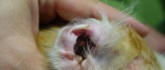
What are the signs to suspect dirofilariasis in cats? Symptoms of infestation may vary slightly depending on the organs in which the parasite is localized. At the first stage, when it enters the subcutaneous tissue layer, the following signs of the disease can be observed:
- Redness of the skin surface.
- The appearance of pustular formations.
- Itching.
Subsequently, as the larvae develop and mature, in most cases symptoms appear such as blue discoloration or obvious redness of the mucous membranes, dull hair falling out in clumps, general weakness and apathy of the pet, and decreased appetite.
However, all these signs appear most often when the invasion is already in the active stage. The main danger of the disease is the absence of visible symptoms at the initial stage. For this reason, veterinarians recommend screening for infestations at least once a year.
Causes
This disease in pets is caused by two members of the family Filariidae:
- Dirofilaria repens. Individuals belonging to this species are localized in the muscles and subcutaneous tissue. Due to parasites, animals develop a skin form of the disease.
- Dirofilaria immitis. These worms, after entering the cat’s body, concentrate in the heart and pulmonary arteries. Dirofilariasis, caused by the parasite Dirofilaria immitis, is very dangerous. At any moment, individuals can block the blood flow. This will lead to the death of the pet.
The carriers of parasites are mosquitoes and fleas. Helminths stay in the intestines of blood-sucking parasites for about 16 days and gradually accumulate in the mouthparts. At the moment of the bite, the larvae move into the capillaries of the skin of their new host. Along with the bloodstream, worms are spread throughout the body.
In cats, this disease occurs mainly in summer and autumn. During the warm season, conditions for the spread of these parasites are optimal. The fact is that mosquitoes appear at the beginning of summer, and the larvae in the bodies of cats travel through the blood after infection for 80-120 days.
Cardiopulmonary dirofilariasis
Difilaria worms can parasitize the heart of an animal, in its right half, for years. With dirofilariasis in the circulatory and respiratory system, symptoms such as:
- Asthmatic symptoms: shortness of breath, cough. For this reason, the disease may be mistaken for asthma; this diagnosis can be refuted with further examination.
- Stiff movements, slowness.
- Loss of body weight, causeless vomiting.
Morphology of alariasis in cats
The front part of the helminth's body is flat, and the back is cylindrical. The parasite is a hermaphrodite; in the mature stage it reaches a length of 2.4-4.4 mm. 1.2-2.1 mm wide. Around the oral sucker, at the anterior end of the body, there are ear-shaped protrusions. The reproductive system of alaria is well developed and is located in the posterior cylindrical part. The eggs are yellowish in color, 0.107-0.131 mm long, 0.063-0.093 mm wide.
The development cycle of the parasite occurs with the participation of intermediate and additional hosts.
Intermediate hosts are freshwater mollusks Planorbis planorbis and Planorbis vortex, additional hosts are tadpoles and frogs. Reservoir hosts - mammals of various species (mice, moles, ferrets, sables, minks, etc.) can also participate in the development of the helminth.
Dirofilariasis in the eyes
The organs of vision are a comfortable environment for difilaria worms: the parasites are equally readily localized in the mucous membrane, in the eyelid, and in the eyeball. The main danger of this type of invasion is that vision deteriorates significantly, and even after treatment, visual functions may not be restored. An eye infection can be easily recognized independently by external signs:
- Watery eyes accompanied by redness of the mucous membrane.
- Visible swelling of the eyelid.
- A noticeable protrusion of the infected area in the eye area. A small bump is formed, which, when pressed, causes pain in the animal.
The development cycle of alariasis in cats
Carnivores infected with Alaria alata release parasite eggs into the external environment with feces, which, under optimal temperature conditions (21-27 ° C), mature for 11-12 days until miracidia form.
The miracidia emerging from the eggs must penetrate the freshwater mollusks Planorbis planorbis and Planorbis vortex. In mollusks at a temperature of 22-24 °C for 37-45 days, and at a temperature of 18-19 °C for 77 days, miracidia turn into cercariae.
Cercariae released from the body of mollusks actively invade additional hosts - tadpoles and frogs, where they develop to the metacercariae stage.
The full development cycle of Alaria alata from egg to mature stage lasts 92-114 days.
Examination and tests for diagnosing dirofilariasis
Dirofilariasis in cats can be confirmed using diagnostic methods such as:
- X-ray examination. Most often it is prescribed to monitor the pet’s condition, as well as to identify regression of body functions and assess the development of the disease. The method is not 100% accurate, but it allows you to make a decision about the admissibility of drug or surgical intervention.
- Diagnosis is serological. Test for the presence of adult difilaria antigens in the body. A negative result obtained after one or two studies also does not guarantee the absence of invasion. Maximum accuracy of results depends on the presence of mature worms.
- Echocardiography. This type of diagnosis is by far the most effective method for identifying dirofilariasis in cats and other carriers of the disease. Examination of the heart using ultrasound allows you to determine existing pathologies and identify the presence of roundworms in the cardiovascular system.
As a rule, in addition to the examination, the veterinarian prescribes a general and complete blood and urine test, which allows an accurate assessment of the changes in the host’s body that have occurred during the parasitism of difilaria.
Prevention
Experts recommend conducting preventive examinations of your pets every six months, especially in endemic regions. This will allow you to timely identify the disease and prescribe treatment. Microfilaricidal agents are used for prevention purposes. They are capable of killing immature worms after infection. These are Milbemax, Stronghold, Advocate and others.
The most effective way to avoid heartworm disease is to limit your cat's exposure to mosquitoes. For this purpose, special collars and other means are used.
Treatment in a clinic setting
To combat Dirofilarifsis infestation, there are two methods prescribed depending on the degree of development of the disease. Treatment often consists of a course of injections. Today, the drugs “Tiacetarsamide” or “Melarsomin” are used. Both substances are arsenic compounds, so they should not be used independently for home treatment.
"Tiacetarsamide" contains a higher concentration of poison, in addition, it always causes complications on the liver and kidneys of the furry patient. For this reason, the drug is prescribed only if there is complete confidence in the presence of the disease. Melarsomin is no less effective, but it belongs to a new generation of drugs. The concentration of arsenic here is not as high as in its predecessor "Tiacetarsamide", so the drug does not cause serious consequences. But it also has a number of side effects, and therefore arsenic-based drugs are prescribed if the benefits from them are greater than the negative consequences.
If the respiratory organs are seriously damaged, prednisolone or other corticosteroids may be prescribed. The entire course, from the start of administration to gradual withdrawal, is also prescribed and monitored by a veterinarian.
Another treatment for heartworm infection may be surgery to remove adult parasites. This method is used in cases where an X-ray examination reveals the presence of adult difilariae - the destruction of such parasites by medicinal methods can lead to blockage of the blood flow. In addition, surgical removal of the helminth is the only way to get rid of the parasite living in the eye area.
After surgery, the cat undergoes a recovery period with a rest regimen and periodic treatment with adulticides that eliminate the remaining parasites.
In addition to the above treatment methods, a number of maintenance therapy procedures can be additionally developed to allow the animal to restore maximum health. Such effects include a course of medicinal solutions to dilate the bronchi, oxygen treatments, etc.
Treatment options
Melarsomin
This drug is the main drug used for heartworms. However, it is toxic because it contains arsenic compounds.
Only a specialist can prescribe this remedy; its administration should be carried out strictly by a veterinarian and preferably in a medical facility. Melarsomin is administered parenterally and deeply.
Usually, a single injection of the drug is not enough, although it has been proven that one injection destroys most parasites. For a favorable result, it is necessary to repeat the administration of the drug after several months.
Pirantel
Studies have proven that this drug and any other products based on it have a good therapeutic and preventive effect. But it does not affect adult parasitic nematodes - only worm larvae die. And the administration of Pirantel once every three months as a preventive measure will protect your pet from exposure to any type of helminths.
Despite the fact that the drug is relatively toxic, the benefits of preventive treatment are much greater than subsequent treatment of helminthic infestation.
Treatment at home
The drug “Stronghold” based on selamectin has excellent preventive and anthelmintic agents. Regular external application of the substance during warm months, when mosquito bites are possible, reduces the number of larvae in the animal's bloodstream. Absorbed through the skin, the active substances of the drug cause paralysis and, as a result, the death of difilaria.
The well-proven drugs “Advocate” (active substance - moxidectin) and “Milbemax” (milbemycin oxime) have similar properties.
It should be noted that the effect of the above remedies is possible only at the initial stage of invasion. The medicine is useless against mature parasites.
To ensure that home treatment for dirofilariasis does not cause even more harm to your pet, the following rules must be followed when providing self-help:
- There is no need to try to make a diagnosis or prescribe a course of treatment on your own. This should be done by a veterinarian.
- During the course of treatment, your pet will need to ensure compliance with the regimen recommended by the veterinarian.
- During the treatment process, regular monitoring by veterinary clinic specialists of the condition of the animal’s lungs, bronchi, and cardiovascular system is required.
Cutaneous form of the disease
The parasite Dirofilaria repens, entering the cat's body, causes a cutaneous type of dirofilariasis. It is characterized by the following symptoms:
- redness of the skin;
- itching;
- hair loss;
- the appearance of purulent blisters.
Many pet owners, having noticed similar signs in their pets, begin to use drugs for topical treatment. However, these remedies do not improve the condition of cats. Topical medications are not able to eliminate parasites from the body.
Alaria symptoms
If the dog has been recently or mildly infected, then outwardly it looks quite healthy. It should be noted that the more alarium the pet has swallowed, the more severe the symptoms of the disease.
The general symptoms of the disease look like this:
- Depressed, lethargic state;
- Poor appetite, thinness;
- Heavy breathing with wheezing in the lungs;
- Increased body temperature;
- Digestive upset: diarrhea or vomiting;
- Retarded growth and development.
Puppies experience the disease especially hard. In some cases, epileptic seizures were observed.
If the infection is severe and there is no proper treatment, the animal may die. After the death of the animal, the source of the disease is determined based on the results of the autopsy.
Videos and Illustrations
Hill's webinar “Dirofilariasis. Part 1".
Dirofilariasis in dogs and cats
Dirofilaria on x-ray
Life cycle
What does a parasite look like?
Organ damage
Heartworms
Current guidelines for the diagnosis, prevention and treatment of heartworm disease in cats
date of last editing 03/16/2016 author Chernov V.N.
Content
- Aspects of morphology and biology of parasites
- Cardiopulmonary form of dirofilariasis Pathogenesis and signs of the disease
- Diagnostic approach
- Laboratory diagnostics
- Special studies
- Prevention
- Treatment
Introduction The main material for the creation of this guide is information from the most authoritative organizations in the fight against parasitic diseases of animals, primarily the recommendations of the American and European Societies for the Control of Dirofilariasis, materials of European and American congresses dedicated to dirofilariasis, results of completed studies, articles of leading experts, to some extent, the opinion of a number of reputable practicing veterinarians and the experience of our clinic.
Aspects of the morphology and biology of parasites Dirofilariasis (Dirofilariasis, from the Latin “diro, filum” - “evil thread”) is a helminthic disease caused by nematodes of the genus Dirofilaria. The length of adult individuals reaches 40 cm, diameter up to 1.3 mm. The usual definitive host for heartworms is domestic dogs and other members of the canine family. The intermediate host is mosquitoes (genus Aedes, Culex, Anopheles). The total number of helminths infecting a dog varies from 1 to 250 individuals. In addition to dogs, other species may be susceptible to infection: wolves, foxes, coyotes, domestic and wild cats, ferrets, muskrats, sea lions, noses, and humans.
In Ukraine and Russia, two types of heartworms are recorded in cats:
— D.immitis — adults of this species parasitize the pulmonary arteries and right parts of the heart, causing a cardiopulmonary form of the disease; — D.repens — adult individuals of this species parasitize the subcutaneous tissue and muscles, causing a cutaneous form of the disease.
Cardiopulmonary form of dirofilariasis
There are significant differences between heartworm disease in cats and dogs. Cats, although susceptible hosts, are more resistant to infestation. During a mosquito bite, infective L3 stage larvae enter the cat's body, develop in the subcutaneous tissue for several months, and then migrate into the systemic bloodstream and pulmonary arteries. Most of the helminths die when they reach the pulmonary arteries, this happens 3-4 months after the onset of infection, at the stage of immature adults. In most cases of infestation in cats, fewer than six adult helminths develop, usually one or two individuals. But, given the small weight of the cat, this is always a serious infestation. In about a third of infected animals, infestation by same-sex helminths is observed.
The duration of the invasion is usually 2-3 years. More often than in dogs, atypical migration of helminths occurs (abdominal and pleural cavities, systemic arteries, central nervous system).
The prevalence of heartworm disease in cats is likely underestimated due to diagnostic limitations, a tendency for episodic signs of the disease and sudden death without confirmation of infection. It is believed that the prevalence of infection among domestic cats living in an endemic area ranges from 5 to 15% of the total number of infected dogs.
Circulating microfilariae are rarely detected, because Infestation by same-sex individuals is common, the lifespan of microfilariae in a cat’s body is relatively short, there is immune-mediated and drug-induced destruction of mirofilariae, as well as suppression of their production in adult female parasites.
Pathogenesis and signs of the disease In all cases of infection, similar histological changes develop in the lungs; in addition to damage to the pulmonary arteries, the bronchi, bronchioles and alveoli are always involved in the pathological process. Specific histological and radiographic changes, together with the characteristic clinical course of dirofilariasis in cats, are combined into a separate respiratory syndrome, heartworm-associated respiratory disease (HARD). In some infected animals, despite damage to the lungs, there are no symptoms of the disease throughout the entire period of invasion. If there are obvious signs of the disease, the symptoms develop in two stages: 1) the arrival of young heartworms in the pulmonary arteries, 2) the death of adult helminths.
The first stage of HARD syndrome develops 3-4 months after infection. The most typical symptoms are shortness of breath and cough. These early signs of the disease are a consequence of an acute vascular and parenchymal inflammatory reaction that occurs due to the arrival of immature helminths in the lungs and the subsequent death of most of them. A number of authors note that during this period, the disease can be misdiagnosed as “feline asthma.” In addition to the similar course of the disease, the symptoms, as with asthma, are easily relieved with corticosteroids. Vomiting, acute or chronic neurological disorders, sometimes anorexia, and weight loss may occur. If there is periodic vomiting not associated with food intake in a cat living in an endemic area, dirofilariasis should be suspected. A number of other disorders such as ascites, hydrothorax, chylothorax, pneumothorax, ataxia, convulsions, and syncope have been reported but are rare.
The second stage of HARD syndrome is caused by the death of adult parasites. Decaying heartworms cause severe inflammation and thromboembolism in the lungs. In many cases, acute pulmonary damage is caused by an anaphylactic reaction, which occurs in response to the death of an adult helminth. Acute severe respiratory failure, ataxia, collapse, convulsions, and hemoptysis often develop. The development of the second stage of HARD syndrome often causes the sudden death of a cat. The average lifespan of cats with symptoms is about one and a half years (until the natural death of one of the adult parasites).
In all cases of infection, cats develop proliferative changes in the intima of the pulmonary arteries, similar to damage to the arteries in canine dirofilariasis. Clinically significant pulmonary hypertension develops less frequently in cats because the number of adult helminths is small, the duration of invasion is relatively short, and damage to the pulmonary arteries is usually localized. Accordingly, right ventricular heart failure is less common in cats than in dogs. Vena cava syndrome due to heartworm disease is also rare. The presence of even one or two helminths can lead to tricuspid insufficiency and the appearance of a murmur if the parasites are located in the right side of the heart.
Diagnostic approach
Dirofilariasis in a cat is a diagnosis that is difficult to confirm; the most informative studies are: immunodiagnostic testing for D.immitis antigen, antibody test, radiography, echocardiography (ECHO). In many cases, studies are carried out repeatedly.
Laboratory diagnostics
Immunodiagnostic tests As in dogs, disposable test systems are used for rapid diagnosis. Interpreting the results of antibody and antigen testing is complex, and in order to use them in a clinical setting, it is necessary to sufficiently understand the diagnostic value and limitations of these test methods.
Antigen testing Antigen tests are highly specific, but their sensitivity is low. Test systems detect a protein secreted by adult female heartworms; in cats, infestation is often found only in males; symptomatic infestation is often found, but with immature individuals. Tests can detect antigenemia 5.5-8 months after the onset of infection. The study is not species specific, i.e. Test systems for dogs can also be used in cats.
If the test result is negative, it is considered that the likelihood of infection is not high. A positive result confirms the presence of adult helminths. Disadvantages of the test: immature worms or males are practically not detected.
Results from a recent study in cats showed that preheating serum (103⁰ C for 10 min) before antigen testing can significantly reduce the number of false negative results. Today, routine heat treatment of whey is not recommended, because This is a violation of the test manufacturers' instructions. In such a situation, when the diagnosis of dirofilariasis is highly likely, but we do not have enough data to confirm it, it is advisable to repeat antigen testing, having previously treated the serum thermally.
Antibody tests The presence of antibodies produced in response to infection with heartworm larvae can be detected as early as 8 weeks after infection. Tests detect infestation by both females and males. Testing, however, does not allow us to judge whether adult worms have developed or whether an infestation currently exists in the animal. A positive test result only indicates that infection has occurred. To date, antibody tests are considered highly specific (98%), with test sensitivity reported in several studies to be 32-89%. Tests from different manufacturers vary in their sensitivity to detect different stages of larval development. In some cases, if the antigen test is positive, the antibody test may be negative. This is because antibody levels in cats decrease over time as adult worms develop. Antibody testing is species specific.
If the antibody test is negative, the likelihood of infection is not high; a positive result makes infection more likely. Disadvantages of the test: a positive result means infection with larvae, but does not confirm the development of adults.
Microfilariae Test Most cats are amicrofilariemic. Concentration testing methods (modified Knott method) are preferable to native blood smears.
Special studies
Radiography Regardless of the results of serological testing, characteristic changes on radiographs in cats living in an endemic area can be used to confirm the diagnosis. Radiography is necessary to assess the severity of the patient’s condition, monitor the progression or regression of the disease. The most typical changes are an increase in the pattern of the pulmonary arteries of the caudal lobes (photo 1) and especially local damage to the peripheral pulmonary arteries. Unlike dogs, the characteristic radiographic picture of dirofilariasis in cats tends to normalize; over time, signs of invasion may completely disappear. In approximately half of affected cats, changes on radiographs are nonspecific. Often, symmetrical or asymmetrical pulmonary inclusions of various sizes and densities are detected, without clear contours, sometimes consolidation of the lung lobes, hydrothorax, pneumothorax, pulmonary emphysema and flattening of the diaphragm. In some cases of infection, x-rays may appear normal. It should be noted that increased bronchial pattern in a cat can also be a sign of dirofilariasis.
Photo 1 From Schafer M, Berry CR: Cardiac and pulmonary artery mensuration in feline heartworm disease. Vet Radiol Ultrasound 36:499, 1995. Enhanced pattern of pulmonary arteries of the caudal lobes.
Photo 2 From Schafer M, Berry CR: Cardiac and pulmonary artery mensuration in feline heartworm disease. Vet Radiol Ultrasound 36:499, 1995. Severe lesion in a cat with dirofilariasis, arrows indicate dilated, thickened, deformed pulmonary arteries.
Photo 3 From A.Plotnick:Heartworm Disease in Cats. https://www.petplace.com Angiogram of a cat with dirofilariasis.
Echocardiography In some cases of infestation, echocardiography allows a definitive diagnosis, especially if the cat has several helminths. Pulmonary hypertension is detected less frequently than in dogs.
Photo 4 Right parasternal approach along the short axis of the left ventricle at the level of the pulmonary artery. An adult heartworm is located in the right pulmonary artery.
Photo 5 From DeFrancesco TD, Atkins CE, Miller MW, et al: Diagnostic utility of echocardiography in feline heartworm disease. In: Soll MD, Knight DH, eds: Proceedings of the American Heartworm Symposium '98.. Batavia Right parasternal short axis approach of the left ventricle, five-chamber view. An adult heartworm is located in the right atrium.
Prevention
In those regions where drug prevention of dirofilariasis is carried out in dogs, it is clearly necessary in cats. According to one retrospective study, of the total number of cats infected with mature D.immitis, 25% were indoor animals whose owners stated that the cats did not go outside.
Table 1 Dosage of macrolides for the prevention of dirofilariasis in cats
Monthly use of one of the drugs presented in Table 1 is a safe and effective way to prevent the disease. The first treatment is carried out at the age of 8 weeks. In adult animals, in the absence of clinical suspicion of infection, testing for antigen and antibodies is not strictly necessary, but is advisable. When using macrolides in cats, severe reactions to the destruction of microfilariae are not expected. Preventive treatments are recommended to be carried out during the mosquito flight period and another month after the end of the flight. Start treatments again a month before the mosquitoes start flying. However, year-round prevention is preferable.
The combination drug Broadline (fipronil, (S)-methoprene, eprinomectin, praziquantel) can also be used for drug prevention of heartworm disease in cats. (Links to drug research results)
Treatment
In animals with a confirmed or highly probable diagnosis, but no clinical manifestations of dirofilariasis, it is recommended not to start treatment, but to monitor the patient’s condition (repeat x-rays, antigen and antibody tests every 6-12 months). This approach to patient management is based on the results of a study that showed that in asymptomatic cats with infestation, spontaneous recovery occurs in 80% of cases.
In cases where infection is confirmed or highly probable and there are characteristic symptoms, corticosteroids are indicated. Empirical prednisolone dosing regimen: 2 mg/kg/day, gradually reducing the dose over two weeks to 0.5 mg/kg every other day, and another two weeks to 0.5 mg/kg every other day. If symptoms recur, the course is repeated; continuous use of corticosteroids is not recommended.
Adulticidal therapy with melarsomin in cats is not recommended. Melarsomin is considered the treatment of last choice for animals that are stable but exhibit disease symptoms that cannot be controlled with corticosteroids.
Aspirin and other NSAIDs do not improve the prognosis and are thought to worsen it.
Low doses of macrolides with prolonged use, as in dogs, have adulticidal properties and reduce the number of adult individuals. However, in cats the problem is not only the number of adult worms, but also the severe anaphylactic reaction that develops after their death. To date, there are no studies showing that macrolides increase survival in cats with heartworm disease. The question of their use remains at the discretion of the doctor.
Animals with acute symptoms of pulmonary embolism and signs of shock are given appropriate supportive therapy (oxygen, corticosteroids, fluid therapy, bronchodilators). Aspirin and other NSAIDs are not indicated.
If you have sufficient experience and technical base, good results are obtained through surgical intervention. Extraction of helminths from the vena cava and right atrium can be performed using flexible foreign body removal instruments. Thoracotomy followed by ventriculotomy allows removal of helminths from the atrium and pulmonary arteries using flexible instruments or even rigid alligator forceps.
In all cases of infection, regardless of the presence of symptoms and treatment tactics, repeat serological testing is carried out every 6-12 months. In cases where invasion has been confirmed by echocardiography or radiography, repeat studies may be useful in monitoring the course of the disease.
Cutaneous form of dirofilariasis
Infection with nematodes of the species D.repens causes cutaneous dirofilariasis in cats. The course of the disease, symptoms and prognosis are similar to D. repens infestation in dogs. The diagnosis is made by chance, during surgery (resection of the affected skin area). In an endemic area, prevention is necessary, incl. due to the possibility of mixed invasion if both forms of the disease occur in the region. The dosage of macrolides for prophylaxis is presented in Table 1.
The recommended treatment is to treat the animal monthly with low doses of macrolides for at least eight months, possibly several years, until the adults die. If skin disorders are present - symptomatic therapy. In case of significant local lesions of the skin and adjacent tissues (subcutaneous tissue, muscles), resection of the affected areas may be necessary.
Toads
In our country, common toads are the gray toad (gray-brown on top and yellowish below), the green toad (its lumpy skin is grayish-olive in tone with green spots), the reed toad (less lumpy, with a light stripe along the back) and the similar Mongolian toad .
There are many poisonous glands on the skin of toads, among them two large suprascapular (behind the eyes) glands, which can “shoot” poisonous secretions at a distance of up to 1 m. Small glands are located on the head, body and legs. They are the first to fire at the moment of danger, releasing an odorous secretion with a bitter taste, causing a burning sensation and vomiting in the attacking predator.
A positive point: toads are nocturnal animals and only occasionally crawl out of secluded corners during the day.
Therapeutic measures
The necessary treatment for dirofilariasis largely depends on its course. When a disease is diagnosed but there are no symptoms, some veterinarians do not prescribe drug therapy, since self-healing is possible with good immunity. In this case, the animal must be supervised. Every six months, a sick animal is recommended to be examined.
The cat must be supervised
In case of acute disease, corticosteroids based on prednisolone for oral administration may be prescribed.
The most common and effective drug in the treatment of dirofilariasis is Melarsomin. The medicine is quite potent, but very toxic. It is intended for preparing an injection. The dosage is selected and administered only by a specialist.
After the first administration of the drugs, the death of most of the parasites is observed. But it is worth considering that at the same time, dead helminths pose a great danger to the animal and can lead to no less serious consequences.
If the pet was infected less than six months ago, then treatment with the drug gives good results and assumes a favorable prognosis.
The medication is administered several times at intervals determined by the doctor. In some cases, surgical intervention is indicated to evacuate dead helminths.