15244Pavel
1
In order for a cat to always be healthy and cheerful, the breeder must carefully monitor the health of his pet. You should not think that the animal does not need full care and can be spared various problems and diseases. Particular attention should be paid to the visual organs, because they are the ones who can primarily tell you about the animal’s health status. Many people often notice a spot on a cat’s eye, but they just don’t know what to do about this symptom. But this may indicate serious pathological processes in the pet’s visual organ.
What it is
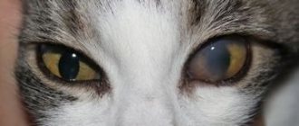
Leukoma in cats is a pathological condition characterized by the appearance of a scar on the cornea, the formation of which can be caused by various ophthalmological diseases.
What does leukoma indicate?
As the disease progresses, changes occur in the organ of vision. The animal begins to behave differently: hide from the light, look for secluded places.
The cat's tear production increases.
Consequences of ignoring the problem
If dangerous changes are not detected in a timely manner, the cataract will degenerate into adipose tissue. This condition can lead to complete atrophy of the eye. In addition, the disease causes a lot of inconvenience to the cat.
The resulting film provokes a decrease in visual acuity, especially when localized on the pupil itself.
Symptoms
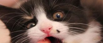
The first sign of keratitis is a cloudy red eye in a cat. Initially, redness and discharge of purulent or serous fluid appear. Then the cornea loses transparency, the eye becomes cloudy, and ulcers or tissue necrosis appear.
An accurate diagnosis is necessary to select the appropriate treatment. It is carried out using a special fluorescent liquid composition. After this, damage to the cornea becomes visible in the light. This type of diagnosis is carried out only in a veterinary clinic.
Types of pathology
An eyesore is classified according to several criteria.
By location type
Highlight:
- peripheral;
- total;
- central.
In the first case, the pupil is not affected; the spot is mainly localized on the side of the eyeball, has a white tint and black inclusions.
With the total type, the cataract completely covers the entire eye.
In the central form, the center of the visual organ is affected, and complete occlusion of the pupil is often noted.
According to the pathological process
Leukoma can be congenital or acquired . The first type of disease is rarely diagnosed and occurs against the background of negative changes in the cat’s body that occurred during the period of intrauterine development.
The second form is more common and can be caused by many factors.
How to help your pet?
Effective therapy is prescribed by a doctor after identifying the causes of the disease. It is very important not to delay visiting the clinic while the disease is at the inflammatory stage and scar tissue has begun to form.
- If keratitis is detected, the fluffy must be treated with drops or ointments (tetracycline or fluxal). The course is at least 14 days.
- To speed up the process of corneal restoration, keratoprotectors are used, which are placed in the cat's eyes 3 times during the day. Course – 14 days.
- If a cat has a spot on its eye due to injury, then the visual organ should be immediately rinsed. solution or miramistin, and then use tetracycline drops.
- In case of severe pain, in order to alleviate the condition of the mustache, a specialist injects a solution of novocaine in combination with hydrocortisone under the eyeball.
- In case of increased pressure inside the visual organ, pilocarpine or brinzolamide will most likely be prescribed.
- The cataract itself is removed through surgery, but only after the inflammation has subsided.
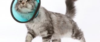
For the therapy to fulfill its purpose, follow all the veterinarian’s instructions. Also, provide the mustache with rest and monitor the condition of the eyes (if it worsens, consult a doctor). If your furbaby is constantly rubbing his eyes, you should put a veterinary collar on him.
Causes of cataracts
Various factors can contribute to the formation of a cloudy film and stain. The problem in kittens is identified in rare cases when the cat suffered from infectious diseases at the time of birth.
Among the main reasons that provoke a thorn are the following:
- dysfunction of the endocrine system;
- eye injury;
- failure to comply with hygiene rules;
- infections;
- unbalanced diet.
Causes of the disease
The main causes of blurred vision in a cat include:
- Diseases of the eye cornea. It loses its shine, becomes whitish, and a bluish tint appears.
- Diseases of the lens. Such pathologies are characterized by clouding of the pupil, the cornea remains transparent and is not affected. When the light is pointed at the organ of vision, the haze narrows. This confirms that the reason lies precisely in the pupil.
Below we will look at the origin of the loss of transparency of the animal's eye in more detail.
Diseases in which ocular leukoma develops
Veterinarians identify some pathologies that can lead to clouding and the appearance of a white spot on the visual organ.
Trachoma
This is a disease of infectious nature. In most cases, eye damage is accompanied by the formation of a cataract.
Inflammation on the cornea
One of the most common provoking factors of the disease.
If veterinary care is not provided in a timely manner, the risk of pupillary clouding increases several times. The superficial form is characterized by a weak degree of severity, and the thorn is almost invisible.
Preventive measures
It is easier to prevent any illness than to treat it later. Therefore, if the visual organ is injured, the mustache should be immediately taken to the hospital for examination.
At the same time, do not forget about scheduled vaccinations and regular preventive examinations of your pet.
Plan your cat's menu - it should include all the necessary macro and microelements, as well as vitamins. Protect your purr as much as possible from all kinds of chemicals and toxic substances.
If you suspect a disease of the visual organ, you should not use medications without first consulting a specialist. Also, do not use drugs intended for humans.
Symptoms of pathology
It is difficult to treat a thorn. It is necessary to pay attention to changes in a timely manner and show the cat to a specialist.
The pathological process is accompanied by the following symptoms:
increased lacrimation;- fear of light;
- swelling;
- redness of the cornea;
- discharge of purulent contents;
- retinal atrophy;
- development of strabismus.
Depending on the type of lesion, the clouding may have a light yellow or red-gray uneven tint. In the ulcerative form of the disease, the thorn protrudes outward.
In most cases, the animal’s behavior changes noticeably: the cat becomes more aggressive and irritable, is unsure of its orientation in space, bumping into objects that appear in its path, washes itself frequently,
Corneal dystrophies
Alexander Konstantinovsky, veterinarian, ophthalmologist, member of the board of directors of the European Society of Veterinary Ophthalmologists (ESVO) Yulia Nikolaeva, veterinarian, VetExpert clinic, Kazan Photos of Alexander Konstantinovsky are used in the article
Corneal dystrophies are hereditary diseases, most often characterized by symmetrical damage to both eyes. With dystrophies, various substances can be deposited in the cornea: lipids, cholesterol, calcium salts, etc. Clinically, dystrophy manifests itself as a local gray, white or metallic-tinged clouding on the cornea or in its thickness. The disease is more common in dogs, very rare in cats. Dystrophies usually debut between the ages of 4 months and 13 years; they are not associated with injuries to the eye and/or cornea, metabolism, or systemic diseases.
The cornea is part of the outer shell of the eye; it is normally transparent and does not contain blood vessels. The cornea in dogs and cats consists of four layers:
1) epithelium - surface protective layer. Consists of several rows of cells, quickly updates and regenerates. The main function is to nourish and maintain optimal moisture of the cornea;
2) corneal stroma - the most voluminous layer of the cornea, contains large quantities of collagen fibers. The upper layers of the stroma are very rich in nerve endings, therefore, with minor damage to the epithelium and irritation of these endings, the animal experiences severe pain and discomfort;
3) Descemet's membrane - separates the stroma from the endothelium. Has high elasticity;
4) corneal endothelium - the inner layer of cells that participates in nutrition and is responsible for the transparency of the cornea. The endothelium is a kind of “pump” that pumps out excess fluid from the cornea. Does not regenerate in dogs and cats. In cases of damage to the endothelium, fluid from the anterior chamber begins to leak into the stroma, causing the formation of vacuoles with fluid (bulls) in it.
Depending on the location, epithelial, stromal and endothelial dystrophies are distinguished.
Epithelial dystrophies
With this type of dystrophy, the upper layer of the cornea is damaged. Damage to the corneal epithelium can be primary (the epithelial cells are directly damaged) or secondary (the corneal epithelium is damaged due to changes occurring in the upper layers of the stroma). Clinically, these lesions manifest themselves as opacities, erosions and ulcers of the cornea. The animal experiences discomfort and corneal syndrome is observed (lacrimation, blepharospasm, photophobia). A distinctive feature of epithelial dystrophies is positive fluorescein staining.
Treatment is most often ineffective, but there are a number of proposed surgical and therapeutic techniques. In therapeutic treatment, proteolysis inhibitors, antibacterial drugs and keratoprotectors are used locally. Surgically, such animals can undergo superficial keratectomy. In the early postoperative period, this operation gives good results (the cornea becomes more transparent, there is no corneal syndrome), however, in long-term periods the frequency of relapses is high. The disease can appear as early as 4 months of age.
Affected breeds: Sheltie, Boxer, Pembroke Welsh Corgi, Boston Terrier.
Stromal dystrophies
This type of dystrophy is characterized by the deposition of lipids, cholesterol, calcium salts and other substances in the stroma. This condition does not cause any concern to the animal, usually progresses quite slowly, and vision suffers only slightly (although there are some exceptions).
| Photo 1. Stromal corneal dystrophy. |
Affected Breeds: Airedale Terrier, Dachshund, Afghan Hound, English Springer Spaniel, American Cocker Spaniel, German Shepherd, Basenji, Poodle, Beagle, Border Collie, Bichon Frize, Briard, Cavalier King Charles Spaniel, Golden Retriever, Irish Wolfhound, Labrador Retriever, Miniature Pinscher, Nova Scotia Retriever, Collie, Samoyed, Siberian Husky, Alaskan Malamute, Lhasa Apso, Mastiff, Pointer.
Among predisposed breeds, Airedale terriers have a specific course of stromal dystrophies. In this breed, corneal dystrophy is sex-linked and appears at the age of 4 months. The disease is characterized by rapid progression, leading to significant weakening of vision up to blindness. A similar “malignant” course of dystrophy can occur in Boston Terriers, Chihuahuas and Dachshunds.
This condition can be easily confused with another group of non-hereditary diseases - corneal degenerations. Corneal degenerations can be a consequence of systemic diseases in the animal (hypothyroidism) or some pre-existing corneal disease. In this case, there may be blood vessels at the site of deposition of lipids, cholesterol or metal salts.
Endothelial dystrophies
These dystrophies are characterized by damage to the inner layer of the cornea - the endothelium. This disease is similar to Fuchs' dystrophy described in humans.
| Photo 2. Endothelial dystrophy of the cornea. |
The pathogenesis is based on an increase in the permeability of the endothelium to fluid from the anterior chamber of the eye, which enters the corneal stroma and causes its edema (turbidity). Endothelial failure can lead to bullous keratitis and secondary corneal ulceration. Bullous keratitis is a serious disease that is difficult to treat. Treatment options are discussed below.
Drug treatment
Hyperosmotic drugs (ointments and drops of 3–5% NaCl or KCl) are used to reduce corneal edema. The use of these drugs can reduce corneal swelling (usually for a short time), but in some cases the use of these drugs can be effective for a long time (in these cases, hyperosmotic drugs are used for life). A more effective treatment option is the use of soft contact lenses.
Surgery
In veterinary practice the following are most often used:
• thermokeratotomy (penetrating keratoplasty);
• laser keratoplasty;
• conjunctival plastic surgery;
• penetrating keratoplasty.
In medical practice, high-tech, complex techniques are used to treat Fuchs' dystrophy, which in recent years have found their application in veterinary medicine. This:
• deep lamellar endothelial keratoplasty;
• endothelial keratoplasty with removal of Descemet's membrane (DSEK);
• Descemet's membrane grafting (DMEK).
Affected breeds: Boston Terrier, Chihuahua, Miniature Dachshund.
Conclusion
Corneal dystrophies are hereditary diseases, therefore it is not recommended to breed animals with epithelial, endothelial and “malignant” stromal dystrophies. It is not advisable to use animals with stromal damage (in the absence of visual damage) for breeding.
SVM No. 4/2013
Diagnostics: techniques and methods
During the initial examination, the veterinarian evaluates the clinical picture and conducts an external examination of the pet, especially the eye area.
The specialist also collects information about when the spot was first noticed, the quality of food, and studies the animal’s history.
Based on the data obtained, a diagnostic examination is prescribed, including the following manipulations:
- clinical and biochemical blood test;
- ophthalmoscopy;
- serological tests;
- determining the pressure in the eye (to assess how likely it is to develop glaucoma or cataracts);
- Seidel test (fluorescein test);
cytological examination of damaged tissue taken from the conjunctiva;- biopsy;
- microscopic examination of scraping;
- gonioscopy.
If for some reason it is not possible to conduct a standard diagnostic examination, the doctor may prescribe an ultrasound examination, which will help identify the true causes of clouding of the eyes.
If concomitant diseases of viral and infectious origin are suspected, additional diagnostic methods may be prescribed.
Diagnostics
If your pet is diagnosed with leukoma, the most correct decision would be to take it to a veterinary clinic, where a specialist will first determine the root of the problem. The veterinarian will work according to the following scheme:
- Examine the fluffy's fundus. Using a slit lamp, he will analyze the structure of the conjunctiva, as well as the condition of the lens, cornea and iris;
- Conduct a fluorescein test - an effective method that excludes or confirms damage to the epithelial layer of the visual organ. If the cause of the problem is mechanical damage or ulceration, then this is the most effective stage of diagnosis;
- Perform a Seidel test, which is necessary to identify penetrating damage to the cornea or fistulas;
- Perform tonometry (to determine intraocular pressure) and gonioscopy (to examine the anterior part of the visual organ);
- If there is a suspicion of viral diseases (hepatitis, immunodeficiency virus or plague), the specialist will prescribe a referral for diagnostic tests;
- If there is a separation from the visual organ, he will take smears to analyze the flora;
- Perhaps he will give you a referral for an eye ultrasound.
Basic principles of therapy
Treatment of cataract requires strict adherence to all the veterinarian’s instructions.
Medicines
Eye drops are used for 14 hours.
This could be Floxal, Tetracycline. In the morning and evening, it is necessary to apply a product with antibacterial properties to the lower eyelid. It is advisable to do the procedure at the same time.
When the provoking factor of the lesion is injury, the eye is first treated with an antiseptic (Miramistin), followed by the use of drops, for example, Levomycetin.
To relieve severe pain, the doctor advises injecting a special solution into the eyeball. For increased eye pressure, Brinzolamide and Pilocarpine are prescribed.
To obtain the desired effect, all medications must be used in combination.
Surgical intervention
If drug therapy does not help, surgery is performed.
During keratoplasty, the cornea is completely replaced. Tarsorrhaphy involves suturing the edges of the eyelid, which will protect the cornea and speed up recovery.
ethnoscience
Treatment of leukoma at home is possible only after a full examination and in combination with medications.
A honey solution is effective. To prepare it, you need to mix honey with water in a small amount and drop 3 drops into your eyes until complete recovery.
Treatment
Therapy at home in advanced cases usually does not bring positive results. But at an early stage, methods from traditional medicine are sometimes used, in particular, water with added honey is poured into the cat’s eye or powdered sugar is poured into it.
Since cat eyesores form on the eyes for various reasons, it is necessary to treat it, taking into account all the characteristics of the body. If it is caused by an infection, then honey will be powerless, and the situation of the sick cat will worsen. It is best to consult a doctor, but if this is not possible right away, you can use tetracycline ointment for treatment over a course of two weeks. It is worth remembering that it is not advisable to prescribe treatment for leukoma on your own and take your cat to a veterinarian as soon as possible.
If the cat is bruised or otherwise damaged the eye, it urgently needs to be treated with saline solution or miramistin, and then drops of tetracycline should be instilled.
Veterinarians, if the cataract is painful, inject the cat with hydrocortisone and novocaine into the area under the eye. In addition, the disease is fought with the help of cornegel (suitable for the treatment of corneal tissue), chloramphenicol, taufon, brinzolamide (for increased pressure inside the eye), gamavit.
So, drug treatment is aimed at:
- fight infection and inflammation;
- corneal healing;
- pain reduction;
- decreased pressure inside the eye.
It is not possible to completely cure an eyesore with medication; it can only be removed surgically. The appointment of an operation is possible when the course of drug treatment has been completely completed: inflammation has passed, there is no swelling, redness, the pressure inside the eye is normal and there is no discharge.
Preventing eye problems in cats
To prevent the development of the disease, it is important to adhere to a number of simple rules:
provide the animal with healthy, nutritious nutrition;- regularly examine your eyes;
- protect your pet from contact with other animals;
- do not let outside unattended;
- carry out vaccinations in a timely manner.
It is also necessary to regularly visit the veterinarian as a preventative measure, which will allow the disease to be identified at the beginning of its development.
Prevention
In order to prevent clouding of your cat’s eyes, you should follow a few simple rules for keeping your pet:
- Vaccinate the animal against infectious pathologies in a timely manner;
- undergo timely examinations by a specialist;
- Consult a doctor immediately after identifying unpleasant eye pathologies.
It should be borne in mind that if the cat already has inflammation of the organs of vision, the animal must be closely monitored. Your pet should be examined at least twice a day. If severe inflammation, redness, or clouding of the cornea appears, you should immediately go to the veterinarian. To prevent the condition from worsening, all stress factors for the animal should be kept to a minimum. Since they are the ones who can provoke a deterioration in the animal’s condition. The owner must also take into account the fact that from the room where the sick cat is located, it is necessary to remove interior items that the cat could hit and suffer.
It should also be understood that any eye pathology can lead to loss of vision in a cat, so you should not leave even the most harmless inflammation of the mucous membrane uncontrolled.
In addition, the owner must provide the sick animal with complete rest and, if possible, include foods with a high concentration of tocopherol and retinol in the cat’s diet. These vitamins have a positive effect on damaged epithelial layers of the cornea and restore the organs of vision.
Eye pathologies
The main pathologies of the eye that cause clouding of the lens or cornea. Of these, the following should be highlighted:
Keratitis
or the occurrence of inflammatory processes in the corneal tissues. In this case, the eye becomes cloudy only on the outside, while no dangerous changes occur inside. But why do inflammatory processes occur? Inflammation often occurs due to injury or infection in the visual organ. The described disorders are often the cause of the development of a disease such as keratitis. Any form of this disease causes significant harm to the health of the pet. This can be explained by the fact that the inflammatory process in the cornea in most cases leads to its death.
Glaucoma
With this disease, intraocular pressure increases, as a result of which the organ of visual perception increases in size. At the same time, the ability to see clearly is lost. If such a disease occurs, you can observe poor coordination of movements in the cat and dilated pupils. As for the cornea of the eye, it completely whitens and becomes less sensitive. Due to the sudden loss of vision, the animal runs the risk of bumping into various objects, which can lead to additional damage. The risk of negative consequences increases if the second eye is also infected. Due to such a serious illness, the tissues of the sensory organ harden, and the eye lens dislocates. It is also worth noting that in some cases this pathology is congenital and often occurs in an acute form.
Cataract
As this disease progresses, predominantly the central part of the eye, in the area of the pupil, becomes cloudy. One or both eyes can become infected with cataracts. As the disease progresses, the lens becomes less transparent, almost white. The disease can occur due to infection, as well as as a result of traumatic exposure or an inflammatory process.
Conjunctivitis
It is characterized by copious transparent discharge from the eyes, which in the most severe stages can become purulent. At the same time, the pet’s general health worsens, body temperature rises, redness and swelling of the eye occurs, and the pupil area often becomes cloudy. If eye pathologies occur, you must immediately contact a veterinarian with many years of experience. Based on the tests, it will help to correctly determine the severity of the disease and prescribe the necessary treatment to prevent negative consequences.
You might be interested in: Why does a cat not open one eye?
How to wash a cat's eyes
- To wash your eyes at home, you can use a solution of furatsilin or ordinary warm boiled water.
- To prevent lint from getting into your eye, it is better to use a cotton swab. You should never use cotton swabs.
- Cats usually don't like the procedure, so it's best to do it with a partner.
- To remove tear or purulent discharge, it will not be enough to simply wipe the eye: you will need instillation or rinsing with a generously moistened cotton pad.
- When instilling, you can use a regular pipette or syringe without a needle: the animal’s head is well fixed, the rinsing solution is injected into the upper outer edge of the eye.
- If your eyes become sour and crusts form in the corners, you need to soak them before washing them.
- The procedure is completed by wiping with a damp disc in the direction from the outer corner to the inner.
Causes
A hereditary breed predisposition to fatty degeneration of the cornea has been identified: Siberian huskies, beagles, American cocker spaniels, Cavalier King Charles spaniels, and German shepherds are at risk. Usually both eyes are affected, and the cause of lipid deposition in the cornea remains unknown.
The disease can also develop against the background of hypothyroidism and concomitant high cholesterol levels in the blood.
Inflammatory eye diseases (dry eye syndrome, uveitis, pannus, etc.) can lead to the development of lipoid corneal dystrophy.