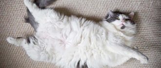
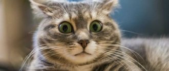
Most “novice cat owners” do not see anything suspicious in a small soft bump on the belly of a new pet. Breeders know that an umbilical hernia in a kitten is most often a factor of poor heredity. Associatively, the diagnosis of hernia implies surgical treatment, which is not entirely correct. Let's figure out what an umbilical hernia is and what the dangers of such a seemingly harmless pathology are.
Causes of umbilical hernia in kittens
The umbilical cord, which serves as a source of nutrition for the kitten during fetal development, is attached to the baby's stomach through the umbilical ring. During childbirth, the umbilical ring narrows, and when the umbilical cord is chewed, it closes completely. Ideally, everything happens exactly like this and the baby’s tummy looks completely flat as soon as the dried umbilical cord falls off.
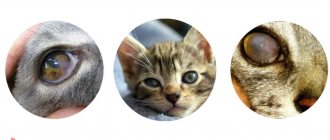
Hernia is a broad concept, implying displacement of an organ into a newly formed area. In the case of an umbilical hernia, a small hole is formed in the kitten’s peritoneum - a hernial orifice, into which the abdominal membrane is squeezed out, and with it part of an organ located in the cavity - a fragment of the intestine, uterus, bladder. The extent of the hernia depends on the muscle tone of the umbilical ring; as can be seen in the photo, the pathology can be insignificant or reach impressive sizes. The hernial orifice is a weakened muscle ring that must close, however, if penetration of the organ has already occurred, the reverse process may follow.
What to do?
Diagnostic measures
Sometimes it is advisable to examine an animal using an ultrasound method in order to accurately diagnose it.
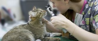
If the owner notices breathing problems in a cat or other undesirable symptoms, it is important not to delay a visit to the veterinary clinic. First of all, the veterinarian interviews the owner and clarifies how long ago the cat’s condition worsened. Then the cat is examined and x-rayed. A routine examination or a technique during which a contrast agent is used is prescribed. If part of the digestive organs is localized in the chest cavity, the preliminary diagnosis of diaphragmatic hernia is confirmed. To detect a diaphragmatic rupture, the cat is prescribed a peritoneogram. Sometimes an ultrasound examination is required.
How is the treatment carried out?
Only surgical intervention can help cope with diaphragmatic hernia in cats. The operation involves opening the chest by a veterinarian. After this, he returns the organs that have moved back to the peritoneum. Then the doctor sews up the diaphragm, but first resorts to plastic surgery. The surgeon places a special mesh on the muscle-tendon plate. In a situation where internal organs are strangulated, their resection is sometimes required.
If after the intervention the animal may develop a bacterial infection, then appropriate medications are prescribed.
12 hours before surgical treatment of a diaphragmatic hernia, the cat is prohibited from giving food. It is not recommended to give water to your pet 2-3 hours before surgery. In most situations, surgery is scheduled in the morning. By evening the animal can return home. The rehabilitation period takes about 14 days on average. After this time, the stitches are often removed. For 2 weeks, the cat's owner will need to give her complete rest and monitor the operated area. Sutures must be treated with medications prescribed by a veterinarian. Sometimes antibacterial treatment is prescribed to avoid infection of the operated area.
Preventive actions
To prevent the occurrence of a diaphragmatic hernia in a cat, veterinarians from the Pride clinic recommend that pet owners be attentive to changes in the cat’s condition. If any problems with digestion or bowel movements are detected, it is important not to delay a visit to the veterinary clinic. Timely diagnosis allows us to identify the possible presence of a congenital hernia and carry out appropriate treatment. If your pet is kept in an apartment on the upper floors, you should not let him sleep on the windowsill or walk along the ledge. Once again, it is not recommended to open the balcony and windows unless necessary. Such precautions are due to the fact that many situations have been recorded when cats, trying to jump onto the windowsill, did not calculate the distance, missed and fell.
Why is an umbilical hernia dangerous?
A small soft tubercle, most often, does not cause discomfort to the kitten, even if you stroke the baby’s belly. Whether complications arise is a matter of chance. It is worth noting that hernias are divided into irreducible and reducible. If you can press the bump with your finger and straighten the skin on the kitten's belly, then the hernia is most likely reducible.
As the animal grows, its internal organs naturally increase in size. This is where the first danger lurks. The umbilical ring, retaining its previous size, catches the prolapsed fragment of the organ, most often the entire intestine, which is fraught with consequences:
- Restless behavior due to abdominal pain.
- Loss of appetite and vomiting.
- An increase in base body temperature as the body’s reaction to the onset of the inflammatory process.
- Poor circulation of the intestinal fragment located in the hernial sac.
- Degenerative changes in intestinal tissue due to oxygen starvation.
- Tissue necrosis.
Important! Necrosis is a rapidly progressive, acute condition accompanied by intoxication of the body, sepsis and shock. Leads to the rapid death of the animal.
The pathology is called a strangulated hernia; keep in mind that once you notice the first symptoms, you should not hesitate or hesitate. If a fragment of the intestine is strangulated, the animal requires professional help, most often surgery.
Treatment method and prognosis
The main way to treat a hernia is surgery. In case of prolapse of organs and their infringement, this is generally the only way to save life. The displaced organs are returned to their place; if the tissue begins to die, they are excised, and the umbilical ring is sutured.
To improve healing and prevent the development of infection, the animal is prescribed drug therapy, including painkillers, anti-inflammatory drugs, and antibiotics.
With timely intervention, the prognosis is positive. There is a risk of re-formation of hernias, so animal care and preventive measures are of great importance.
Treatment of umbilical hernia in kittens
If you find an umbilical hernia, do not rely on luck and take your ward to the veterinarian. By palpation, the doctor will determine how mobile the hernial orifice is and determine what to do next. In the most positive case, the kitten’s hernia is repaired by tucking a fragment of intestine behind the abdominal wall. A coin is applied to the tubercle and secured with a bandage or a moderately tight bandage. To stimulate contraction of the umbilical ring, heating is used, which is carried out under the supervision of a veterinarian.
A more traditional, effective and guaranteed method is to remove an umbilical hernia in a kitten through surgery. Reduction is carried out under general anesthesia in several stages:
- Opening the hernial sac and analyzing the situation.
- If the fragment of the intestine or other organ is not damaged, it is set into the abdominal cavity, and the umbilical ring is narrowed or sutured.
- When necrosis begins, the animal is placed under general anesthesia. Dead tissue is removed, and healthy tissue is fused. The abdominal wall is sutured with a self-absorbing thread. After the procedure, the cat will need a bandage and rehabilitation therapy.
If necrosis was detected during the operation, the veterinarian will instruct you on how to treat the animal in the postoperative period; most often, antibiotics, immunostimulants and agents to maintain intestinal microflora are prescribed.
Note! After the operation, in order to avoid relapse, the kitten will be given an injection to stimulate the tone of the umbilical ring.
Veterinary clinic of Dr. Shubin
Description
Peritoneo-pericardial diaphragmatic hernia is a congenital defect in which in animals in the prenatal period a pathological connection of the abdominal cavity with the cavity of the heart sac (pericardial sac) is formed through a hole in the diaphragm (the membrane separating the thoracic and abdominal cavities). The pericardial sac fuses with the hole in the diaphragm, creating a kind of tunnel through which abdominal organs can move into the pericardium (the sac surrounding the heart) and back.
Clinical signs
In most animals, there are no signs of peritoneo-pericardial hernia; this occurs in cases where the abdominal organs slide freely from the abdominal cavity to the pericardium and back. In such animals, the hernia may be detected as an incidental finding during radiographic (x-ray) examination.
Some animals experience intermittent vomiting, decreased appetite, weight loss and diarrhea. These signs may be associated with movement and some compression of the stomach and intestines in the cardiac sac (pericardial sac). In another part of animals, when internal organs are displaced into the cardiac membrane, compression of the heart and lungs may occur, which is accompanied by shortness of breath and cough. Difficulty in breathing (dyspnea) is the most characteristic sign of peritorneo-pericardial hernia in cats; in dogs, signs of gastrointestinal dysfunction are more often observed.
Diagnostics
Peritoneo-pericardial diaphragmatic hernia is usually diagnosed by radiographic (x-ray) examination and identification of the stomach, intestines or liver in the cavity of the pericardial sac. In some cases, a radiographic examination with contrast is recommended; the animal is given barium and a series of photographs are taken to identify the abdominal organs in the cardiac sac. Cardiac ultrasound can help differentiate peritoneo-pericardial diaphragmatic hernia from cardiac enlargement due to heart failure (usually differentiated from hypertrophic cardiomyopathy in cats).
Treatment and prognosis
The only effective treatment for peritoneo-pericardial diaphragmatic hernia is surgical correction. Reconstructive surgery is indicated for all animals with signs that have developed due to displacement of organs into the cardiac membrane, as well as for all young animals, regardless of the symptoms that have developed. Older animals experience some intraoperative difficulties due to the fusion of prolapsed organs with the cardiac membrane, but if they have clinical manifestations, the best treatment method is still surgical correction. In older animals with asymptomatic peritoneopericardial hernia, periodic monitoring of the condition is acceptable, and surgical correction is carried out only if clinical signs appear.
The prognosis after surgical correction of a hernia is often favorable, complete recovery is noted, and the disease can no longer affect the animal’s life expectancy. In asymptomatic animals, the likelihood of developing clinical signs in the future has not been determined.
Veterinary clinic of Dr. Shubin, Balakovo.
How to prevent the formation of an umbilical hernia in kittens
General anesthesia is always a risky and globally harmful treatment measure. Naturally, preventing an umbilical hernia in a kitten is much safer than surgery. Even if a kitten has a hereditary tendency to form an umbilical hernia, you should not give up. First, you need to invite a veterinarian who will examine the newborn kittens. With minimal risks of pathology, babies are prescribed a tummy massage and gluing the umbilical cord with a bandage.
Note! At an early age, with proper care, spontaneous reduction of the umbilical hernia is possible.
Kittens are monitored; it is also worth monitoring babies who begin to play and try themselves in the role of adult cats - excessive tension in the abdominal wall can provoke the opening of the umbilical ring. If preventive measures do not help, it is recommended to perform surgical reduction at an early age, which significantly reduces the risk of necrosis.
Which breeds are more susceptible
There are no differences between purebred and outbred cats and dogs. Both the highest-bred offspring of multiple prize-winners and “nobles” picked on the street can easily get the defect. Since the defect is caused by genetic pathologies, it can be found in kittens and puppies of any breed.
Main causes of pathology
There can be many reasons, from an infectious disease to a congenital malformation. We will briefly look at the main ones:
- As we have already said, a diaphragmatic hernia is most often a congenital disease . One can only guess about its reasons. This can happen both due to some genetic anomalies in the organisms of the parent individuals, and under the influence of toxic/infectious factors directly on the pregnant cat.
- Inflammation of the pericardium of the effusion type can be caused by a wide range of different causes. The pathology may be due to the presence of the same diaphragmatic hernia, the effusion accumulates due to right-sided heart failure, and the possibility of developing cysts cannot be ruled out . Fluid leaks into the pericardium during hypoalbuminemia (when there is little albumin in the blood), which often happens with various infectious diseases. In particular, pericarditis in cats may well be a consequence of canine distemper . Bleeding in the pericardium can occur due to tumors, trauma, or bleeding disorders . Sometimes the reasons remain unidentified (idiosyncrasy).
- Pericardial stenosis most often develops as a result of its inflammation, which often happens in infectious diseases; cases of stenosis that appeared against the background of oncological pathologies have also been described.
Classification of species
Umbilical hernia varies in severity and can be reducible or irreducible. The first type is characterized by the absence of pain and easily returns to the peritoneum with slight pressure. If the kitten is turned onto its back, such a hernia will retract on its own, and the belly will become flat.
An irreducible tubercle causes pain and discomfort. The formation is hot, if touched, almost motionless. Due to the inflammatory process inside, it may gradually increase in size.
Reference! A reducible hernia can develop into a second type of hernia due to the onset of inflammation inside the formation. Additionally, adhesions and pinching may form.
Symptoms
Externally, the disease manifests itself as a lump in the navel area. The pain depends on the degree of development of the pathology: in the early stages the swelling is painless, noticeable only when the baby walks, and disappears during sleep.
Strong seal
Other symptoms appear if organs trapped in the hernial sac are pinched: pain, loss of appetite, and possible vomiting. Later, signs of inflammation appear: increased temperature, problems with peristalsis and stool, necrosis of tissue around the navel.
You should know that there are two types of hernias: reducible and unreducible. You can determine the type yourself: you need to carefully press in the seal. If the skin straightens easily, then conservative treatment is possible. Reducible hernias are usually painless and are not noticeable when the baby lies on his back. But we must remember that a mild form of this pathology can progress quite quickly to the point where conservative therapy becomes impossible.
Sometimes prolapse and pinching of internal organs does not occur even in the case of a fairly large hernial sac. But this is no reason to calm down: the pathological process can progress, and the increasing compaction, if it does not pinch the intestines, will constantly put pressure on it and other organs, preventing their normal functioning.
In any case, treatment and diagnosis should be carried out by a specialist.