When a pet has health problems, the first thing the cat is sent for tests, including a urine test. This study is carried out without fail; this analysis is required if kidney pathologies are suspected. Urine collection can be carried out in a veterinary clinic by a veterinarian, or the owner can independently collect urine from the pet. In the future, it will need to be taken for laboratory analysis. In this article we will introduce you in more detail to the indicators that are present in a urine test.
Color: normal from rich yellow to orange or even light brown
Shades of yellow and orange are given to urine by the end products of the biotransformation of hemoglobin and other blood pigments: urobilin, urochrome A and B, urorosein, etc. Urine can become brownish as a result of the oxidation of these urinary pigments during long-term storage. The color of urine largely depends on its density. Urine can become colorless (for cats this is almost always a pathology) due to diabetes mellitus and diabetes insipidus, heavy drinking, impaired renal concentration function (including CKD) and the use of loop and osmotic diuretics. Urine and urinary foam acquire a saffron-yellow and greenish-brown color with an increased concentration of conjugated bilirubin in it. Red color of urine is observed with gross hematuria, the cause of which may be acute and subacute (malignant or rapidly progressive) glomerulonephritis, babesiosis, venereal sarcoma, autoimmune hemolytic anemia, urolithiasis, neoplasms of the bladder and urethra. Urine acquires the color of meat slop with a suspension of dirty brown flakes in acute pyelonephritis, hemorrhagic and acute urocystitis. With urinary tract infections caused by Pseudomonas aeruginosa, the urine usually turns a dirty green color. Rifampicin (contained in the animal drug Dorin) turns urine brick-colored, and the endogenous interferon inducer Cycloferon, widely used in domestic veterinary medicine, and its chemical formula Anandin, which is similar to it, give urine an opalescent bluish-violet or amethyst hue.
Symptoms of pathology
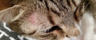
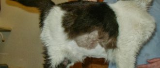
Signs of infection of the body depend on the location of the bacterium, and the severity of the disease is determined by its type and the state of the body’s defenses. And yet, the presence of staphylococcus can be suspected by the presence of high body temperature, frequent headaches, weakness and general exhaustion in the patient. On the skin, the bacterium causes the development of rashes, acne and eczema. In the gastrointestinal tract it causes dyspepsia, diarrhea and nausea, and in the urinary system it causes cutting pain when urinating and itching in the genital area. They often provoke the development of sore throats and tonsillitis in children.
Transparency: normal clear to cloudy
In healthy animals, urine should be clear immediately after collection. Its slight clouding when viewed in transmitted light is possible due to the presence of a small amount of epithelial cells, hyaline casts or mucus. At the same time, the presence of a large amount of salts in the urine of cats normally causes it to become cloudy during storage and transportation, especially at low temperatures. If cloudiness of urine is associated with aggregation of urates and uric acid, then heating it to 60–70 ° C, as a rule, leads to clearing. If the turbidity is associated with the presence of a large number of phosphates, then the same effect can be achieved by adding 10% acetic acid to the urine, and if oxalates, then hydrochloric acid. If the urine remains cloudy after all of the above measures, this may indicate bacteriuria. Pathological turbidity of urine usually occurs with leukocyturia and/or bacteriuria (urocystitis, pyelonephritis, pyonephrosis), hematuria (urinary and nephrolithiasis, acute pyelonephritis and glomerulonephritis, malignant neoplasms of the kidneys, bladder and urethra, prostatitis, babesiosis, hemolytic anemia), severe proteinuria (chronic pyelonephritis and glomerulonephritis, amyloidosis, diabetic nephropathy) and a large amount of epithelium in the urine (pyelonephritis, urocystitis).
Specific gravity: norm 1030–1035 (1085) (depending on the volume of liquid consumed)
An indicator assessing the ability of the kidneys to concentrate urine. It depends on the amount of substances dissolved in it: urea, uric acid, creatinine and salts. Glucosuria, leukocyturia, proteinuria and bacteriuria, as well as the administration of large doses of drugs that have preferential elimination through the kidneys, can increase urine density, and the use of loop and to some extent osmotic diuretics significantly reduces this indicator (other diuretics have little effect on urine density, since they affect areas of the tubules and/or collecting ducts, the reabsorption capabilities of which are insignificant). However, it must be taken into account that with many glomerulonephritis, for example, proteinuria, on the one hand, increases the density of urine, and the pronounced tubular component, which is an integral part of it, reduces it. Hyposthenuria (decreased urine density) is also a consequence of diabetes insipidus (deficiency of antidiuretic hormone), chronic kidney disease (due to dystrophy and atrophy of tubular epithelial cells and a decrease in the amount of aquaporin protein in the ascending segment of the loop of Henle), heavy drinking or infusion therapy. In dogs, hyposthenuria or hypersthenuria can be established only if 2–4 portions of urine have been examined over several days. In cats, 1–2 samples are usually sufficient for this.
Treatment
Proteinuria is most often treated on an outpatient basis. Therapy directly depends on the disease that caused the appearance of proteins in the urine. The most common cause of protein in the urine is kidney pathology. If this is a functional disease, the cat may be prescribed ACE inhibitors to eliminate renal failure: Benazepril, Imidapril, Lisinopril, Ramipril.
Preparations containing fatty acids ALA, EPA and DHA (omega-3 group) help improve the condition of kidney blood vessels. These unsaturated acids are taken for a long time, and it is recommended to give them to older animals constantly. For inflammatory processes in the kidneys or urinary tract (pyelonephritis, cystitis, urethritis), antibiotics of the penicillin or cephalosporin group (Penicillin, Carbenicillin, Amoxicillin, Cefepime, Cefotaxime), as well as sulfonamides (Sulfene, Sulfadimethoxine), are prescribed.
Antibiotic therapy with tetracycline drugs is used if a cat is diagnosed with ehrlichiosis, an acute infectious disease transmitted by ticks. If it is determined that the cat suffers from hypertension, he is prescribed a course of treatment with antihypertensive drugs (Losartan or Telmisartan) and/or potassium-sparing diuretics (for example, Spironolactone). As an additional means of treatment and prevention, a diet limited in fat and salt is used.
For anemia not associated with blood loss (hemolytic, hypoplastic or nutritional), the animal is prescribed drugs that increase hemoglobin. These are preparations of iron, copper, cobalt, as well as B vitamins. Often nutritional anemia with a decrease in the level of red blood cells and hemoglobin in the blood is observed in young cats and kittens due to improperly organized nutrition or impaired absorption of iron by the body.
In such cases, the veterinarian will recommend introducing a product such as animal liver into the cat’s diet. The intensity of proteinuria, even if it is caused by severe pathology, can be effectively reduced by limiting protein-rich foods in the cat's menu and increasing the amount of Omega-3 and Omega-6 fatty acids in it.
The state of the animal’s immune system is also important. To increase its resistance, a course of immunomodulators is recommended for a cat who has recovered from proteinuria; veterinarians usually prescribe Gamapren, Gamavit, Vetozal or Immunovet.
Sources:
https://kinpet.ru/belok-v-moche-u-koshek-prichiny-i-lechenie/
https://petshoptop.ru/ru/pet/cats/health-nutrition/povyshennoe-soderzhanie-belka-v-moche-koshek-333
https://usatiki.ru/belok-v-moche-u-koshek-prichiny-i-lechenie/#i-5
Urine microalbumin, mg/l: normal 0–30 Protein, mg/dl: normal - negative.
Urine containing a large amount of protein foams heavily, and the foam persists for a long time. Normally, during high physical activity, hypothermia, allergic reactions and some other conditions, a larger amount of albumin may be present in the urine (functional hyperalbuminuria). In addition, proteins produced by tubular epithelial cells can also be detected in the urine. The cause of persistent hyperalbuminuria requires clarification (especially with concomitant persistent hyposthenuria), since this condition usually serves as one of the first markers of the development of severe nephropathies (diabetic nephropathy, primary chronic glomerulo- and tubulointerstitial pathies). Globulinuria (proteinuria) is always a pathology and can be associated with both infectious and non-infectious diseases of the kidneys and urinary tract.
Bilirubin, mg/dl: normal - negative.
In cats, bilirubin is not conjugated in the renal tubules. Therefore, the detection of bilirubinuria in cats (even in a mild and/or transient form) requires clarification of the causes of its occurrence. Bilirubinuria occurs in many liver pathologies. With mechanical (prehepatic) jaundice, bile with the bilirubin it contains enters the intestinal lumen in small quantities. For this reason, the bulk of bilirubin remains in the blood and is excreted from the body in urine, which gives it shades of color ranging from saffron-greenish to greenish-brown. At the same time, the content of urobilinogen and urobilin in the urine is significantly reduced, or these substances are not detected in it at all. In hemolytic jaundice caused by intensive breakdown of red blood cells (including babesiosis), urine, on the contrary, contains large amounts of urobilinogen and stercobilinogen, and bilirubin is usually absent in it.
Urobilinogen, mg/dl: normal - Traces
A colorless substance, a product of bilirubin reduction, formed in the intestines, including. and under the influence of bacteria. Urobilinogen is formed from direct bilirubin that enters the intestine as part of bile and is partially absorbed back into the blood. Oxidation of urobilinogen in urine leads to the formation of urobilin. This reaction causes urine to darken during storage. With hemolytic jaundice, toxic and inflammatory liver lesions, enteritis and constipation, the level of urobilinogen increases significantly. When the bile duct is blocked (obstructive jaundice), urobilinogen is usually absent in the urine.
Glucose, mg/dl: normal - negative.
Any level of glucosuria requires clarification and further examination of the patient, since it can be one of the symptoms not only of diabetes mellitus or Cushing's syndrome, but also of TIN and CGN with a tubular component. Normally, the tubular epithelium completely reabsorbs glucose from primary urine, and glucosuria not associated with diabetes mellitus indicates its severe damage. Glucosuria can be transient in nature during pregnancy and excessive feeding of various sweets to dogs, and especially cats, whose insulin reserves are small.
Red blood cells (reaction to hemoglobin), units/µl: normal - negative.
Microhemoglobinuria is characterized by the content of red blood cells in the urine up to 100 cells per field of view and is not reflected in its color. The cause of urine staining in a red-brown color of varying intensity - macrohematuria - may be hemoglobinuria (if the pathological process that led to it is sufficiently severe, the blood plasma also turns red, clinical symptoms of oxygen deficiency increase, but red blood cells are not detected in the urine sediment), erythrocyturia (the blood plasma remains straw-yellow, and fresh or leached (dysmorphic) red blood cells are found in the urine sediment), as well as myoglobinuria, the cause of which is most often rhabdomyolysis. During the day, 2–3% of red blood cells are destroyed in the body. Even if for some reason parts of the hemoglobin protein and its compounds enter the primary urine, they are completely metabolized and reabsorbed. This oxygen-consuming and highly energy-intensive function is one of the priorities for tubular epithelial cells and, when its physiological capabilities for its implementation are exceeded, it begins to be carried out even to the detriment of its own interests of metabolism and energy. Therefore, any conditions characterized by massive, and especially prolonged, breakdown of erythrocytes (blood parasitic infections, transfusion of incompatible blood, extensive hematomas), as well as muscle fibers during rhabdomyolysis, for example, can result in degeneration and atrophy of tubular epithelial cells, which, in turn, turn, can lead to TIN. In this regard, it is nephropathy, and not liver damage, as is usually believed (hepatocytes, unlike tubular epithelial cells, are capable of intensive regeneration after the disappearance of the damaging factor), are a very significant complication, for example, of babesiosis, and conditions characterized hemoglobinuria or hematuria, necessarily require clarification of its occurrence and, if necessary, subsequent nephroprotective treatment. In addition, hematuria is characterized by many kidney lesions of an autoimmune (most GN) and toxic nature, as well as infectious and oncological diseases of the urinary system and urolithiasis. The presence of acanthocytes (red blood cells with uneven contours, resembling a maple leaf) in urine sediment is a highly specific sign in determining glomerular hematuria. And the presence in the urine, in addition to acanthocytes, also agranulocytes, as well as leukocyte and granular casts, gives good reason to give the patient a clinical diagnosis - chronic GN. Hypocoagulable hematuria is also identified, which is not associated with diseases of the MVS organs.
How to properly collect urine for analysis?
Failure to follow the basic rules for collecting analysis can result in unreliable results. Cocci and other bacteria can get into the urine from a poorly washed potty or from an unsterile container in which the owner tests the cat. Collect urine for research as follows:
- Prepare the animal's tray - wash it thoroughly without using chemicals and dry it dry.
- Avoid using sand and other types of cat litter.
- Wait until the kitten goes to the toilet and carefully pour the urine into a sterile container: a special container or syringe with a volume of at least 20 ml.
- Deliver the test to the laboratory within 2 hours after collection. Long-term storage of the sample leads to distortion of the indicators.
The resulting sample must be decrypted immediately. It is impossible to independently suspect signs of illness from a urine test in a cat. The only obvious factor is color. Normally it should be yellow. All other indicators are determined by microscopic examination.
What to do if a laboratory test reveals staphylococcus in the urine? This is perhaps the most common microbe that poses difficulties in treatment, as it quickly develops resistance to many antimicrobial medications. The pathogenic microorganism, which is staphylococcus, enters the body in two ways - through the urethra or through the blood from infected foci. If treatment is untimely, it actively multiplies and the patient begins to observe the corresponding symptoms.
pH: normal - acidic
In cats, as obligate carnivores, urine is normally acidic. The predominance of plant components in the diet of dogs can change the pH of urine in the alkaline direction. The pH of urine is also affected by the level of acid-base balance in the blood, since the kidneys actively participate in its maintenance and remove excess hydrogen ions through the filtration process. Pathological causes of urine acidification (pH<5): respiratory or metabolic acidosis, hypokalemia, profuse diarrhea, anorexia, diabetes mellitus, prolonged hyperthermia. Long-term use of aspirin and methionine can also lead to urine acidification. Pathological causes of alkalinization of urine (pH>7): severe chronic renal failure, when the kidneys are no longer able to remove excess hydrogen ions from the blood (tubular acidosis), hyperkalemia, compensated or uncompensated hyperphosphatemia, hyperplasia of the parathyroid gland (hyperparathyroidism), prolonged vomiting, cancer kidneys or bladder.
Staphylococcus and its varieties
Types of infection detection:
- a general or detailed blood test can detect inflammation in the body;
- delivery of biomaterial – sputum, feces, secretions;
- general urine analysis.
Indirect indicators of infection of the genitourinary system are excess protein and leukocyte standards.
To accurately identify the pathogen, the microflora and its sensitivity to antibiotics are examined in urine culture.
A high frequency of detection of staphylococcal infections in women who lead an active intimate life. Infection develops cystitis - inflammation of the bladder. With timely treatment, further spread of bacteria through the genitourinary system and kidney disease can be avoided.
Nitrites: normal - negative.
The appearance of nitrites in urine is possible under the influence of enzymes of certain bacteria that enter it both from the kidneys and urinary tract, and when it is contaminated from the outside. Therefore, nitrituria in MJ is only indirect evidence of infectious diseases of MW. On the other hand, not all uropathogens are capable of fermenting nitrites from nitrates. Therefore, if nitrites are not detected even in long-term stored urine samples, this does not necessarily exclude a urinary tract infection.
Diagnostics
If staphylococcus is suspected, the veterinarian will prescribe a series of tests. The symptoms of this infection resemble those of many other diseases. Therefore, for differential diagnosis it is necessary to conduct the following studies:
- blood test for bacterial culture;
- microscopy of scrapings from the affected area of skin;
- smears of discharge from the genital tract.
A general blood test for staphylococcal infection is not informative, since pronounced changes are noted only at the stage of sepsis.
Leukocytes, units/μl: normal - negative.
Leukocyturia, especially weakly or moderately expressed, does not always (primarily in cats) indicate an infectious disease of the urinary system, although it is the most common reason for the unjustified prescription of antibiotic therapy. Most chronic GN and CKD are characterized by intense focal and/or diffuse infiltration of the renal parenchyma by agranulocytes, which are involved either in autoimmune inflammation or in the processes of destruction of sclerotic areas of the parenchyma. On the other hand, infectious (septic) leukocyturia is a characteristic sign of acute pyelonephritis. However, during remission or during a latent course of chronic pyelonephritis, it may be absent. Therefore, in most cases, an infectious disease of the urinary system is recognized only if a urine sample correctly obtained by transperitoneal urocystocyntesis during bacteriological examination reveals a microflora that is pathogenic and not saprophytic for the skin or gastrointestinal tract. However, when assessing negative results of bacterial culture (urine is sterile), one should take into account the intermittent (variable) nature of bacteriuria in chronic pyelonephritis (this is not observed in pyonephrosis), as well as difficulties in identifying certain forms of pathogenic bacteria.
How to get tested for staphylococcus in urine
To get more reliable results, you need to follow some rules when collecting it.
It is better to collect material for research in the morning and on an empty stomach. Women are not recommended to perform the procedure during menstruation. It is customary to choose either the previous period or 2-3 days later. You should also stop taking diuretics 2 days before.
Before carrying out collection manipulations, it is necessary to prepare.
- Touching only with clean hands, prepare a glass jar scalded with boiling water or buy a sterile container at the pharmacy.
- Gently wash with boiled warm water (better without using soap or gel), wipe off the moisture with a clean napkin.
- Select the middle portion of urine, releasing the first and last into the toilet.
- It is advisable to cover the vaginal opening with a sterile swab at the time of collection.
It is best to provide urine in a laboratory setting using a securely sterilized (preferably disposable) catheter.