A type of mechanical injury in domestic cats and dogs is a bruise. The damage is characterized by a violation of the integrity of blood and lymph vessels, as a result of which subcutaneous hemorrhage develops, but the skin remains, in the vast majority of cases, completely untouched.
The location of the bruise swells, the animal clearly shows anxiety, feeling discomfort and pain. The bruised limb is not used by the animal; the cat avoids stepping on the paw, heals and licks it.
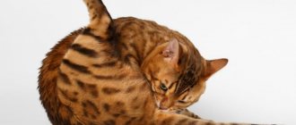
A feature of domestic cats is increased mobility and curiosity, especially in small kittens. Kids always play actively, performing mind-blowing stunts that often end in mild to moderate injuries. In some cases, the injury can be caused by another cat or cat, as well as a dog.
The situation is much more serious if the pet has free access to the street. In this case, the risk of serious injury to the cat increases. A caring cat owner should know how to distinguish a normal bruise of a pet's body part from more serious injuries. What symptoms are typical for bruises of the limbs and body, and also in what cases it is necessary to contact a veterinary clinic for help as soon as possible.
Various fractures and serious dislocations are dangerous for the animal and cannot be avoided without the intervention of a specialist.
Causes of hematoma
The main cause of this type of injury is mechanical damage. It could be a fall from a height, a fight, an accident.
Hematomas occur with bruises, fractures, closed head injuries, as well as with improperly closed postoperative wounds.
It is also worth considering the causes of auricular hematoma, which is most common in cats.
- Ear mite
- Fleas.
- Fungal infections.
- Allergic reactions.
- Insect bites.
The skin of the auricle in cats is very thin and has an extensive capillary network; even minor damage can lead to bleeding and the formation of a large hematoma.
With all of the above diseases, there is severe itching, which forces pets to scratch their ears, damaging the blood vessels.
Platelet dysfunction
Platelet dysfunction can occur when platelet numbers decrease or do not function properly. The number of platelets becomes smaller when there is insufficient production of them in the bone marrow, or when they are destroyed. Platelet dysfunction can be either hereditary and congenital, or acquired.
When your cat has bruises or bleeding and you, not knowing what to do, are looking for advice on this topic on the Internet on forums, we recommend not to self-medicate or experiment on your beloved cat. The fact is that there are many reasons for hematomas and bleeding in an animal, and the consequences of your experiment may disappoint you and your family.
The following disorders cause a decrease in platelet count:
- Medicines that poison the bone marrow.
- Bone marrow diseases caused by bacteria, viruses or rickettsia (microorganisms that carry the characteristics of bacteria and viruses, for example ehrlichiosis)
- Immune destruction of bone marrow
- Bone marrow cancer
- Myelofibrosis or myelophthisis, which causes scarring and loss of bone marrow cells.
- Excessive production of estrogen in the bone marrow
The following disorders cause active removal of platelets from the circulatory system:
- Ehrlichiosis (tick-borne disease)
- Vasculitis (inflammation of the walls of blood vessels)
- Parasites
- Disorders of the spleen
Factors that affect platelet function:
- Congenital disorders of platelet function
- Some medications
- Infections
- Kidney failure
- Liver failure
- Some types of leukemia
Vascular diseases often lead to bleeding because
The walls of blood vessels weaken. High blood pressure can also cause bleeding. Disorders that cause blood vessel fragility:
- Vasculitis – inflammation of blood vessels
- Hyperadrenocorticism is a syndrome in which the adrenal glands produce too much cortisone.
- Diabetes
- Uremia – poisoning of the body as a result of kidney failure
- Hereditary factors leading to hemophilia
- Warfarin poisoning (rodent poison)
- Excess vitamin K
- Liver diseases
- Disseminated intravascular coagulation (DIC) - impaired blood clotting
- Von Willebrand's disease (hereditary disease) – spontaneous bleeding
Types of hematomas
Hematomas are divided according to their location and the nature of the bruise itself.
By location there are:
- Subcutaneous - can be localized anywhere, most often occurs as a result of injury and is visible on the surface of the body.
- Retroperitoneal - formed in the retroperitoneal space with abdominal trauma and fractures of the pelvic bones, spine or ruptures of internal organs.
- Intermuscular - forms between the fascia of the muscles, often leading to extensive abscesses.
- Intraorgan - occurs when an organ is damaged by an impact, and may be accompanied by its rupture. The most difficult to diagnose without the use of special equipment.
- Intracranial - has the most serious consequences, accompanied by symptoms of damage to brain tissue.
Also distinguished along the stream:
- Arterial - appear when arteries rupture, have a bright red color.
- Venous - obtained when veins are ruptured or damaged, have a blue-violet color, and are hard to the touch.
- Mixed - the most common types of hematomas, are formed when arterial and venous blood flows.
- Pulsating - with this type, the hematoma is compressed by the vessel and, as a result, the flow of blood is prevented. It can only be treated surgically due to the risk of arterial bleeding.
Stages of development
Veterinary specialists note the following stages of the pathological process in the development of hemorrhages in the area of the outer ear in animals.
| Hematoma stage | Period | Short description |
| Stage 1 | On the first day | At this stage of the disease, blood from damaged blood vessels fills the interstitial space. Red blood cells are destroyed and hemoglobin is oxidized. This leads to a change in the color of the blood - it turns blue. The animal has a red or purplish-bluish swelling and mild pain when touched. The degree of pain reaction largely depends on the size of the damage. As a rule, the first stage of hematoma develops within 24 hours after mechanical damage to the soft tissues of the organ. |
| Stage 2 | 2 - 3 days after injury | Along the edges of the hematoma, color changes to a yellowish-blue tint are noticeable. Yellow spots can be observed across the entire surface of damaged tissue. There is moderate pain and swelling in the area of injury. |
| Stage 3 | After 4 - 5 days | The swelling on the skin decreases, there is practically no pain. If the hemorrhage was extensive, then the swelling “slides” to the base of the ear. The swelling becomes greenish in color. |
Such stages are typical for hemorrhages uncomplicated by infection. In case of extensive release of blood from the vessels, failure to provide assistance to the animal in a timely manner at any stage of the disease, an inflammatory process, infection and suppuration of the injury may develop.
The formation of a hematoma begins with damage to blood vessels without compromising the integrity of the skin. As a result, the flowing blood begins to stretch the surrounding tissues due to pressure. This process occurs until the pressure inside the vessel is balanced by the resistance of the stretched tissues.
Small hematomas resolve on their own, but large ones without treatment form a subcutaneous blood clot, which is later replaced by connective tissue. Further, calcium salts may be deposited in this area, causing a cyst to form. When infection penetrates, an abscess or phlegmon forms at the site of the hematoma.
READ Symptoms and treatment of vitamin deficiency in cats
Symptoms
Superficial hematomas are quite easy to detect in your pet. Due to the covering with fur, its color is rarely visible, but often subcutaneous hemorrhage is accompanied by significant edema or swelling, which can be either soft or hard to the touch.
A hematoma of the auricle is characterized by the following symptoms:
- The swelling quickly grows along the auricle and has a soft consistency.
- Painful to the touch.
- The cat is restless, shakes its head and tilts it towards the damaged organ.
- Often red in color and hot to the touch.
In cases of stagnation, the hematoma may take on a bluish tint, fester, and there may also be an increase in overall body temperature.
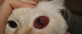
If an eye injury is accompanied by a hematoma, then it is characterized by bleeding inside the eye. The eye becomes bright red and watery.
With bruises of internal organs, characteristic signs do not appear immediately, but as the damage increases. Accompanied by characteristic symptoms of damage depending on the location.
Symptoms of Cranial Hematoma
This type of injury requires resuscitation measures. As a result of the injury, damage to brain tissue occurs, which is accompanied by swelling and contusion.
The most common symptoms of such injury are neurological:
- Oppression, blindness (most often one-sided), behavioral disturbances.
- Possible impairment of consciousness up to coma, impaired breathing and motor functions (paresis, paralysis, hemiplegia).
- Changes in gait (walking in a circle), drooping of the head in the direction of the lesion, stiffness (tension) of the muscles of the back of the head
How to call a veterinarian at home?
What questions will need to be answered? In order to call a veterinarian, you need to:
- Call the operator at the numbers specified in the Contacts section;
- Tell what happened to the animal;
- Provide the address (street, house, front door, floor) where the veterinarian will arrive;
- Specify the date and time of the doctor’s arrival
Call a veterinarian at home and he will definitely help you. At home, as they say, walls can be healed.
Any mechanical damage, regardless of the cause, and a bruised paw in a kitten, is stressful for the animal and often causes serious problems later. In some cases, even a slight bruise of the first and second degrees does not go away without a trace for the cat.
If a kitten or adult cat has injured its paw and is limping, then a timely visit to the YA-VET veterinary emergency center will help deal with the injury as quickly as possible without consequences and expensive treatment. Correct diagnosis and qualified assistance from veterinarians who have extensive experience working with cats of any age guarantee that the injury will go away as soon as possible, and the pet will enjoy excellent health and the same activity!
Call us and find out how much emergency veterinary care costs in order to order a visit from specialists without overpayments!
If you find an error, please highlight a piece of text and press Ctrl Enter.
Diagnosis of hematoma
Hematomas are often confused with tumors, allergic diseases and dermatitis.
Usually the veterinarian asks the owner about the root cause of the disease; if it was an injury, then in most cases a hematoma is diagnosed.
It is also necessary to conduct an ultrasound of the internal organs and a plain X-ray of the bones, especially if there is a history of a fall from a height or a car accident.
If the tumor is located on a limb, it is also necessary to take an x-ray to examine the integrity of the bone.
For a closed head injury, an MRI or CT scan of the head, as well as an X-ray of the chest and spine, must be performed. It is also necessary to undergo a cerebrospinal fluid puncture, ECG, ECHO of the brain, general clinical tests, fundus examination and neurological monitoring.
How to treat injuries?
The treatment regimen for the cat is determined by the veterinarian; self-medication is prohibited. Most severe injuries are treated with surgery using osteosynthesis instruments, blood transfusion, and thoracentesis are performed. The broken limb is fixed with a splint. Depending on the type of injury, antibiotics, sedatives, painkillers, and antiemetics are prescribed. If the musculoskeletal system is damaged, after treatment, physiotherapy and rehabilitation exercises are prescribed. If the cat does not walk, a wheelchair is required.
Treatment of hematoma
- If your cat is injured, then even in the pre-hospital stage, apply ice to the injury; it should be wrapped in plastic and a clean, dry cloth. If possible, give an injection of painkiller - analgin or ketofen.
- For small hematomas, conservative therapy is used. The bruise site can be warmed with paraffin applications, resorbing ointments - ichthyol, turpentine.
- With extensive effusion, treatment of hematomas requires surgical intervention. To do this, a puncture is performed, followed by evacuation of the contents, rinsing the cavity and applying an antibiotic ointment (mainly levomekol).
- In case of hematomas of the auricle, its opening is mandatory, since a long course of the disease leads to the formation of scar tissue and a change in the shape of the ear. The cavity is cleaned of fibrin and blood clot, then a fixing suture is applied.
- If there is a tick-borne or fungal infection of the ear, it is necessary to carry out etiological (antiparasitic or antimycotic) therapy along with the treatment of the hematoma.
- If the hematoma is pulsating, then during the operation a ligature is applied to the damaged artery and the wound is sutured.
- A cranial hematoma requires complex treatment with resuscitation measures and medical supervision.
As a wound healing therapy after puncture, you can use ointments - levomekol or synthomycin. If a purulent complication is suspected, bandages with Vishnevsky ointment are applied.
First aid: what to do?
Any injury threatens the life of the animal, so the owner must urgently take the pet to the veterinary clinic. Depending on which part of the body is injured, the owner’s pre-medical actions should be as follows:
Before visiting the doctor, the pet owner can use a cold compress.
- Provide your pet with complete rest.
- Apply a cold compress to the injured part of the body.
- Treat the wound with hydrogen peroxide, being careful not to touch bone fragments in open injuries.
- Place the cat on a hard, flat surface, securing it with a wide belt in case of a spinal injury.
- Secure the limb with planks, placing a cotton-gauze pad between the paw and the fixator.
- Wear a protective collar or secure the jaw with a bandage.
- In case of chest injuries, leave the animal in the position in which it is comfortable, placing it on a hard surface.
Return to contents
Ear hematoma in cats
Hematoma of the auricle (we studied this disease in detail and consider it more correct to call it a false aneurysm of the auricle) looks like a moderately painful, limited hemispherical tumor on the ear cartilage.
The skin at the site of the hematoma may be red and hot. A hematoma can be of different sizes - from a small seal on any part of the ear, to a completely swollen ear over the entire area. The ear itself is swollen and hanging. This causes anxiety to the cat; the head is often tilted towards the sore ear, the animal tries to scratch it and very carefully shakes its head.
External view of a cat's ear with otohematoma. A focal pronounced enlargement of the auricle is visible. When exposed to light there is a clearly defined dark spot. The contents of the hematoma contain blood, so it is perceived through the skin as a darkening.
Otohematoma of the auricle usually occurs as a result of mechanical damage to the walls of the subcutaneous vessels. The main reasons for this are: – itching that accompanies otitis media of various etiologies, and, as a result, scratching; – exposure to ectoparasites – fleas, ticks, etc.
, causing itching and scratching; – as a result of acute allergic reactions, causing a strong rush of blood to the auricle and, again, scratching; – after insect bites or when foreign objects get into the ear canal, which forces the dog to scratch its ears intensively; – skin diseases of any origin on the head – dermatitis, eczema, etc., provoking the animal to scratch its ears; – due to external mechanical trauma (bite of relatives, hit against a wall, etc.); – oncological formations of the ear canal or nearby tissues. As we see, most of the reasons come down to the fact that the animal itself damages its own ear. However, this is not always what happens. Often, the accumulation of fluid in the ear occurs suddenly. It would seem that a couple of days ago the ear was normal, but today it suddenly became swollen, drooping, soft and hot to the touch.
In addition to the mechanical causes of hematoma, there are also factors predisposing to this disease. The main ones are: liver disease, old age, systemic blood diseases. Therefore, before, during, or after treatment of otohematoma, it is important to donate blood for a biochemical analysis in order to diagnose functional disorders of internal organs. It is clear that before starting treatment, all factors that force the animal to scratch its ears should be eliminated: allergies, flea itch, otitis, etc. If it is impossible to quickly eliminate the provoking factors, you should, at a minimum, protect the ears from damage, namely, wear a protective collar.
Treatment of hematomas in cats:
- Conservative methods. Suitable for small hematoma sizes and the absence of inflammatory tissue reactions in it.
- Surgery. They are used for extensive hematomas accompanied by severe inflammation.
Conservative methods of treating ear hematoma:
Aspiration using a surgical needle . This method is suitable for cases where complex surgical intervention can be avoided. The hematoma is punctured with a needle and the liquid is sucked out using a syringe.
Hormones and antibiotics are injected back into the cavity through the same needle into the hematoma cavity. In general, the method is not reliable, and a positive result is not always achieved. And the aspiration procedure, as a rule, needs to be repeated from 3 to 7 times.
Installation of capillary drainage into the hematoma cavity. Possible with some perseverance from the patient. The inner surface of the auricle is treated with a topical anesthetic, then local anesthesia is performed. The hematoma is opened and a silicone strip is inserted into its cavity - drainage.
Through it, all newly formed fluid is removed from the hematoma. The swelling cannot form again and gradual fusion of the cavity occurs. The method is effective only in the case of constant compression on the area of the hematoma, so with this method of treatment you always have to tightly press the ear to the head with elastic bandages for the entire healing period.
This is not always technically possible, and without this condition the method does not work well.
Ointments and compresses . This is perhaps the most unreliable method of treating otohematomas. It is used only in cases where it is impossible to contact a veterinarian.
It works sometimes, but more often it is used to delay the development of a hematoma and relieve inflammation from it, until it can be examined by a doctor. Ointments containing hormonal, cooling, vasoconstrictive and capillary blood supply components are applied to the swelling.
Plus, hot compresses containing dioxidin, dimexide, novocaine and hormones are applied to the hematoma for 20-30 minutes 2 times a day.
Surgical methods for treating ear hematoma:
Cutting the auricle (carrying out incisional drainage). This operation is performed under general anesthesia. The hematoma is opened along its entire length, the cavity is washed. The edges of the wound are sutured, and the ear cartilage itself is stitched through. The seams are staggered.
This method is effective, but the pinna, after healing, is usually moderately deformed due to fibrosis and thickening of the ear.
Therefore, this technique is not always used due to insufficient cosmetic effect after surgery, which is especially important for show or decorative cats.
Stitching with a hemostatic sponge. This is the most highly effective way to solve the problem. With this technique, under general anesthesia, the ear is stitched with a special matrix hemostatic sponge. With its help, all the factors of successful healing are ensured: fluid drainage, compression, antibacterial effect.
The auricle is stitched onto the sponge with a slight stretch, this ensures a high cosmetic result after the operation. In most cases, no trace remains after the operation. Another significant advantage of the hemostatic sponge method is the minimal participation of the animal owner in the postoperative care of the animal.
You don't need to do anything other than wearing a collar. The sponge itself will do everything. But you need to understand that this is not just an ordinary hemostatic sponge, but a special sponge for the treatment of otohematomas. Over a long period of using this method, we have not noticed any cases of relapse - the appearance of a second hematoma on the operated auricle.
This technique is definitely recommended by us.
The method of treating otohematoma is chosen by the veterinarian individually depending on the situation, the complexity of the disease and technical capabilities!
Blood suction
The operation should be performed immediately after detection of a large hematoma, while the blood has not yet clotted. The blood is sucked out using a syringe. Due to the low invasiveness, the cat is not subjected to anesthesia, it is not even necessary to perform anesthesia, but it is important to firmly fix the animal so that the needle does not cause additional damage.
The needle is inserted into the upper part of the ear, and the blood is sucked out carefully so as not to create excessively low pressure in the hematoma cavity. After suctioning the blood, the needle is not removed, but the syringe is disconnected from it and a solution of antibiotics and novocaine is injected through the same needle. After the operation, a pressure bandage and collar are applied to prevent the animal from trying to remove it.
[custom_ads_shortcode3]
Surgery to remove blood
Suction does not always give results, so if after 5-7 suctions no effect is observed, then an operation is prescribed to open the hematoma and suture the vessel. Surgery is also recommended to remove a blood clot when the blood in the hematoma has already clotted. Surgery is performed under general anesthesia.
Anesthesia Before anesthesia, a fasting diet is required for 8-12 hours; 2-3 hours before surgery, the cat should not be allowed to drink. There are two common anesthesia mixtures:
- Premedication . A 1% solution of atropine sulfate is injected subcutaneously in an amount of 0.5-1 ml/kg of animal weight. After 15-20 minutes, cats are injected intramuscularly with a 2% solution of Rometar in an amount of 0.1 ml/kg body weight. In the same dosage, the ketamine-containing drug ursotomin can be used intramuscularly. When using Rometar, it is recommended to administer a 1% ketamine solution intramuscularly at a dose of 0.1 ml/kg.
- Thiopental sodium anesthesia . Fast-acting type of anesthesia. A thiopental sodium solution is used in an amount of 0.025-0.03 g/kg of cat weight, the drug is administered intravenously at a concentration of 5-10%. It is especially important to administer the drug slowly to preserve respiratory and heart function. The action occurs instantly - after the introduction of the first drops. The anesthesia itself lasts 20-40 minutes, and post-anesthesia sleep lasts 1-4 hours.
Operation technique First, treat the skin with any available antiseptic. Then an incision is made along the ear in the center of the hematoma. If there is heavy bleeding from a damaged vessel, then it is blocked by pinching it with your fingers. Liquid blood is removed using a syringe, and blood clots are removed using tweezers; however, blood clots should not be torn off by force; you must act carefully so as not to create a new wound.
If necessary, the vessel is sutured with a ligature. After all manipulations, the hematoma cavity is washed with an antiseptic solution (0.02% furatsilin). Usually the doctor stitches the ear through, this leads to complete healing of the ear, but it may lose its natural shape and become thicker than normal. Therefore, it is recommended to use a rolled suture, especially for competition cats. The stitches are removed after 10 days.
Rolled suture technology Used on tissues with high tension, such as stretched skin due to a hematoma. For stitching, short gauze rolls, one centimeter long and half a centimeter thick, should be prepared in advance. There should be at least twice as many of them as the required number of stitches.
The skin is pierced with a needle at a distance of 0.5-1.5 cm from the edge of the incision; with strong tissue tension, the distance can be doubled. In this case, the needle is not passed simultaneously through two edges of the wound, but first the first edge is pierced, the needle is taken out, and only then the other edge is stitched. It is especially important that when puncturing the wound surface, the thread passes over the very bottom of the wound.
To create a beaded seam, use a thick silk thread, threaded so that the two ends of the threads are of equal length. When the seam is created, the needle is removed, resulting in a loop at the first edge, and hanging threads at the opposite edge. At both edges, rollers are inserted between the threads, and then tied tightly.
Symptoms and Disease Identification An otohematoma, or pinna hematoma, is a blood “pouch” that forms on the outside of the pinna, between the skin and cartilage in your pet's pinna. As a rule, otohematoma is caused by damage to the ear due to scratching or occurs as a result of too intense and frequent shaking of the head, which leads to injury. Both dogs and cats suffer from ear hematomas, although dogs (especially those breeds that are prone to allergies and ear infections) are more susceptible to the disease. Otohematomas are usually caused by some kind of self-inflicted trauma - for example, when the pet aggressively scratches and scratches the ears, or shakes its head, causing the ears to be injured when they hit the head. This injury can lead to bleeding, vascular damage, and blood pooling in the space between the skin and cartilage. In general, there are underlying causes of ear scratching and excessive head shaking:
- ear mites
- bacterial and/or fungal infections in the external auditory canal (otitis media)
- allergic skin diseases are considered one of the main causes of bruises - dogs that suffer from allergies are much more susceptible to ear infections
[custom_ads_shortcode1]