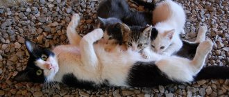
Providing first aid and treating bruises in cats is of great importance for the future health of the pet. Veterinarians diagnose several types of injuries. Most often the head, jaws, fore and hind limbs, chest, abdomen, internal organs, spine and tail are injured. Any injury can become fatal if the owner does not show the pet to the doctor in a timely manner.
According to veterinarian statistics, 15% of cats are injured due to the owner’s carelessness when falling from a height.
A little anatomy
The cat's spine consists of many small vertebrae, between which are elastic intervertebral discs. The openings in the vertebrae form a canal - a container for the spinal cord. In cats, the spinal cord reaches the end of the sacrum, and its continuation, the filum terminale of the nerve substance, still stretches to the 3rd caudal vertebra. Further, in the tail, the vertebrae do not have a cavity, they are just elongated small bones.
Spinal injuries are dangerous precisely because they are almost always accompanied by damage (rupture) or pinching (compression) of the spinal cord and its roots.
Types of fractures
In veterinary practice, it is customary to distinguish the following types of destruction of the spinal column:
- Compression fracture of the dorsal vertebral body
Compression fracture of the dorsal vertebral body. Damage most often occurs due to the vertical direction of the mechanical impact, as well as when the animal’s back is sharply bent. This damages the vertebral body, intervertebral discs and ligaments. - Distraction fractures. Destruction is characterized by the destruction of the intervertebral disc due to excessive stretching of the spinal column. Subluxation and dislocation of bone structures are also observed.
- Rotational damage. A fracture is characterized by the destruction of all anatomical elements of the animal’s spine with obvious neurological symptoms.
The most severe are distraction and rotational fractures, characterized by damage to the spinal cord, a pronounced neurological picture and an unfavorable prognosis for the animal.
Causes of spinal injuries
Typically, spinal injuries are caused by severe mechanical trauma. Much less often, the functioning of the spinal cord is disrupted by tumors, infections and hereditary defects.
The following incidents are typical for adult cats:
- falling from height;
- auto injury;
- dog bites;
- gunshot wounds.
In addition, objects that fall on top of them are dangerous for kittens: babies are playful and curious, and stick their noses everywhere. For example, the spine can easily be broken by a cutting board falling from the table.
And for kittens suffering from nutritional dystrophy (hyperparathyroidism), even minor injuries are dangerous. Their spinal fractures can be spontaneous, actually under their own weight.
Disturbances in the functioning of the spinal cord are possible even when the integrity of the spinal column is preserved, for example, with severe bruises. When the vessels of the dura mater are damaged, a hematoma occurs, which compresses the spinal cord.
Symptoms
The degree of neurological impairment depends on the severity of the spinal injury and location. If the vertebra is broken without displacement or it is not the body but the process of the vertebra that is damaged, then the cat retains the ability to walk. In such cases, the main symptoms will be pain and decreased motor activity (no running, no jumping).
In case of serious injuries, paralysis of the limbs occurs:
- If the cervical or thoracic spine is damaged, all four legs are removed.
- In case of lumbar injury - only the posterior ones.
In addition to being unable to walk, an injured animal may lose the ability to empty its bowels and bladder. Another ominous symptom is the lack of pain sensitivity in the paralyzed area.
When spinal injuries occur in animals, spinal shock develops. The cat can die from problems with blood circulation and internal organs. Clear signs of shock:
- mucous membranes are pale (white) or blue;
- rapid shallow breathing;
- pulse weak, frequent, uneven;
- body temperature decreases (sometimes to 33-35 o C and below);
- the pet is indifferent to external stimuli and loses consciousness.
The state of shock progresses quickly and requires immediate medical intervention.
Features of internal bleeding
Internal bleeding is the release of blood from damaged vessels into the body: into the abdominal or pleural cavity, into the space between muscles, into subcutaneous tissue, as well as inside hollow organs, for example, into the bladder, trachea or stomach.
The degree of danger to the pet's life depends on the rate of bleeding. With chronic, relatively small blood losses, oxygen starvation of the body tissues begins, anemia develops, metabolic processes are disrupted, which entails cell death. The animal’s activity decreases, the quality of its fur deteriorates, and the pet loses weight to the point of anorexia. In rare cases, lethargy develops.
Bleeding may stop due to the formation of clots that block the breakthrough, and then resume again due to their displacement. In this case, periods of weakness in the animal will alternate with normal health.
A blood clot can block a damaged vessel, but this improvement will only be temporary
With severe internal bleeding, the cat can die both due to a drop in pressure and oxygen starvation, and due to blood squeezing vital organs, such as the heart or brain. Without qualified assistance, death in this case occurs within a few hours or minutes, depending on the type of injury.
Diagnostics
Neither successful treatment nor a reasonable prognosis for the affected cat is possible without an accurately established diagnosis.
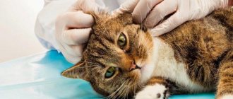
On the examination table, the doctor examines and carefully palpates the injured animal. Particular attention is paid to the preservation or absence of motor reflexes and pain sensitivity. Already at this stage, it is possible to approximately assess the location and extent of damage to the spinal cord.
Severe fractures of the spine are visible to the naked eye (back deformation, unnatural body position), but the doctor always sends a cat suspected of such a diagnosis for an x-ray examination.
The good thing about X-rays is that the images can be taken without anesthesia and quickly provide additional information about the injury. Digital X-rays are not available everywhere, but not a single decent veterinary clinic can do without a conventional device.
The images will show tumors, fractures and vertebral displacements. In case of severe injuries, based on X-rays, it can be confirmed that the spinal cord is ruptured (a combination of fracture and dislocation of the spine). However, the nerve substance itself cannot be seen on an x-ray. Therefore, if the owners are ready for surgery and want to do everything possible, the cat will have to undergo a special diagnosis.
To make the spinal canal visible on x-rays and determine the location of its compression, a contrast agent is injected between the membranes of the spinal cord. The pet must be under anesthesia. After contrast is administered, a series of x-rays are taken at regular intervals.
The advantage of such research is its wide availability (no special equipment is required) and information content. However, compared to CT or MRI, myelography is more dangerous because it is an invasive diagnostic method. In 1-2 animals out of 100, it causes complications associated with increased intracranial pressure or accidental damage to a vessel with a needle and the formation of a hematoma.
The gold standard for diagnosis for any spinal injuries is MRI, as it makes it possible to see in detail the nervous tissue of the spinal cord, the area and degree of damage, and compression. This study is performed under general anesthesia. An MRI should precede all spinal surgery (and helps determine whether such surgery is advisable at all).
Urinary Tract Trauma in Small Animals
Accumulation of urine in the abdominal cavity, retroperitoneum, or subcutaneous tissue of the perineum and thighs most often occurs as a result of blunt or penetrating trauma to the abdomen or pelvis. The incidence of urinary tract injury in small animals has not been recently determined. According to 1975 data, urinary tract injuries occurred in 15 of 600 (2.5%) road traffic accidents. In a 1982 study of 100 consecutive cases of dogs with pelvic trauma, 39 dogs (39%) were diagnosed with associated urinary tract injuries. Urinary tract injuries included bladder rupture (7%), urethral rupture (5%), ureteral avulsion (4%), bladder entrapment (5%), hydroureter or hydronephrosis (3%), and bladder lining abnormalities or thrombi (15%). Surgery due to abdominal urine retention was necessary in 16 of 39 (41%) dogs diagnosed with urinary tract injuries. Urinary tract injury was not clinically detectable in one third of these dogs.
The kidneys and ureter are anatomically located inside the retroperitoneal space, separated from the peritoneal cavity. If the retroperitoneum is not compromised by trauma, leakage of urine from the kidney or ureter may result in urine pooling only within the retroperitoneum without any connection to the peritoneal cavity. Fluid collection within the retroperitoneum alone may present a challenge to initial diagnosis based on clinical examination. The presence of urine within the abdominal cavity and retroperitoneum often delays the onset of clinical symptoms associated with uremia and peritonitis. Clinical symptoms may be subtle and may be attributed to diseases of various organ systems. It is important to pay attention to the patient's posture, appetite, and ability to urinate. However, patients with unilateral renal or urethral injury, bladder rupture, or partial urethral injury may demonstrate normal voiding with or without hematuria. In the absence of complete rupture of the urethra, urination is usually observed. Dysuria, stranguria, and anuria are the most commonly reported clinical symptoms systematically associated with urinary tract trauma.
A complete and thorough clinical examination of the patient is essential. You should carefully palpate the abdomen if the muscle structures of the abdominal wall are intact - external swelling may indicate the presence of hernias. A common symptom is abdominal tenderness, which may interfere with deep internal palpation of the abdominal organs, including the kidneys and bladder. It is important to remember that the presence of a palpable bladder has no bearing on whether urinary tract injuries exist. The subcutaneous tissues of the abdomen, perineum, and thighs should be examined for hematomas or swelling. Urine leakage into the subcutaneous tissue can lead to an intense inflammatory reaction. An increase in body temperature may be the result of subcutaneous inflammation, peritonitis or sepsis.
The presence of urine in the peritoneal cavity has serious metabolic consequences, but is not usually associated with bacterial peritonitis. Urine is not considered an adjuvant compound for peritonitis. Chemical peritonitis due to the presence of urine is minimal. Urine can remain in the peritoneal cavity for months without causing irritation, as long as it is sterile. Analysis of a sample of abdominal fluid is important to make a definitive diagnosis of the presence of urine in the abdominal cavity.
Elevated concentrations of urea nitrogen and creatinine, as well as increased concentrations of potassium in the abdominal cavity, cause non-acute peritonitis as a result of urine hyperosmolarity. The urea molecule is a relatively small molecule and is able to pass through the peritoneal membrane and re-enter the circulation - an increase in serum urea nitrogen concentration occurs as a result of urine accumulation in the peritoneal cavity. The creatinine molecule, however, is a larger molecule and cannot pass through the peritoneal membrane efficiently and remains much longer in the peritoneal cavity. As a result, serum creatinine concentration does not rise as quickly as serum urea nitrogen in response to urine accumulation in the peritoneal cavity. A definitive diagnosis of the presence of urine in the peritoneal cavity can be made when the creatinine concentration in an abdominal fluid sample exceeds the peripheral blood concentration. Serial monitoring of serum urea nitrogen, creatinine, and potassium concentrations in patients with suspected urinary tract leakage is not a valid or evidence-based diagnostic tool—these values may be elevated in cases of trauma, renal failure, hypovolemic shock, dehydration, and urinary tract obstruction.
Immediate surgery is contraindicated in patients who have been diagnosed with urine in the peritoneal cavity—dehydration, azotemia, and hyperkalemia make the patient metabolically and hemodynamically unstable for general anesthesia and definitive treatment. Uremic patients are particularly sensitive to the effects of most anesthetics, analgesics and sedatives. In hyperkalemic patients, bradycardia and idioventricular rhythms are possible.
Although repair of a urinary tract leak is not considered an emergency procedure, drainage of urine from the peritoneal cavity is considered an emergency.
Dehydration, azotemia, and electrolyte imbalance can be addressed by aggressive administration of appropriate fluid resuscitation. Saline solution quickly eliminates hyponatremia, hypochloremia and hyperkalemia associated with the presence of urine in the abdominal cavity during the formation and excretion of urine. The rate of administration of infusion solutions should be calculated to eliminate dehydration during the first 4-6 hours and establish a diuresis 2-5 times higher than the maintenance rate. Once the patient is stabilized, urinary drainage from the abdominal cavity should be established. Until X-ray contrast studies are performed, the specific location of the urinary tract discharge is usually unknown. Retrospective studies of urinary tract injuries in dogs and cats have shown that the bladder is the most common site of leakage after blunt abdominal trauma and pelvic injuries. Despite the rupture of the bladder wall, urine often continues to accumulate in the bladder. Inserting a urethral catheter into the bladder may help keep the bladder in a decompressed state and may reduce the flow of urine into the peritoneal cavity. Commercially available percutaneous peritoneal dialysis catheters can be aseptically inserted through the ventral abdominal wall into the peritoneal cavity to provide continuous drainage of urine. Multiple fenestrations of peritoneal dialysis catheters prevent blockages and allow more efficient drainage of fluid from the peritoneal cavity. If a dialysis catheter is not available, a Fogarty catheter may be surgically inserted into the peritoneal cavity to provide continuous drainage of fluid. The ventral part of the abdomen is aseptically prepared from the navel to the pubic bone and to the side to the area of the folds of the lateral surface of the body. General anesthesia should be avoided in these patients, but narcotic analgesia is indicated. In addition, a local anesthetic is injected into the skin, subcutaneous tissue, and body wall at a distance of 2 to 3 cm caudal to the umbilicus at the ventral midline.
Once the patient is rehydrated and stabilized, diagnostic radiography is performed to determine the location of the urinary tract leak. Plain abdominal radiographs may suggest urinary tract injury but are rarely diagnostic. Changes in retroperitoneal appearance, including dilation, increased density, and heaviness, have been shown to be the most consistent finding on plain radiographs of urinary tract injury. Contrast radiography procedures are indicated to determine the specific location of urine leakage. Given the increased frequency of reports of lower urinary tract injuries, positive contrast enhancement of the urethra and bladder is usually performed first. Evaluation of the upper urinary tract required intravenous administration of contrast agent—excretory urography is usually contraindicated in patients with dehydration or azotemia, as it may result in nondiagnostic examination or traumatic effects on the kidney. Penetration of the contrast agent into the peritoneal cavity, retroperitoneal space or subcutaneous tissue positively determines the defect.
The main effective treatment is based on the location of the urinary tract injury. General anesthesia and surgical repair can usually be performed in patients who respond appropriately to active drug stabilization within 24 to 48 hours. The defect in the bladder is treated and closed with a one- or two-layer appositional or eversion suture. It is recommended to check the bladder for leakage to determine the presence of other defects by administering sterile saline. If the blood supply to the bladder is compromised or in cases of severe tissue hematoma, the suture line may be reinforced with a serous patch of the adjacent small bowel and postoperative bladder decompression may be supported by a urethral catheter.
Incomplete urethral tears (even large ones) can be healed without surgery, provided that the urethral mucosal strip remains intact and urine is drained. The intraurethral catheter must remain in place for at least 3 weeks. Strictures usually develop.
Treatment for complete urethral transection is resection of the damaged tissue and anastomosis to prevent stricture formation. Injuries to the intrapelvic urethra may require pelvic osteotomy for repair. Osteotomy repair usually involves the use of wires. Incomplete bony union was observed 4 months after pelvic osteotomy, suggesting possible prolonged healing. The presence of an intraurethral catheter facilitates identification of the urethral lumen, and it has been suggested that the results of urethral anastomosis are significantly improved in the presence of a catheter during surgery. Direct (via cystotomy) and retrograde passage of a urethral catheter can help identify the ends of the urethra in cases of complete dissection. The traumatized urethral margins are excised (minimally) and primary repair is accomplished using an interrupted absorbable suture with 4-0 or 5-0 monofilament suture. Magnification with a surgical loupe can make suturing easier. It is important to perform sufficient dissection to prevent tension during the anastomosis, since tension promotes stricture formation. Postoperative urinary diversion can be continued by leaving the repair catheter in place for 1 to 2 weeks, although it is unclear whether this prevents or promotes stricture formation. Urethral stricture is the most common complication of urethral trauma. The presence of a stricture does not always lead to blockage. However, there is certainly a greater risk of blockage for the patient if the stricture progresses or stones form.
Traumatic urethral injury often leads to nephroureterectomy in patients with normal contralateral renal function. The decision to remove the kidney and urethra is often influenced by financial and humane considerations in an attempt to eliminate the many potential complications associated with primary repair procedures. Ureteral anastomosis (ureteroureterostomy) is the preferred method for proximal ureter injuries. This procedure, however, is extremely difficult and should not be attempted without magnification (loupe or operating microscope). Despite precise, atraumatic tissue management, the incidence of complications, including stricture, dehiscence, and obstruction, can be high. The ends of the ureter have wide, rounded edges, and the anastomosis is performed with an interrupted 5-0 or smaller absorbable suture. A ureteral stent may be placed to even out the area of the anastomosis, providing a shape around which the ureter can heal and preventing urine leakage. If suture line tension is a problem, the kidney can be mobilized and the capsule can be sutured to the lumbar muscle fascia at a location caudal to the renal fossa (renal prolapse). If the length of the ureter is insufficient and does not reach the bladder, but is sufficient to cross the midline without tension, another option for repair of proximal ureteral injury is transureteroureterostomy. In this procedure, the remaining proximal segment of the ureter is passed through the midline and anastomosed to the renal pelvis or ureter on the contralateral side (end-to-side anastomosis) with a 5-0 or less absorbable suture. Replantation of a ureter that has been removed from the renal pelvis is a technically difficult operation and significant tissue damage often occurs. Nephroureterectomy is considered due to the complexity of this type of injury in the presence of normal contralateral renal function. Treatment of injury to the distal third of the ureter is usually performed by neoureterocystoneostomy. The ureter is reimplanted into the bladder anywhere along the dorsal or apical portion of the bladder to relieve tension. Kidney drop or psoas “lift” may also be used to further relieve tension. The technique of “lifting” the psoas muscle is based on the fact that stretching the bladder does not affect function and gives 3-5 cm of ureter length. Traction is used to move the bladder cephalad and the dorsal surface of the bladder is then sutured to the psoas tendon. For extensive mid- or distal-third segment injuries resulting in a ureteral defect that cannot be reattached, ureteral replantation and psoas muscle “lift” may be considered. Bladder flaps, such as the Boari flap, are most commonly used with psoas muscle elevation to treat midureteral defects and offer a possible 15 cm increase in ureteral length. In this technique, a dorsolateral full-thickness bladder flap is created, the bladder defect is closed, and the edges of the flap are sutured together to form a tube, and the end of the tube is sutured to the distal end of the ureter.
Treatment
Immediately after an injury, the task of doctors is adequate pain relief and anti-shock therapy. While the cat's condition is unstable, most diagnostic procedures can lead to deterioration. A spinal fracture may not be the only problem, but may be accompanied by a bruise or rupture of internal organs, abdominal bleeding, or accumulation of fluid or air in the chest cavity.
Once an accurate diagnosis has been established, the doctor recommends conservative treatment or surgery. The final decision is made by the owner based on the information provided about the cost of treatment, possible complications and prognosis for recovery.
Drug treatment involves the use of large doses of steroid hormones:
- metipred,
- dexamethasone,
- prednisolone.
These substances relieve swelling of the spinal cord as much as possible. The animal is limited in mobility.
Conservative treatment is justified if there is no pronounced displacement of the vertebrae, and pain sensitivity and reflexes of the limbs are preserved.
The surgeon's goal is to relieve spinal cord compression and stabilize the vertebrae. For this purpose, a wide variety of metal structures are used. After the intervention, the cat receives:
- antibiotics,
- painkillers,
- anti-inflammatory drugs.
Regardless of the chosen treatment method, an acute period after injury and a recovery period are distinguished. Rehabilitation of a cat with a back injury can take a long time. During this period, doctors prescribe:
- various physical therapy;
- B vitamins;
- basic care required for a paralyzed animal.
Treatment of hematoma
- If your cat is injured, then even in the pre-hospital stage, apply ice to the injury; it should be wrapped in plastic and a clean, dry cloth. If possible, give an injection of painkiller - analgin or ketofen.
- For small hematomas, conservative therapy is used. The bruise site can be warmed with paraffin applications, resorbing ointments - ichthyol, turpentine.
- With extensive effusion, treatment of hematomas requires surgical intervention. To do this, a puncture is performed, followed by evacuation of the contents, rinsing the cavity and applying an antibiotic ointment (mainly levomekol).
- In case of hematomas of the auricle, its opening is mandatory, since a long course of the disease leads to the formation of scar tissue and a change in the shape of the ear. The cavity is cleaned of fibrin and blood clot, then a fixing suture is applied.
- If there is a tick-borne or fungal infection of the ear, it is necessary to carry out etiological (antiparasitic or antimycotic) therapy along with the treatment of the hematoma.
- If the hematoma is pulsating, then during the operation a ligature is applied to the damaged artery and the wound is sutured.
- A cranial hematoma requires complex treatment with resuscitation measures and medical supervision.
As a wound healing therapy after puncture, you can use ointments - levomekol or synthomycin. If a purulent complication is suspected, bandages with Vishnevsky ointment are applied.
Forecasts
If the paralysis is caused by blood pooling under the dura mater (for example, after a contusion or needle puncture), the prognosis is good. After 1-2 weeks, the hematoma resolves even without treatment, and the animals gradually recover until full recovery.
If bone fragments press on the spinal cord, the prognosis largely depends on how quickly the diagnosis and surgical intervention were carried out. With spinal injuries, time is sometimes counted by the clock. If nerve fibers have been compressed for too long, their function may not recover, or the recovery period may take months or even years. Often, with successful spinal surgery, urination is completely restored within a few months.
If a spinal cord rupture occurs, neurological impairments remain for life. No operation, no timely and competent treatment will lead to restoration of limb function. Another question is that under the care of a loving owner, even a paralyzed cat can live a long, fulfilling life.
Causes of hemorrhages
Internal bleeding may occur for the following reasons:
- Mechanical injuries.
- Entry of a foreign object into the respiratory tract.
- Ingestion of sharp objects by a cat, including fish or tubular bones.
- The rectum can be injured by hard feces.
- Neoplasms. The tumor may ooze slowly or rupture and cause severe bleeding. In some cases, it grows into a blood vessel, destroying it.
- Coagulopathies are blood clotting disorders. There are several reasons for their appearance, one of the common ones is poisoning with rat poison. Blood diseases, jaundice, and sepsis are also possible.
- Uterine bleeding can occur due to birth injuries, remaining placenta, or even a dead fetus. Possible tumors and inflammatory processes caused by infection, a large number of empty heats, or drugs to prevent them.
- Ulcers of the stomach and esophagus. Most often they manifest themselves by vomiting. In the first case, the blood will be dark and coagulated, in the second - scarlet.
- Infestation with parasites.
- Hypertension. Pressure above 160 over 100 can cause nosebleeds. Usually accompanied by irritability and impaired coordination of movements.
My relatives' Persian cat had a nosebleed once. The cause turned out to be inflammation of the tooth, but given Malkin’s advanced age, the veterinarian advised measuring his blood pressure at least once every six months by placing the cuff of an ordinary human tonometer on his paw or the base of his tail. It turns out that hypertension is quite common in older cats and can lead to serious complications. According to the same specialist, such measurements are best carried out at home - in a veterinary hospital, animals are nervous, so the results are almost certainly inaccurate. Now I also measure my Siberian daughter’s blood pressure every few months - she’s still young, but caution won’t hurt. As a rule, Irma endures the procedure with some surprise, but calmly.
It is useful for a cat owner to know the basic techniques of providing first aid.
Popular belief states that a cat has nine lives. True, this is just a belief. Specialists from the American Veterinary Insurance Society compiled a rating of the reasons for visiting clinics with cats. All kinds of wounds and injuries were on the seventh line of the gloomy “hot ten”. Not a single cat is immune from these diseases, and it is useful for the owner to know at least the basic rules and techniques for providing first aid to an injured pet. We talked about them with Doctor of Veterinary Sciences, Professor Sergei Aleksandrovich Yagnikov.
Most often, domestic cats suffer from wounds received in fights with fellow cats or dogs, as well as from the consequences of falling from a height. A non-specialist, based only on external signs of damage, can very rarely correctly assess the true nature of the injury and its danger to the life and health of the animal. Therefore, a responsible owner should not ignore any injury that occurs, even if it seems to him that the cat escaped with a few scratches and feels quite normal.
First aid and treatment of hematomas
Immediately after injury, ice should be applied to the skin. No ice? Open the freezer, take any food, wrap it in a towel and bag. Calm the cat and cool the injury for 10 to 20 minutes.
Important! The cat needs to be injected or given a painkiller.
If the wound is open, the edges are treated with iodine. On the first day or before arriving at the doctor, a pressure bandage is applied. A closed injury is covered with an elastic bandage. Be careful when applying pressure bandages, use appropriate force and check local skin temperature. If you suspect internal bleeding, apply ice and go to the doctor. If a limb is injured, we pull the paw above the bleeding wound and record the time. The tourniquet is removed after a maximum of 25–30 minutes.
Absorbable ointments Levomekol, Sintomycin, Ichthyol and Vishnevsky are used locally (for suppuration). The animal is kept warm and placed on an easily digestible diet. In case of extensive hemorrhages, the hematoma is opened and “cleaned” surgically using local anesthesia.
Important! If your cat develops a hematoma for no apparent reason, contact your veterinarian. Swelling may be associated not only with vascular injury, in which case symptomatic treatment is a waste of time.
source
Cat wounds
Most often, cats have bitten, lacerated “battle wounds” received in fights with other cats or dogs. The most obvious sign of such wounds is damage to the skin, which requires appropriate treatment. First of all, the wound must be washed with a disinfectant. A regular solution of furatsilin is suitable for this. If you don’t have a ready-made solution, you can prepare it yourself by dissolving two tablets in a glass of warm boiled water. The surface of the wound is treated with this solution. If possible, remove fur from the area around the injury to prevent hair from sticking to the wound.
When the owner is unable to immediately deliver the injured cat to a veterinarian, she should be given an antibiotic. If the owner does not have a special veterinary antibiotic at hand, it can be replaced with a human drug, for example, Augmentin (amoxiclav). The antibiotic is given in a dosage per 1 kg of animal body weight twice a day. That is, an “average” cat that weighs 3 kg should receive about 50 mg of antibiotic at one time. And, of course, the animal needs to be shown to a veterinarian as soon as possible.
If the cat received a cut wound and started bleeding, then you need to do the following: take a clean, or better yet, sterile napkin, apply it to the surface of the wound and bandage it with moderate force. The pressing effect will reduce bleeding. If a large artery is damaged, it will most likely have to be sutured. Bleeding from small vessels can stop on its own, but treatment of such a wound still requires the supervision of a specialist.
Eye injuries are also common in cats and, unfortunately, in this case, the owner can do practically nothing himself. The damaged eye should be carefully washed with furatsilin solution and immediately consult a doctor.
Cats also have so-called abscesses. They usually develop as complications if the wound has not been washed with an antiseptic. Pathogenic microorganisms enter it and begin to develop, causing a cavity with pus to appear under the skin - an abscess itself. Externally, an abscess manifests itself as swelling, a “bump” on the skin and painful sensations when touched, since the cavity compresses the nerve endings. Sometimes the abscess opens spontaneously and pus is released from the wound. However, even in this case, it is better not to hope for the animal’s independent recovery. An abscess requires mandatory intervention by a veterinarian, because antibiotics alone cannot suppress a purulent focus. It is necessary to open and wash this cavity, install drainage in it and maintain it until the pathogenic process stops.
Diagnosis and causes of hematomas in practice
Cats tend to lick their fur and, if necessary, wounds. Even a closed injury, which the cat constantly “massages” with its tongue, resolves faster. However, the animal cannot lick the back of its head, ears and muzzle. The only way to deal with discomfort is to scratch with your paws, which essentially injures you even more.
The “leader” in terms of frequency of occurrence and number of possible causes is auricular hematoma. Subcutaneous and ear mites, fleas, allergies, fungal skin diseases, injuries after fights are just some of the reasons for scratching your ears. The cat can tear its skin into blood or cause a head injury. Ear hematoma in cats is a consequence of the destruction of the thinnest blood vessels, which are protected by a thin layer of skin. Naturally, the injury must be treated, but the main task is to eradicate the root cause and relieve the animal from itching.
Note! With a subcutaneous tick, the animal scratches its head so much that a hematoma on the ear is not the most negative complication of the disease.
Postoperative hematoma in a cat can be considered a complication or a normal phenomenon, depending on the clinical picture. A slight swelling and extensive bruising in the area of the sutures is a normal state of affairs; the animal requires comprehensive postoperative care. Increased temperature, sharp pain, severe redness, discharge of pus from under the sutures against the background of a hematoma are bad symptoms indicating infection of the wound. The situation is possible in three cases:
- Not a sterile environment during surgery.
- Poor cauterization of dissected tissues.
- Damage to the sutures is most often observed after sterilization if the cat does not wear a blanket or collar. The animal bends over to lick the seam on its stomach (eventually licks it), and pulls the threads with its teeth.
Injuries in cats
Cats often fall from heights and are injured. Many ordinary people tend to consider a broken limb to be the most “terrible” of such injuries, but in fact, fractures in cats are well treated, and the animal has every chance of making a full recovery. True, fractures are also different. For example, an animal may suffer an intra-articular fracture, in which a crack runs along the surface of the “hinge” of the joint. Such fractures are the most complex, they require long-term treatment and sometimes lead to the development of arthrosis.
Very often, after a fall, the animal begins to drag its hind legs. This may be a sign not so much of paw fractures as of some kind of neurological injury, for example, a fracture of the spinal column. It occurs quite often in cats, and most often occurs at the level of the last thoracic and first lumbar vertebrae. Such a fracture is fraught with serious injury to the spinal cord and, unfortunately, the prognosis in this case is unfavorable.
A rupture of the spinal cord may be indicated by the absence of pain. This is easy to check even at home. You need a suitable tool, such as tweezers, to squeeze the skin between the toes of the hind paw quite firmly. In a normal situation, such manipulation will cause pain - the cat will scream, try to scratch or bite. In this case, the prognosis will be favorable. But if the animal does not react in any way to such pain, then, most likely, its spinal cord is seriously injured. Even if the cat jerks its paw when squeezed, this does not mean anything. This is how the flexion reflex manifests itself, which can persist even with a complete rupture of the spinal cord. In case of spinal injury, only the feeling of pain can be considered a favorable factor that gives the animal a chance to recover.
Often, injuries associated with a fall from a great height lead to rupture of the diaphragm (the so-called partition between the chest and abdominal cavities). When it ruptures, part of the internal organs of the abdominal cavity, in particular the intestines and liver, can move into the chest cavity, where the lungs are located. Externally, such damage is expressed in the fact that the animal develops rapid breathing and cyanosis of the mucous membranes, because the lungs can no longer fully expand. The cat loses its appetite, and due to the pain caused by the injury, stiffness appears in its movements. But an accurate diagnosis can only be made using radiographic examination. If the diaphragm ruptures, the animal requires surgery, which can only be performed in a veterinary clinic.
How to identify a bruise
If a cat is bruised, the owner is unlikely to remain unaware. The resulting injury in the area of the hind or forelimbs leads to the fact that the cat has a pronounced limp, holds the affected paw in a taut state, trying as much as possible not to let the paw come into contact with a hard surface.
The cat stops stepping on the limb, and if it does, with mild degrees of damage, it becomes noticeably lame, automatically transferring the weight of its body to other supports. There are several main signs of a bruise in a cat that will help the owner suspect a pathological condition and take the animal to a veterinary clinic.
The main symptoms of bruises are:
- swelling when a paw is bruised in the area of the affected limb (swelling develops against the background of accumulation of lymphatic fluid and blood from ruptured vessels into the subcutaneous fatty tissue);
- hematoma (bruise) at the site of the lesion;
- disturbance of the cat’s normal gait and severe lameness;
- the animal anxiously tries to lick the sore spot, stimulating microcirculation for speedy resorption of the bruise;
- an increase in temperature at the site of damage, but not locally;
- aggression of the pet when the owner tries to touch the damaged area.
At the same time, against the background of a bruise, the cat should not have any significant damage to the integrity of the skin, as well as violations of the joint and bones on the paw.
Severe or minor bruises of the first and second degree under normal conditions disappear within a week.
Swelling gradually decreases, gait normalizes, and the animal becomes normal. If the bruise is associated with damage to ligaments or muscle fibers, consultation and treatment cannot be avoided.
It is important to know how to correctly distinguish a true bruise from a possible fracture or dislocation. So, with fractures in a cat, a visual change in the structure of the limb or other damaged part of the body is observed. The pet experiences severe pain at the site of injury, plaintively asking for help.
By gently palpating the affected limb, the owner will be able to feel for instability of the bone. In addition, with fractures, edema develops not only in a certain place, but is distributed over large areas.
With dislocations, the clinical picture has the following characteristic feature. At the site of dislocation, the boundaries of the exit of the head of the bone from the articular capsule are clearly defined. The limb weighs like a whip, and the cat cannot use it. In such cases, you cannot set the bone yourself; you must contact a specialist.
Pain relief for cats
Most injuries cause the cat to experience severe pain. The owner can independently reduce it by using pain relief therapy. Special veterinary drugs ketofen or nalbuphine are well suited for this purpose. But it is better not to give such popular painkillers as baralagin and analgin, especially in tablet form, to a cat, because they cause severe salivation in the animal. As a last resort, you can inject analgin by injecting 0.4 ml intramuscularly (dose for an average cat).
Also, in case of injury, traumatic shock is possible, one of the manifestations of which is vasospasm. This spasm has a very bad effect on the liver and kidneys. It can be removed with anti-shock therapy. The affected animal should be given corticosteroid drugs, for example, prednisolone intramuscularly). But injections should only be considered as an emergency aid. At the veterinary clinic, anti-shock therapy will be continued using drip infusion.
Pain is a physiological reaction to certain disturbances in the normal functioning of the body’s vital systems. This is a signal to take action to help the pet. In some cases, feline pain medication should be given immediately.
In the article, in addition to drugs for pain relief, we also look at the causes of pain and give advice in which cases a cat really needs to be given medicine. We do not recommend skipping these sections, but if you want to know only about the best drugs and their prices, then refer to the contents.
Rehabilitation of a cat after a fracture
The recovery program for a pet after surgery depends on the severity of the injury and the degree of manifestation of the neurological disorder. The complex of rehabilitation measures includes physiological procedures (massage, darsanvalization), the use of drugs that improve tissue regeneration, and vitamin therapy.
The duration of the rehabilitation period ranges from several months to a year.
We recommend reading about a broken jaw in a cat. You will learn about the types of jaw injuries, the causes and symptoms of this fracture, diagnosis and treatment options. And here is more information about dislocation in a cat.
Signs of pain
Usually cats tolerate pain patiently, but in some cases it is a signal for an urgent visit to the veterinarian.
Signs that your cat is in pain:
- unusual behavior: excessive activity or lethargy;
- unjustified aggressive reactions;
- lack of appetite;
- desire for solitude;
- uncharacteristic apathy and drowsiness;
- refusal of the litter tray;
- drooling and vomiting;
- cardiopalmus;
- heavy or difficult breathing, shortness of breath.
When is pain medication needed?
Pain syndrome can be acute and chronic. In case of severe injury or cancer, delay in providing assistance to your pet can lead to painful shock and even death.
Injuries most often occur during fights with dogs and other cats, when the animal is hit by a car, when falling out of a window or simply from a height.
Typically, pain in cats is accompanied by:
- Oncological diseases.
- Injuries - superficial extensive wounds and cuts, fractures of the paws and spine, bruises and torn ligaments.
- Postoperative period after surgery.
- Recovery after castration or sterilization.
- Chronic diseases in the acute stage - pancreatitis, cystitis, urolithiasis, others.
- Kidney or stomach colic.
- Disruption of the nervous system.
- Tooth and jaw pain (usually when enamel is destroyed).
Prognosis for the animal
Destruction of the spinal column in domestic cats is one of the most severe injuries in veterinary practice. The prognosis depends on a number of factors: the timeliness of first aid and qualified surgery, the success of surgery, as well as the subsequent rehabilitation period.
A spinal fracture in a cat is a serious and life-threatening injury that requires immediate qualified assistance. The owner must have an idea of how to properly transport a sick animal to the clinic. The professionalism and experience of the surgeon give a chance to bring your pet back to life. Recovery after surgery is a long and complex process, requiring patience and boundless love for the pet from the owner.
Similar articles
- Dislocation of the hip joint in a cat, anterior or...
The pathology is complex, accompanied by intra-articular fractures, ruptures of ligaments, tendons. Symptoms of a dislocation in a cat. Read more - Tail fracture in cats and cats: symptoms and treatment
A broken tail in a cat can happen for a variety of reasons. ... The tail of a domestic cat is an anatomical continuation of the spine with the presence of small vertebrae, blood vessels and nerve endings. Read more
- The cat can’t pee: what to do, how to help the cat or...
Causes of rectal prolapse in cats and methods of treatment. A cat has an ingrown toenail: how to help your beloved furry friend? How does phlegmon manifest in cats, how dangerous it is and how to treat it. Read more
- How to treat a cat’s wound (open, purulent) after...
How to treat an open wound in a cat depends on the further development of the traumatic process. ... Tail fracture in cats and cats: symptoms and treatment. Read more
- The cat is limping on a paw (back or front), the paw is swollen...
If a cat's paw hurts or is swollen, it is advisable to first find out the reason, for example, after an injection or a fall. … To learn how osteosynthesis is used to treat a cat’s tibia fracture, watch this video Read more
Types of veterinary drugs for pain
Cats differ from humans and most other domestic animals in their response to pain medications. The list of approved products that do not have a high toxic effect on your pet is very narrow.
Types of analgesics used in veterinary medicine and allowed for cats:
- Narcotic. Prescribed only under the supervision of a veterinarian in exceptional cases - in case of severe injuries and oncological pathologies. These are Omnopon, Fentanyl, Tramadol, Codeine.
- Non-steroidal anti-inflammatory drugs (NSAIDs). Unlike the first group, they are not addictive and not only relieve pain, but also have a therapeutic effect. In acute attacks they are ineffective. These are Ketonal, Ketofen, Quadrisol, Rimadyl, Meloxicam, Vetalgin.
- Other. Some medications intended for treating humans are also suitable for cats. These are Papaverine in the form of injections, Baralgin, some others as prescribed by a doctor.
How to give a pill to a cat and give an injection
All injections that the owner can give to the animal himself at home are usually done subcutaneously at the withers.
To do this, you need to fix the cat on a hard surface (with the help of a second person), pull up the skin at the withers and insert the syringe needle under the skin, the drug is infused slowly. Gently rub the injection site.
The tablet can be given using a special device, crushed and inserted into the oral cavity from a syringe without a needle with water or liquid food.
You can also take the animal by the scruff of the neck, secure it well on the floor, open its mouth and place the tablet on the root of the tongue. Then hold the mouth tightly with one hand, and with the other hand, carefully move it several times along the animal’s neck in order to evoke a swallowing reflex.
Symptoms of hidden bleeding
Hidden bleeding in a pet is indicated only by indirect signs: lethargy and apathy, paleness of the mucous membranes of the mouth, rapid breathing and shortness of breath, and sometimes fever. The tips of the ears and the skin on the pads of the cat's paws become sharply cold, and clouding of the pupils is observed. Fainting is possible.
If blood flows into the abdominal cavity, the volume of the abdomen increases. Sometimes bruising becomes visible under the skin. Shortness of breath begins due to pressure on the diaphragm.
Hidden intrauterine hemorrhage may be indicated by inflamed external genitalia and abdominal consolidation. In this case, the cat usually licks its belly frequently or assumes a posture as if for urination.
Hematomas feel like elastic swellings.
Hematomas often form on cats' ears due to infection or injury.