Causes
For cats, discopathy is an extremely rare disease; usually older cats suffer from this disease. Another reason for the development of spinal diseases may be the genetic characteristics of the breed - for example, in the Kuril Bobtail, due to a short tail, changes occur in the sacral region, which can lead to discopathy.
It is possible that the flexibility of the disc may be impaired during rickets, which during the subsequent growth of the kitten leads to deformation and protrusion (bulging) of the intervertebral disc. Another common cause can be spinal injuries - bruises, jumping from great heights, as well as infectious diseases.
What you need to know about intervertebral discs
These are a kind of shock-absorbing “pillows” that are located between two vertebrae. The disc consists of fibrous tissue, cartilage and a special lubricant that has a protective function. Essentially, it is a fibrous ring that has a fibrous coating inside, and a nucleus pulposus located in the center. With degenerative changes, the main function of the cartilaginous layer decreases, compression of the spinal cord occurs, and in advanced cases, spina bifida develops.
Basic information
The disease is an age-related, degenerative disorder (especially in the case of cats). What are the reasons? In rare cases, pathology can also occur in young animals that, during growth, lacked nutrients, vitamins and microelements. Sometimes this outcome is caused by certain infectious diseases, in which the tissue of the intervertebral discs becomes inflamed and hardens, but more often this is associated with age. The disease can occur in one of three possible scenarios.
Types of discopathy
There are three types of discopathy. They differ from each other in the speed of development of the process and clinical manifestations.
Type I
It is characterized by an acute onset and almost immediate development of the disease. It is extremely rare in cats. Typically, this type of damage occurs due to an infection that hardens the disc, interfering with the normal movement of the spine. During rotation, deformation of the intervertebral layer occurs, which leads to its displacement and gradual destruction.
Symptoms of the lesion can vary greatly in manifestation - from not clearly expressed stiffness of movement to the development of paralysis of the animal.
Type II
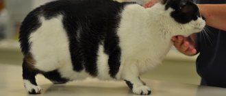
In general, its manifestations are similar to the first type, but does not have a lightning-fast onset. The disease develops gradually, and its early manifestations are difficult to notice in a cat. Typically, older cats are susceptible to the second type of discopathy. The disease usually begins with minor restrictions in movement and decreased physical activity. With further progression, a violation of posture may occur - stooping appears.
III type.
The most rare type of disease in cats. Usually develops due to serious damage to the spine. A distinctive feature of the third type is the sudden manifestation of the disease against the background of complete well-being. The contents of the intervertebral disc are strongly pressed into the spinal cord, causing the formation of a hernia.
Sharp pain occurs and the animal becomes immobilized. There may be uncoordination of movements and a “drunk” gait, and partial or complete paralysis may develop. The outcome of the third type of discopathy usually ends in the death of the cat from respiratory arrest.
A little anatomy
The cat's spine consists of many small vertebrae, between which are elastic intervertebral discs. The openings in the vertebrae form a canal - a container for the spinal cord. In cats, the spinal cord reaches the end of the sacrum, and its continuation, the filum terminale of the nerve substance, still stretches to the 3rd caudal vertebra. Further, in the tail, the vertebrae do not have a cavity, they are just elongated small bones.
Two types of “roots” depart from the spinal cord: sensory and motor nerve fibers. Without them, the work of internal organs and all muscles is simply impossible.
Spinal injuries are dangerous precisely because they are almost always accompanied by damage (rupture) or pinching (compression) of the spinal cord and its roots.
Symptoms of the disease
- One of the first signs of the development of intervertebral disc pathology is a decrease in the pet’s activity and the appearance of stiffness in movements.
- Lameness appears. Depending on the area of the spinal cord injury, limping on one or both legs may be observed.
- Pain. The animal begins to meow for no reason and finds no place for itself. When stroking the back it can show aggression.
- Disorientation in space and changes in gait.
- Convulsive syndrome is possible.
- When the lumbar and sacral spine is affected, defecation and urination are impaired.
Typically, with the development of intervertebral disc diseases, several symptoms from the list are observed at once. The sooner you contact a veterinarian, the better the outcome of the disease and the subsequent restoration of lost functions.
Diseases of the nervous system: list and characteristics
Pathologies associated with disruption of the nervous system are diverse and have extensive symptoms. They may concern damage directly to the nervous tissue or as secondary signs, a consequence of other diseases (hepatitis, endocrine pathologies).
- Aggressive behavior.
A common symptom of neurological diseases. Expressed in behavioral disorders not associated with natural physiological states (estrus, pregnancy, lactation). Often, symptoms of aggression go away on their own or are corrected with the help of hormone therapy or sterilization (castration). A sudden attack of aggression requires the attention of a veterinarian and may be a symptom of rabies.
- Neurotic conditions.
They are considered reversible pathologies and develop when higher nervous activity malfunctions. The impetus for neuroses is cruel treatment by the owner, stress, frequent exhibitions, and conflicts between pets. In rare cases, infectious diseases. Neuroses are a collective image that is not characterized by a single symptom; the following come to the fore: depression, neurasthenia, hysterics. Manifestations can be directly opposite in terms of symptoms - on the one hand - aggression, bright, non-standard reactions to completely harmless, everyday phenomena. On the other hand, it is characterized by apathy, lethargy, anorexia. Treatment of nervous disorders consists of eliminating the stress-provoking factor and stabilizing the nervous system with the help of mild, sedative drugs.
- Kay-Gaskell syndrome.
Dysotonomia has no specific causes and is rarely diagnosed in cats. The mechanism of development and methods of its treatment are also poorly understood. The syndrome affects the PNS and almost all internal organs. Treatment is ineffective, the nervous system and vital functions (respiratory, urinary, gastrointestinal tract) suffer. In cats, there is: alternating constipation and diarrhea, prolapse of the third eyelid, mydriasis, dry mucous membranes, incontinence of feces and urine.
- Acute cerebrovascular accident.
Diseases – stroke, heart attack. Against the background of ischemia, a heart attack develops. The vessels become clogged with a blood clot, and the area of the brain left without blood supply dies. Hemorrhage occurs during hemorrhagic strokes, against the background of a violation of the integrity of the vessel. In cats, strokes and heart attacks are diagnosed in adulthood, nervous symptoms depend on the area of the lesions. More often: paralysis, paresis, disruption of physiological processes.
- Discopathy.
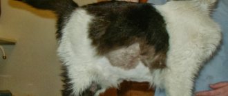
A collective term indicating secondary lesions of the spinal cord, often due to trauma. In kittens, the pathology is associated with congenital anomalies of the development of the skeleton and bone tissue (rickets). Symptoms are combined with the location of the lesions; sometimes the curvature of the spinal column or protrusion of the vertebrae is visually visible. There are: pain, inability to move freely, paresis, paralysis, involuntary release of feces and urine.
- Meningoencephalitis.
More often, nervous pathology develops against the background of viral infections that cause serous meningoencephalitis. Inflammation affects the meninges and brain. If detected early, it can be treated and goes away almost without leaving a trace. With a bacterial infection, purulent meningitis develops; it is difficult to treat with extensive residual effects that cannot be corrected.
Symptoms: impaired consciousness, mood swings (drowsiness or aggression), vomiting, muscle tension, convulsions, breathing problems, etc.
- Hepatargy.
Another name for nervous pathology is hepatic encephalopathy, which is expressed in disruption of the liver during sepsis and severe infections. The role of a provoking factor is assigned to an increased amount of hydrogen nitride, which has a neurotoxic effect. When it acts on the brain, foci of softening are created, and ischemic encephalopathy is diagnosed. Neuropsychic behavioral pathologies are observed, progressing gradually. Without treatment, hepatic coma occurs.
- Concussion.
Is the result of injury. Cats lose consciousness, coordination of movements is impaired. There is vomiting, no appetite, rapid pulse.
- Loss of mobility.
Paresis and paralysis develop against the background of injuries, as a consequence of stroke and pathologies that lead to deterioration of cerebral circulation. A cat’s paws can be very toned, or “lifeless,” flabby, or atrophied.
- Epilepsy.
It is characterized by organic lesions of the brain, most often the frontal lobe. It can be true or false, the latter develops against the background of the underlying disease. Symptoms: convulsions, seizures. Neurological disorders are irreversible; treatment is only supportive, aimed at increasing the time between attacks.
- Eclampsia.
Diagnosed only in lactating cats or in the last stages of pregnancy. It is characterized by the release of calcium from the body, the formation of edema, and the presence of tonic-clonic convulsions. In cats, the heartbeat quickens, blood pressure rises, coordination of movements is impaired, vomiting and diarrhea occur.
Diagnostics
One of the leading diagnostic techniques is radiography. It allows you to see a clear picture of changes and distinguish discopathy from injuries and damage to the spine. X-rays can show destruction of the discs, their narrowing or hardening, as well as damage to the spinal canal. For a more effective and revealing picture, it is possible to inject a contrast agent into the cerebrospinal fluid.
It is also possible to use computed tomography and magnetic resonance imaging. These methods are rarely used for animals because the equipment is quite expensive. Typically, these studies are carried out in large veterinary clinics and research centers.
To exclude an infectious process, it is necessary to undergo a puncture of the cerebrospinal fluid to determine its composition and bacterial culture.
An ultrasound of the spine can be performed to clarify the location of the lesion.
Electrodiagnostic methods
Electrodiagnostic methods allow early detection of nerve root damage. This is due to the fact that even a slight decrease in the number of motor units (a complex of structures consisting of α-motoneurons, axons, synapses and muscle fibers that are closely connected functionally) will affect the results of the study, especially in comparison with the contralateral side. However, electrodiagnostic research methods do not provide an answer about the causes of the lesion.
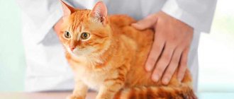
Cats, being born hunters, are distinguished by their mobility and flexibility. Watching these charming animals is a pleasure, but sometimes the owner notices that his four-legged pet begins to move with difficulty, stops jumping on the sofa or other furniture, loses most of its natural grace and hunches its back.
The most likely reason for a change in the behavior of a pet that was previously active and inquisitive is discopathy.
Treatment
If you notice signs of discopathy in your cat, you must ensure complete rest. It is best to move the bed to a warm place, make it soft and comfortable. If incontinence occurs, keep the bedding clean and dry. Move the feeder closer to the sleeping area.
Depending on the severity of the lesion, therapy is possible conservatively and surgically.
Drug treatment uses injections of sedatives, anti-inflammatory and painkillers. Painkillers can be used before contacting the veterinarian to alleviate the animal's condition. For this, use descaphot 0.1-0.2 ml, papaverine with diphenhydramine - 0.3 ml.
As an anti-inflammatory agent, you can use steroid drugs - dexomethasone 0.4 ml.
In case of pronounced destructive changes in the intervertebral discs, traumeel in ampoules is used as homeopathic therapy.
During the first days of the acute period, the drugs must be administered intramuscularly 3 times a day.
If the disease is advanced or medications do not provide the necessary relief, surgical excision of the damaged discs is possible. Implantation of artificial substitutes can also be carried out. Once paralysis develops, complete restoration of function is often no longer possible.
Description of pathology, general information
Osteochondrosis is a disorder of osteogenesis (the process of bone formation).
The cartilage tissue in the joints becomes inflamed, thickening and destruction occurs. Because of this, the surfaces of the bones begin to “dry” slide against each other, which leads to a very strong pain reaction. In practice, it has been found that in cats this disease is often inherited.
The development mechanism is quite simple. If for some reason (hormones or food characteristics) an animal’s body receives an excessive amount of calcium, it first goes into the bone tissue. But when osteocytes can no longer take it, the body sends calcium to the joints.
Their cartilage first ossifies, thickens, and then begins to collapse.
As a rule, clinical signs begin to appear at one year of age. It should be remembered that the process begins long before this, but by this time the body’s reserves are simply exhausted.
However, you should also not think that after a cat reaches the age of one and a half years, nothing threatens him: osteochondrosis affects all animals, regardless of age and physiological condition. However, cats get sick 50% more often than cats.
It is likely that the development of the disease is closely related to the characteristics of certain hormones.
Approaches to diagnosing a pelvic fracture in a cat
A fracture of the pelvic bone is noticeable by the altered posture of the animal. The cat's spine does not form a straight line, but curves below the lower back. Most often, the limbs are placed apart or turned back. If the cat does not try or cannot stand up, then there is reason to suspect a spinal fracture. True, in animals such a diagnosis without damage to the spinal cord does not sound as threatening as in humans.
The only evidence-based method is an x-ray. Required conditions:
- the cat is under the influence of muscle relaxants (Sedatsil, Xyla, Sedazin, etc.), which allows the doctor to do the correct styling without creating unnecessary tension on the muscles;
- two or more image projections were obtained.
An x-ray will indicate the location of the fractures. Only those in which the possibility of the formation of bone calluses (fusion of bone fragments) is preserved due to their position and the small distance between them can be treated non-surgically. In this case, the functionality of the excretory system should not be impaired and sensitivity in the pelvic limbs should be preserved.
If the head of the femur has fallen out of the acetabulum, then you need to decide on surgery or be prepared for disability, which manifests itself in pulling up the hind limbs when moving quickly. A slowly swaying cat will be able to rely on its hind limbs even if the ligaments are torn.
It can be difficult to check the functions of the excretory system in the first days after injury: the absence of urination and bowel movements, as well as their excessive frequency, can be caused by tissue swelling or pain. A bad sign is the lack of attempts to take the appropriate position when defecating - the cat may simply not feel these processes.
Anxiety, an attempt to stand up (even unsuccessfully) before going to the toilet “under oneself” is a sign of preserved sensitivity and normal innervation of the urinary and intestinal tract.
The next step is to evaluate the remaining functions. This is easy to do at home. You will need to use a needle to apply several tangible injections into the area of the pads of the limbs. If the animal reacts to them and withdraws its paw, then sensitivity and conductivity are preserved.
Often owners, having in their hands a cat that is characteristically “refracted” in the lower back, are unable to take an x-ray. In this case, it can be recommended to monitor the preservation of urination and defecation functions, check sensitivity in the limbs and begin treatment, making a preliminary unconfirmed assumption of a pelvic fracture.
The effectiveness of the method was proven by English veterinary traumatologists during a study conducted in 1978. 75% of the observed animals completely or mostly recovered the ability to fully lean on their pelvic limbs, jump and jump.
Timely intensive therapy can relieve tissue swelling, stop internal bleeding, if any were present or not excluded), relieve shock and eliminate inflammation of soft tissues caused by bone fragments. After this, for 3-6 weeks you need to carry out treatment aimed at:
- stimulation of fusion of bone processes;
- preventing rupture of the primary callus;
- prevention of intestinal atony;
- fight against excess weight gain.
The owner will need a cage that limits the animal's mobility. A closed cardboard box will not work because the cat, unable to see what is happening, will try to get out. The cage should be large enough to accommodate just the cat and litter box. The less the animal moves now, the better. An important point is convenient access. Ideal cages for small rodents and rabbits, from which the top is completely removed. When grooming and during injections, it is advisable not to remove the animal from the cage.
A lying lifestyle reduces intestinal motility. The cat is susceptible to situational constipation and may refuse to go to the toilet in unusual conditions. The owner's task is to provide light food. If the animal is on natural feeding, then it is worth giving chicken, fermented milk products (for example, yogurt and cottage cheese), boiled fish, and vegetables in broth.
For cats on dry food, a good solution would be to switch to a sensitive digestive diet. Preventing excess weight is an important task for the owner of an injured animal. You need to feed 60-70% of the cat’s usual diet. It’s not possible for an animal to get better in terms of health faster, but it’s easy to get better in size. Excess weight is detrimental to fragile bone callus. Staying slim during the recovery period is a necessary necessity.
As for medications, starting from the first day, a course of injectable B vitamins can be recommended for 10-14 days, after which oral forms will be required. Traumeel (or its veterinary analogue Traumatin) will help quickly restore tissue after injury. Calcium gluconate is useless, and painkillers are even harmful.
It is very important to improve the quality of the diet by adding drugs containing substances for bone healing. Products from the German manufacturer Canina have proven quality. The supplements are well tolerated by cats. To avoid a gastrointestinal reaction in the form of a disorder, it is worth introducing the drugs gradually, adding one drug every 4-6 days.
Improves bone metabolism and restores ionic balance. This promotes the formation of primary callus. You can connect it to it, enriched with calcium hydroxyapatite. The course of treatment is at least 1.5-2 months. After which, especially in the case of an adult animal, you can switch to a drug that maintains the good condition of the cat’s bones with the help of important macro- and microelements.
It will be a good choice for a long-term oral course of B vitamins. And hemp oil will support not only the balance of amino acids, but also the cat’s intestinal motility, which is reduced after a fracture of the pelvic bone. All drugs are combined with each other and can be used as part of one treatment course.
READ Do-it-yourself drinking bowls for cows
In conclusion, I would like to cancel that modern methods of surgical intervention in the treatment of a fracture of the pelvic bone make it possible to “collect” almost any fragments. In some cases, the animal will recover faster. However, in the absence of the opportunity to visit a specialized veterinary clinic, the owner must know how to help the cat.
A dog's tail can be considered the end part of the spinal column because its anatomical structure is similar to that of the spine. The bony basis of the tail are the vertebrae, most often there are 20-23, less often they range from 15 to 25. The first two to three caudal vertebrae are well developed and have all the characteristic anatomical formations for a typical vertebra.
The remaining caudal vertebrae gradually decrease in size, their parts change in such a way that the last caudal vertebrae look like small, blunt cones. Such anatomical changes are due to the fact that, unlike other parts of the spine, the dog’s tail does not bear much load.
The vertebral bodies are connected to each other by intervertebral discs, which are cartilage. In addition, the caudal vertebrae are connected to each other by ligaments (tendons) of three types. The vertebral arches are connected by interspinal ligaments. The spinous processes of the vertebrae are connected by interspinous ligaments, and the transverse processes are connected by intertransverse ligaments.
In ordinary long-tailed cat breeds, the length of the tail is varied and individual, it can vary from 20-23 cm to 40 cm from the rump to the very tip. The number of vertebrae is also different: there are from 20 to 27, within the same breed there are general trends: Persians have shorter tails, Maine Coons and Orientals have longer tails. The tail has the following parts, between which there are no clear boundaries: the root of the tail, the stem, or the tail itself, and the tip of the tail.
The root of the tail consists of 4-6 vertebrae, starting from the sacrum, consisting of fused indivisible and flattened vertebral bodies, which, together with the adjacent pelvic bones, create a bony ring for attaching the limbs. The vertebrae adjacent to the sacrum are also short, wide and flattened, but already the fifth and sixth ones tend to acquire a cylindrical shape, losing the barely noticeable remains of the spinous and transverse processes.
The middle part of the tail, or stalk, consists of 10-15 vertebrae, which have elongating cylindrical smooth bodies with prominent intervertebral cartilage and slight thickening towards the articular surfaces, giving them the appearance of antique thread spools. The joint spaces between them are filled with a jelly-like substance, which determines the mobility of the vertebrae around the long axis of the tail. This determines sufficient mobility of the tail as a whole.
When mineral metabolism is disturbed, accompanied by a lack of calcium or its increased excretion, the vertebral bodies have an insufficient mineral component and look thin, while the organic cartilaginous matrix of the articular parts grows, and the tail takes on the appearance of rosaries or beads. If the cartilaginous part of the joints thickens and the jelly-like substance - lubricant - loses its qualities, when the tail moves, a “sticking” or clicking sound appears, which can serve as a symptom of insufficiency of the chondroxide system of the body as a whole.
Signs of discopathy in dogs and treatment
Almost every person has experienced back pain, so they can imagine what kind of discomfort this problem brings and how severe the pain is.
Even though our beloved dogs walk on all fours, they can also suffer from spinal disorders. Since the spine plays an important role in the dog’s body, its damage entails a lot of serious consequences. One of the most complex diseases of the spine is discopathy in dogs.
Most often, discopathy in dogs is called a herniated disc, but this approach to the problem is not entirely correct, since this term hides a number of defects of the spinal column and intervertebral discs.
Do you know how the intervertebral disc works? It consists of a fibrous ring, inside of which there is a gel-like substance - the core. The purpose of the disc is to perform shock-absorbing functions between the vertebrae so that they do not come into contact with each other when moving.
When metabolism is disrupted, the nutrition of the intervertebral discs is also disrupted, as a result of which the internal contents of the disc harden, calcium salts are deposited in it and it becomes fragile. Pressure from the vertebrae causes the disc to flatten.
In the future, the situation may develop in different ways:
- protrusion may occur - bulging of the disc into the intervertebral canal while maintaining the integrity of its shell;
- Disc extrusion may occur, in which the disc ruptures and the internal contents of the disc leak into the medullary canal.
In both cases, pressure is placed on the spinal cord, causing swelling and inflammation.
With discopathy in dogs, the processes of changes in the structure of the intervertebral discs are mainly recorded, and subsequent protrusion or extrusion can lead to the occurrence of a hernia and its infringement. Herniated disc space is less common than discopathy in dogs.
Types of discopathy
Depending on the location of damage to the spinal discs in a dog, there are:
- cervical discopathy (or cervical);
- spinal discopathy (or thoracolumbar).
From the names it is clear that in the first case, the problem with intervertebral discs exists in the neck area, in the second - in the area of the sternum or lower back of the dog.
Discopathy also has a second name - Hansen's disease. Based on the mechanism of development of this process, there is another classification of the disease:
- The first type of Hansen's disease is characterized by degeneration of the fibrous membrane of the disc and increasing mineralization of its core, resulting in extrusion of the disc. Pressure on the spinal cord increases, causing swelling and inflammation of the nerve. This type also occurs in young dogs and can develop quite quickly, as it has been recorded in animals under the age of 3 years.
- The second type affects dogs aged 7 years and older. In this case, all processes proceed more slowly, and as a result of degenerative processes, disc protrusion occurs, which causes the nerve to be compressed and the dog experiences pain. In this case, one or two vertebrae are affected.
The symptoms of discopathy in dogs are quite eloquent and when they appear, you should immediately contact a veterinarian in order to detect the disease as early as possible. The disease does not manifest itself in any way at the earliest stages; the dog can be sick for several years and still be active and cheerful.
As degenerative changes in the intervertebral discs increase, its pressure on the spinal cord increases. Then discopathy in a dog has the following symptoms:
- When the cervical spine is affected, the dog tries to move its head less, and stiffness of movements is observed.
- With disease of the lumbar-thoracic intervertebral discs, the dog experiences back pain, almost does not sit down and does not allow itself to be petted.
- The back may be unnaturally arched.
- The animal moves on half-bent legs.
- The dog begins to squeal and whine when trying to change his body position.
- The dog tries to limit his movements.
- The dog has a limp on one or more legs.
- The animal has a tense, constrained gait.
- There are convulsions and trembling of the limbs, and in some cases, the tail.
- The dog may experience uncoordinated movements.
- Abdominal tension and abdominal muscle soreness may occur.
- There is increased sensitivity to touch and an inadequate reaction to it.
- In severe cases, if the dog's paws fail, paw paralysis may occur.
Symptoms rarely appear immediately and pass quickly or occur only with increased stress. For example, if a dog jumped up for a ball and then fell to the ground with a squeal, then one can suspect that it has a spinal disease.
If your pet occasionally exhibits the above symptoms, contact your veterinarian as soon as possible, since the success of treatment depends on the stage of the disease at which it began.
To make a correct diagnosis, the veterinarian conducts a number of studies:
- Analysis of biochemical blood parameters.
- General analysis.
- Neurological tests:
- with a calm step, the doctor assesses the position of the animal’s head, back, limbs, and the tone of its muscles;
- the ability to run, jump, climb up and down from elevated positions is assessed;
- reflexes are checked;
- the pain threshold is examined by pinching the skin between the phalanges of the fingers, while the dog must withdraw its paw and bite; if it only whines, this means that sensitivity is reduced.
- An X-ray of the spine will show the location of the lesion and its extent. When a contrast agent is injected under the meninges, the X-ray result will be as reliable as possible.
- An MRI or CT scan is the most accurate diagnostic method, but is not available in all clinics and is quite expensive.
We invite you to familiarize yourself with the Open Day in kindergarten. Kindergarten teachers, school teachers and educators
In addition to tests and instrumental studies, the veterinarian will ask the owner in detail about the symptoms that the dog has, and by combining all the results obtained, he will be able to make the correct diagnosis.
Treatment tactics for discopathy will depend on the stage of the disease, the age of the dog and its general health.
If the disease is in the initial phase, its symptoms are not very pronounced, then therapeutic treatment is used. The goal pursued in this case is to relieve inflammation and reduce pain.
The following medications are commonly used in drug therapy:
- Steroid medications, such as dexamethasone, to reduce inflammation.
- Pain medications can help relieve your dog's condition. Gabapentin is used to relieve neuropathic pain.
- Muscle relaxants are used to relieve muscle spasms.
- Sedatives.
- B vitamins improve metabolism at the cellular level.
This treatment regimen is used when the process has not yet gone too far. Therapy does not eliminate the problem completely, but only helps relieve acute manifestations of the disease. After this, it will be possible to carry out physiotherapeutic procedures, which also give good results.
If the intervertebral disc is compressed very strongly or it has ruptured and part of the nucleus has entered the spinal cord, then only surgery can help the dog. In this case, you need to hurry, since irreversible changes occur in the spinal cord, which is without blood supply for a long time, leading to paralysis of the dog’s paws.
During surgery, veterinarians try to correct the situation by:
- removal of the part of the intervertebral nucleus that has entered it from the spinal cord;
- eliminating pressure on the spinal space;
- removal of the affected part of the intervertebral disc.
After the operation, the dog is advised to rest completely and take anti-inflammatory and painkillers for at least 1.5 months. To ensure immobility in the surgical area, you can put a special corset on the animal.
If the dog is very weak and cannot move independently, then the owner will have to help it - turn it from side to side so that there are no bedsores, massage the abdomen for normal functioning of the gastrointestinal tract. A prerequisite is a nutritious and varied diet with a reduced fat content.
Once your pet feels better, you can take him outside for a short time, making sure to wear a brace to avoid re-displacement of the discs at the surgical site.
The life prognosis for dogs with this diagnosis is very good. Veterinarians assure that timely started therapeutic treatment “puts about 70% of sick animals back on their paws” without surgery. The main thing is to strictly follow the doctor’s recommendations and take medications according to the schedule. If the animal is overweight, then it is necessary to get rid of it, as this leads to a relapse of the disease.
In case of deterioration in the movement of limbs or their paralysis, literally hours are counting, so a quick visit to the doctor and timely surgery give your pet a chance for recovery.
To prevent relapses, it is useful to do physiotherapeutic procedures, massage and make your pet swim, as this forms a good muscular frame in the animal.
A healthy spine means a vigorous, active and cheerful dog!