When the submandibular or other lymph nodes of a cat are enlarged, this indicates an inflammatory reaction in the pet’s body or other pathological processes. Against the background of an increase, elevated temperature, weakness and lethargy of the pet, and a small tumor in kittens in the groin, on the neck or under the jaw may also be recorded. Experts remind you that you should not ignore this unpleasant symptom; you need to show the cat to a veterinarian as soon as possible, who will find out what caused the inflamed lymph nodes and prescribe the necessary treatment.
What lymph nodes does a cat have?
A cat’s lymph nodes are located in different parts of the body:
- Under the jaw. Localized below the rounding of the jaw.
- Elbows. They can be felt in the area of the front paws.
- Popliteal. Placed on the hind legs on the inside of the knees.
- Under the arm. They can be felt on the front legs in the axillary region.
- In the groin. Hidden under a small layer of fat on the abdomen in the area of the inner thigh.
Functions of lymph nodes in a cat's body
Lymph nodes are an important part of the lymphatic system in the body of our smaller brothers. They are peripheral organs of the lymphatic system.
Functions of lymph nodes in a cat’s body:
- Protective. Lymph nodes are a kind of barrier that prevents foreign agents and bacteria from penetrating other organs and systems of the body.
- Conductive (homepoietic). Conducting lymph from tissues into the venous bed.
- Immune, activation of the immune response. Lymph nodes contain T- and B-lymphocytes - cellular structures that form immune defense.
Thus, the main function of lymph nodes is a biological filter. Lymph flows through them, which comes from the internal organs and systems of the body.
Depending on their location, lymph nodes are classified into: regional (submandibular, inguinal, occipital), superficial (located above the fascia) and deep (subfascial).
Symptoms of the disease
At the onset of inflammation, the kitten exhibits the following symptoms:
- affected areas become hot;
- paired nodes that are close to the source of inflammation increase in volume;
- enlarged lymph nodes in the neck cause sore throat;
- loss of appetite, severe thirst;
- apathy: the animal constantly lies down, reacts poorly to stimuli.
When foci of inflammation are localized in the axillary, groin area, lameness is observed.
Symptoms and first signs of inflammation
The main sign of lymphadenitis in cats is an increase in the size of one or more lymph nodes and their pain. Acute pain occurs due to rapid overstretching of the node capsule.
Symptoms and first signs of inflammation:
- increase in size of lymph nodes;
- pain on palpation and examination;
- sudden increase in temperature;
- change in behavior;
- depression, apathy, anxiety;
- sluggish reaction to stimuli;
- pallor, anemic mucous membranes;
- loss, decreased appetite;
- drowsiness, weakness;
- swelling of tissues at the site of localization of the lesion, inflammation;
- faded wool;
- rapid pulse, palpitations;
- discharge from the eyes, nose;
- increased thirst;
- weight loss;
- swelling in the area of development of inflammation and damage.
Important! Lymphadenitis may not be accompanied by acute pain. It all depends on the root cause of development. Acute pain upon palpation of the node may indicate the active stage of the disease.
When palpated, the cat experiences pain and discomfort. Both general and local temperatures rise. Animals often experience itching. A small red rash and blisters filled with clear liquid appear on the body. Lymph nodes become elastic. They can grow to the size of a small pea.
Lymphadenitis often affects the lymphatic vessels. Lymphangitis and acute intoxication develop.
If oncology is suspected, the affected nodes have a bumpy surface and a dense structure. Lymph nodes are enlarged unequally in size. The node affected by the tumor grows faster than the other. Temperature is within normal limits. Until the appearance of metastases and the transition of neoplasia to the final stages, the cat leads a normal lifestyle.
Causes of the disease
The following reasons are identified that cause the development of bacterial infections:
- strains of fusobacteria, pasteurella;
- causative agents of tularemia, plague;
- some varieties of Bartonella;
- leukemia
The development of fungal diseases is provoked by the following pathogenic microorganisms:
- blastomycetes;
- Sporozoans;
- sporostrics;
- cryptococci;
- Histoplasma capsulatum.
Under the influence of viruses, the following pathologies can occur in the animal’s body:
- plague;
- calcivirosis;
- infectious peritonitis;
- enteritis;
- gastroenteritis;
- colitis;
- various pathologies of the gastrointestinal tract (mesenteric lymphadenitis);
- toxoplasmosis;
- chlamydia.
Symptoms and causes
Contrary to popular belief, even lymphadenitis during distemper does not always cause an enlargement of the affected organs to such an extent that they can be noticed with a simple visual examination of the body of a sick animal. However, the very fact of their inflammation is easy to determine, since upon palpation the animal becomes very painful, and it demonstrates this circumstance in every possible way. The cat may have a fever, no appetite, or other signs of a general infection. In this regard, inflammation of bacterial etiology is very dangerous: if lymphadenitis in a cat turns into a purulent form, with the formation of abscesses, then some microbes are probably to blame for this, since fungi or viruses rarely give such effects.
Diagnostic measures
It is impossible to determine at home that the cause of enlarged lymph nodes was lymphadenitis. You need to seek help from a veterinary clinic.
To make a diagnosis, the doctor collects an anamnesis and conducts an examination, including palpation of all lymph nodes located on the surface.
Additionally, the following diagnostic measures are carried out:
- biochemical, general blood tests;
- Ultrasound of the peritoneum;
- radiography;
- virological, bacteriological, mycological examination;
- biopsy of affected tissue.
Products for external use
In addition to ingesting various formulations prepared on the basis of medicinal herbs, you can use medicinal plants to prepare ointments, rubs, and compresses. If the lymph nodes in the neck are inflamed, it is not always possible to heat them. This is more dangerous than useful. Therefore, before you begin to perform procedures involving the use of topical products, you should definitely consult with an experienced doctor. A qualified specialist will tell you what can be done to eliminate the disease, how to treat an inflamed lymph node, and how many procedures are allowed to be performed.
The peculiarity of compresses that are approved for use for cervical lymphadenitis and relieve pain is the use of medicinal plants rather than alcohol infusions:
- Common dandelion stems are crushed in a blender, the pulp is removed from the bowl and placed on double-folded gauze. Squeeze out the juice and, after soaking a linen napkin with it, apply it to the inflamed lymph node. Fix the napkin with a bandage for 3-5 hours.
- Peppermint leaves are crushed using a blender or meat grinder, placed on gauze and applied to the site of obvious inflammation. The top is covered with gauze, bandaged or wrapped in a light scarf. This lotion stays on for 3 hours.
- Dried mistletoe flowers are poured with a small amount of boiling water, infused for 15 minutes, a natural linen napkin is soaked in the infusion and applied to the site of inflammation. Leave the bandage on for 5 hours.
External procedures also include rinsing, during which it is forbidden to swallow the prepared solution. This can be a mixture of soda and salt taken in equal quantities (0.5 tsp). More often they use furatsilin or saline solution.
The use of traditional medicine in the treatment of inflammation of the cervical lymph nodes at home does not exclude the need for traditional therapy with medications. The duration of treatment and the procedure for using certain drugs are indicated only by the attending physician, who determines their compatibility, the level of interaction and eliminates the possibility of allergic reactions.
Lymph nodes are part of the body's cleansing system, performing the function of drainage, filtering blood that has passed through the organs and tissues of the body. An equally important function of the lymph nodes is to contain the spread of infection, that is, if there were no lymph nodes, any infection that entered the animal’s blood would affect the entire body in one blood circulation cycle. Inflammation of the lymph nodes in cats is a secondary phenomenon and a symptom indicating the presence of an inflammatory process.
Basic therapy methods
It is necessary to assess the pet's condition. The following points are analyzed:
- appetite;
- features of the defecation process;
- general state.
Experts recommend not taking any measures at the beginning of treatment, but giving the cat’s body time to cope with the inflammation on its own. If the condition does not return to normal, medical attention is needed .
Treatment largely depends on the cause of the disease.
When bacteria are identified, antibiotics with a wide spectrum of action are prescribed, and the level of sensitivity of pathogenic microorganisms to the drug is determined.
When a fungal infection develops, antimycotic agents are used.
In case of virus penetration, the veterinarian selects medications on an individual basis. The choice of medication directly depends on the condition of the animal and the causative agent of the inflammatory process.
If the pet’s condition worsens, breathing problems are noted, hyperthermia and apathy appear, then hormonal drugs are prescribed.
When fistulas or abscesses form on the affected organ, surgical intervention is performed to remove the tumors. Such actions help prevent the development of sepsis. Conservative methods of therapy in advanced forms are ineffective.
Tip 1: How to treat swollen lymph nodes in a cat
It is important for a loving owner not only to caress the cat, but also to feel it for inflammation. Behind the thick fur, you can notice the appearance of bumps and enlarged organs. Especially if the general condition of the animal does not raise any questions. Treatment of inflamed lymph nodes in a cat requires care.
Causes and symptoms of inflammation of the lymph nodes
The term lymphadenopathy is used to refer to inflamed lymph nodes. Inflammation can occur due to infection and cancer.
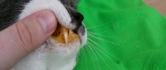
If problems with the lymph nodes are due to infection, then this is a disease called lymphadenitis. When inflammation begins in the body, the lymph nodes adjacent to the affected organ often become enlarged. For example, if a cat’s gums are affected, then the submandibular lymph nodes and tonsils usually become enlarged.
If the animal’s lymph nodes are massively enlarged, then you need to donate blood. With leukosarcoma and leukemia, there may be such symptoms, but without tests these diseases are not recognized in any way.
Other symptoms depend on the organ or part of the body in which the disease develops. With a sore throat, your cat may be reluctant to eat, cough, and drool. Some of these signs may be due to swollen lymph nodes because they interfere with swallowing.
If a cat's paws are infected, there will be an increase in lymph nodes along the animal's paws. Lameness may occur. Moreover, the reason for this will not be so much pain in the limbs themselves, but rather a painful reaction of the inflamed lymph nodes.
Lymphadenitis is generally not dangerous for cats. Naturally, subject to timely detection and treatment. When the disease is cured, the lymph nodes return to their normal state.
Treatment of lymphadenitis in cats
It is possible to choose effective treatment only by accurately establishing the causes
Source
Treatment taking into account the location of inflammation
When choosing the appropriate treatment tactics, it is necessary to take into account the area in which the lymph nodes are enlarged. Treatment measures taking into account this factor will differ radically.
Inflammation in the neck
The submandibular lymph nodes become inflamed for various reasons. Therapy is aimed at eliminating the factors that caused such changes.
Lumps behind the ear often signal diseases of the oral cavity. Treatment of diseased teeth, abscesses, stomatitis, and gingivitis is carried out.
Otitis media can provoke negative changes. Inflammation of the middle ear is detected at home: you need to lightly press the base of the auricle with your thumb.
The animal will show by its behavior that it is in pain; in advanced cases, pus may be discharged. Treatment is prescribed only by a specialist.
Inflammation on the abdomen
You can determine that a lump formed on the abdomen is lymphadenitis by the following signs:
- oblong shape, dense structure;
- immobility of the tubercle;
- soreness of the affected area with light pressure;
- rise in temperature.
Inflammation develops for the following reasons:
- mastitis;
- pathologies of the genitourinary system;
- leukemia;
- plague;
- hypothermia;
- gynecological pathologies.
Any disease must be treated urgently. The animal is prescribed anti-inflammatory drugs, antibiotics, vitamin and mineral complexes that help activate the body's protective functions.
If mastitis is detected, therapy is as follows:
- novocaine blockade;
- physiotherapy;
- surgery (in advanced cases);
- applying compresses;
- taking antibiotics.
Hello student
cat's lymphatic system
The lymphatic system of the cat was first studied in detail by Sugimura/Kudo/Takahata (1955, 1956, 1958, 1959 1960). Meier (1989) paid special attention to palpable lymph nodes in her study. The researcher draws attention to the fact that the palpability of physiological nodes largely depends on the degree of fatness of the cat. In the following, lymph nodes are described as palpable. which are found on a thin animal.
Palpable lymph nodes
Rice. 19. Palpable lymph nodes of a cat, schematized (according to Meier, 1989)
1 Inn. mandibulares; 2 Inn. retropharyngei late-rales; 3 Inn. cervicales superficiales dorsales; 4 In. axillaris proprius; 5 Inn. axillares accessorii; 6 In. inguinalis superficialis; 7 Inn. epigastrici caudales; 8 In. popliteus superficialis.
1. Palpable lymph nodes on the cat's head
Mandibular lymph nodes, Inn. mandibulares are permanent and palpable. In most cases, these are 2 large nodes, which are located behind the uncinate process of the maxillary bone and are laterally adjacent to the lingual facial vein. Dimensions of a separate unit: 12—15×8—10×6—8 mm.
Area of lymph flow: oral cavity and tongue, lips, nose, chin area, cheek glands, eyelids. Transient lymph is taken from the parotid lymph node and lateral retropharyngeal lymph nodes.
Outflow: the efferent vessels go to the accessory mandibular lymph nodes, the medial retropharyngeal lymph node and the ventral or dorsal superficial cervical lymph nodes.
Lateral retropharyngeal lymph nodes, Inn. retro-pharyngei late rales, are permanent and palpable. A group consisting of 1-3 (maximum 7) nodes lies under the base of the auricle on the caudal auricular vein. The lymph node is partially covered by the caudal edge of the parotid salivary gland. Dimensions: 0.5—15x 1—3x0.5 -3mm.
Area of lymph flow: auricle, orbital and frontal areas; parotid salivary gland; back of the head
Outflow: mainly to the medial retropharyngeal node, but also to the mandibular lymph nodes and to the dorsal superficial cervical lymph nodes.
2. Palpable lymph nodes in the neck and lateral chest wall of the cat
Superficial dorsal cervical ganglia, Inn. cervicales superficiales dorsales
3), are constant and palpable in young slender cats. In most cases, these are 2 nodes located under the cervical part of the trapezius muscle at the anterior edge of the scapula Dimensions 8 - 20 x 5 - 6 x 3 - 4 mm.
The area where lymph flows is: the dorsal region of the neck and shoulder, and partly also the thoracic limb. Receives transient lymph from the parotid lymph node and from the lateral retropharyngeal lymph nodes.
Outflow: either into the ventral superficial cervical lymph node and further into the venous angle, or directly into the jugular trunk
Proper axillary lymph node, In. axillaris proprius, is permanent and in most cases palpable. It lies at the level of the shoulder joint in the angle formed by the lateral pectoral and axillary veins. Sometimes there are two nodes Dimensions: 3 - 6 x 4 -5 x 2 - 4 mm.
Area of lymph flow: medial surface of the forelimb, palmar surface of the paw; lateral chest wall and mammary glands. Receives transient lymph from the accessory axillary node.
Outflow: either into the non-permanent axillary lymph node of the first rib, or, in its absence, directly into the venous angle.
Accessory axillary lymph nodes. Inn. axillares accessorii, are permanent and palpable. In most cases, these are two elliptical nodes located at the level of the III - VIII ribs, along the lateral thoracic vein in the angles formed by the outgoing branches. Dimensions: 5 - 10x 2 -3 x 2 - 3 mm.
Area of lymph flow: lumbar region, lateral thoracic and abdominal wall; caudodorsal region of the shoulder girdle and the inner surface of the shoulder and forearm.
Outflow: into the own axillary lymph node, partially past it into the venous angle and into the non-permanent axillary lymph node of the first rib.
3. Palpable lymph nodes in the groin area and on the pelvic limb of the cat
Superficial inguinal lymph node In. inguinalis superficialis, is permanent and occurs in very slender (young) cats. If, as in most cases, it is surrounded by adipose tissue, then palpation is difficult. This single node is located in the interfemoral gap and is adjacent to the external pudendal artery and vein. The position is the same for both males and females. Dimensions. 5—15 x 3—5 x 2—3 mm.
Area of lymph flow: medial surface of the thigh; in females the lymphatic vessels are from the mammary gland, in males from the scrotum. Receives transient lymph from the caudal epigastric lymph nodes and from the superficial popliteal lymph node.
Outflow: efferent vessels pass through the inguinal cleft into the abdominal cavity and reach the sacral lymph nodes.
Caudal epigastric lymph nodes, Inn. epigastrici caudales, is permanent and palpable in slender young cats. Along the superficial caudal epigastric arteries and veins, 1 to 5 nodes are successively located, surrounded by a thick layer of adipose tissue. Dimensions: 3 -8x2 -5x 1-3 mm.
Area of lymph flow, ventral abdominal wall, subcutaneous fat of the thigh; in females the mammary gland.
Outflow: efferent vessels go from one node of the chain to another and reach the superficial inguinal lymph node, and through the inguinal gap they reach the sacral lymph nodes.
Superficial popliteal lymph node, In. popliteus superficialis, is permanent and palpable. It lies in the popliteal fossa next to the lateral saphenous vein in the fatty tissue just under the skin. Dimensions: 6 - 7 x 4-5 x4 mm.
The area where lymph flows is the skin and subcutaneous fat and the muscles of the lower leg and paw.
Outflow: the efferent vessels pass through the inguinal fissure along with the efferent vessels of the inguinal lymph nodes and go to the non-permanent iliofemoral lymph node or directly to the sacral lymph nodes. Along with this, there is a facultative drainage into the sciatic lymph node (if present).
Physiologically nonpalpable lymph nodes
1. Other lymph nodes on the cat's head and neck
Parotid lymph node, In. parotideus, is constant, but not palpable, except in cases of pathological enlargement. It is located behind the temporomandibular joint on the superficial temporal vein. The lymph node may be partially covered by the cranial edge of the parotid gland.
While the mandibular lymph nodes are palpable, the accessory mandibular lymph nodes located behind them, Inn. man-dibulares accessorii, are unstable and cannot be palpated. A group of 1-3 nodes is located lateral to the maxillary vein.
Medial retropharyngeal node, In. retro-pharyngeus medialis, permanent, adjacent to the pharynx (in the form of a pair of nodes). It collects lymph from the mouth and pharynx and is a secondary node for other lymph nodes in the head.
Ventral superficial cervical lymph node, In. cervicalis superficialis ventralis, is almost constant, but is palpated only when it is pathologically enlarged. It lies in the fork between the external jugular vein and the superficial jugular vein before entering the thoracic cavity.
Deep Cervical Lymph Nodes, Inn. cervicales profundi, are represented by an unstable single middle node and an almost constant partially paired, partially unpaired caudal node. They are adjacent to the trachea on the ventrolateral side.
2. Other lymph nodes on the lateral chest wall and in the chest of the cat
Axillary lymph node of the first rib, In. axillaris primae costae, unstable. It occurs in every third animal. It is adjacent to the first rib on the lateral side and receives transient lymph from palpable axillary lymph nodes and accessory axillary lymph nodes.
Aortic thoracic lymph nodes, Inn. thoracici aortici, fickle, found in every second animal. 1-5 nodes are adjacent to the bodies of the thoracic vertebrae, sometimes only on one half of the body.
Intercostal lymph node, In. intercostalis, is only exceptionally found at the dorsal end of one of the intercostal spaces.
Cranial sternal lymph node, In. sternalis cranialis, is permanent. It is located next to the internal thoracic artery and vein at the level of the cartilaginous part of the II or IV rib.
Only every fourth cat has a caudal sternal lymph node, In. stemalis caudalis, adjacent to the sternum at the level of the apex of the pericardium. Cranial epigastric lymph node, In. epigastricus cranialis, found in the area of the xiphoid cartilage only in rare cases.
Diaphragmatic lymph node, In. phrenicus, is found irregularly near the opening of the vena cava in approximately every fourth animal.
Cranial mediastinal lymph nodes, Inn. mediastinales craniales are permanent. They form the largest group of permanent lymph nodes. 2 - 8 nodes are located at the level of the 1st or 2nd rib; on the right side, in the area up to the confluence of the right azygos vein, there are 1-3 more nodes.
Right, left and middle bifurcation lymph nodes, Inn. bifurcationis dexter, sinister et medius, are permanent, located around the bifurcation of the trachea.
Pulmonary Lymph Nodes, Inn. pulmonales are found in approximately every third cat in the extrapulmonary portion of the main bronchus.
3. Lymph nodes in the abdominal and pelvic cavities of the cat and other lymph nodes in the pelvic area
Aortic lumbar lymph nodes, Inn. lumbales aortici, located on both sides of the abdominal aorta to the origin of the deep circumferential iliac artery. A group of approximately 2-5 nodes located cranial to the renal artery occurs constantly. The caudal group has 3-7 nodes, but they may be absent.
Further along the abdominal aorta, between the origins of the deep circumferential iliac artery and the external iliac artery, there are large ribbon-like medial iliac lymph nodes, Inn. iliaci mediates, which are permanent. They collect lymph from the pelvic cavity and hind limb and direct it to the lumbar trunks.
Sacral lymph nodes are constantly found at the beginning of the internal iliac artery, Inn. sacrales. Following the further course of the vessel under the vault of the pelvic cavity, in approximately every third cat, other small lymph nodes under the same name can be found.
Along the course of the external iliac artery, before its entrance into the inguinal fissure, the iliofemoral lymph node occurs very irregularly, In. iliofemoralis. The femoral lymph node is also unstable, In. femoralis, which may be located at the exit of the femoral canal between the tensor fasciae lata and the sartorius muscle. In exceptional cases, the subiliac lymph node can be found in the knee fold, In. subiliacus. Sciatic lymph node, In. ischiadicus is permanent; it is located adjacent to the caudal gluteal artery and vein.
Rice. 20. Schematic representation of the lymph nodes and collector lymphatic vessels of a cat without lymph nodes of the internal organs of the abdominal cavity
Av is the place where lymph flows into the left venous angle, angulus venosus; CC cisterna chyli; Dt ductus thoracicus; Tj truncus jugularis; Tl truncus lumbalis; TV truncus visceralis
1 In. parotideus; 2 Inn. mandibulares; 3 Inn. mandibulares accessorii (non-permanent); 4 In. retropharyngeus lateralis; 5 In. retropharyngeus medialis; 6 Inn. cervicales superficiales dorsales; 7 In. cervicalis superficialis ventralis (almost permanent); 8 In. cervicalis profundus medius (non-permanent); 9 In. cervicalis profundus caudalis (almost permanent); 10 In. axillaris primae costae (fickle); 11 In. axillaris proprius; 12 Inn. axillares accessorii; 13 Inn. thoracici aortici (fickle); 14 In. intercostalis (non-permanent); 15 In. sternalis cranialis; 16 In. sternalis caudalis (fickle); 17 In. epigastricus cranialis (non-permanent); 18 In. phrenicus (fickle); 19 Inn. mediastinales craniales; 20 Inn. bifurcationis et pulmonales (the latter is unstable); 21 Inn. lumbales aortici; 22 Inn. iliaci mediales; 23 Inn. sacrales; 24 In. subiliacus (fickle); 25 In. iliofemoralis (non-permanent); 26 In. femoralis (fickle); 27 In. inguinalis superficialis; 28 Inn. epigastrici caudales; 29 In. ischiadicus; 30 In. popliteus superficialis.
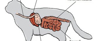
Rice. 21. Lymph nodes of the abdominal organs of a cat and their collecting lymphatic vessels, very schematically: the intestine is shown from the ventral side, the stomach and spleen are thrown forward CC cisterna chyli; Dt ductus thoracicus; Tl truncus lumbalis; TV truncus visceralis
1 Inn. lumbales aortici; 2 Inn. lienales; 3 Inn. gastrici; 4 Inn. hepatici; 5 In. pancreaticoduodenalis; 6 Inn. jejunales; 7 Inn. caecales; 8 Inn. coli; 9 Inn. mesenterici caudales.
For internal organs in the area covered by the celiac artery, lymph drains through the following lymph nodes. Splenic Lymph Nodes, Inn. lienales, are located at the site of branching of the splenic artery and vein. Gastric Lymph Nodes, Inn. gastric, located along the lesser curvature of the stomach. Hepatic Lymph Nodes, Inn. hepatici, can be especially large; they are located on both sides of the portal vein. Pancreatic-duodenal lymph node, In. pancreaticoduodenalis, adjacent to the cranial pancreaticoduodenal vein. All named lymph nodes are permanent; their number may vary slightly.
The following lymph nodes are constantly found in the small and large intestines: A particularly large group is formed by the jejunal lymph nodes, Inn. jejunales. They extend from the cranial root of the mesentery into the mesentery of the jejunum. Lymph nodes of the cecum, Inn. caecales, lie on both sides of the cecum. Lymph nodes of the colon, Inn. colici are located in the mesentery of the ascending and transverse colon. In the mesentery of the descending colon there are caudal mesenteric lymph nodes, Inn. mesenterici caudales.
Lymph flow and collector lymphatic vessels of the cat
The lymph nodes of the head are connected to each other by a network of efferent vessels. The jugular trunk, truncus jugularis, begins from the medial retropharyngeal lymph node. It receives lymph from the lymph nodes and then drains into the venous angle. Lymphatic vessels carrying lymph from the thoracic limb, lateral chest wall and thoracic parts of the mammary gland drain directly into the venous angle.
Lymph from the lateral surface of the hind limb enters the pelvic cavity through the popliteal and sciatic lymph nodes. Lymphatic vessels from the medial surface of the hind limb and the abdominal parts of the mammary gland also enter the pelvic cavity through the femoral canal. Both streams pass through the sacral and medial iliac lymph nodes. From here begin the lumbar trunks, trunci lumbales, which flow into the lumbar cistern, cistema chyli. From the ventral side, the visceral trunk, truncus visceralis, flows into the lumbar cistern.
The lumbar cistern is a sac-like cavity located on the dorsal side of the aorta between the crura of the diaphragm. From it, the lymph enters the thoracic duct, ductus thoracicus, which, after passing through the aortic opening of the diaphragm, forms a vascular system resembling a rope ladder. When passing to the left side in the precordial mediastinum, the thoracic duct forms a single trunk, which, before flowing into the left venous angle, again divides into several branches. The right lymphatic duct, ductus lymphaticus dexter, is formed in the same way as in a dog.
Literature used: Anatomy of a dog and a cat (Coll, authors) / Transl. with him. E. Boldyreva, I. Kravets. - M.: “AQUARIUM BUK”, 2003. 580 pp., ill. color on
Download abstract: You do not have access to download files from our server. HOW TO DOWNLOAD HERE
Prevention measures
Prevention, proper, proper care of the cat will prevent lymphadenitis and reduce the risk of the pet contracting dangerous viral-bacterial and parasitic diseases.
- Timely vaccination, revaccination;
- Balanced diet;
- Reducing contact with street animals;
- Strengthening the immune system with vitamins and mineral complexes with a natural diet.
- Systematic inspection.
- Timely treatment of inflammatory processes, systemic, viral, bacterial diseases;
- Treatment against ecto-endoparasites.
Features of inflammation depending on location
Clinical manifestations depend on where the lesion is located. Let us consider the features of inflammation depending on the localization of pathological processes.
On the front and hind legs
If there is an inflammatory process on the front and hind legs, in addition to weakness, lethargy, and high temperature, lameness, stiff gait, discomfort due to increased tension in the muscle structures, and pain when walking are noted.
The axillary, supraclavicular, ulnar lymph nodes, and inguinal nodes are affected during inflammation of the hind limbs. Causes: infections, injuries.
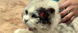
Damage to the lymph nodes in the neck
The lymph nodes in the neck become inflamed due to viral or bacterial infections, or when eating low-quality, stale meat. Noted for toxoplasmosis, respiratory infections (rhinitis, tracheitis, bronchotracheitis, laryngotracheitis, sinusitis, tonsillitis).
The nodes are painful and greatly enlarged. The animal is lethargic, apathetic, and refuses food. Depression gives way to anxiety. A white coating is noticeable on the tongue. An unpleasant odor emanates from the mouth.
Submandibular lymph nodes, on the cheeks
Submandibular nodes and lymph nodes on the cheeks become inflamed due to viral and bacterial infections, dental diseases (stomatitis, gingivitis), injuries to the oral mucosa, and penetration of pathogenic agents into regional lymph nodes. Located under the angular rounding of the jaw near the salivary glands, in the cheek area (lymph nodes on the cheeks).
Cats have difficulty eating and drinking water. The oral mucosa is hyperemic and edematous. The temperature is increased by 1-2 degrees from normal. Loss of appetite causes your cat to lose weight.
Treatment of abscesses
Before the lump on the cat's neck bursts, you should immediately begin treatment. In this case, only a veterinarian can help.
There is no preliminary treatment for such an abscess. After the examination, the cat is immediately sent to the surgeon. He opens the seal with a scalpel and cleans the cavity of pus. After this, antiseptic treatment of the wound is carried out. If necessary, the cat is given stitches and prescribed antibiotics to prevent the re-development of inflammation.
If left untreated, the abscess can open on its own. However, this is very dangerous for the animal, since it can lick the pus flowing from the wound. In addition, there is a high risk of infection.
Risk group
As stated above, older cats are most likely to develop lumps on their necks. At the same time, there are the following risk factors that can cause the development of such formation in younger individuals:
- the presence of endocrine disorders;
- the presence of parasites in the body;
- excess body weight.
Only a veterinarian can accurately determine the cause of the development of a lump under a cat’s neck. He also prescribes a treatment regimen, since it is strictly forbidden to independently treat any formations on the animal’s body.
What is lymphadenitis
Lymphadenitis is an inflammation of the tissues of the lymph nodes, which is accompanied by an increase in their size due to an increase in the number of lymphocytes and the migration of leukocytes into them. In animals, as in humans, the inflammatory process in the lymph nodes develops due to primary inflammation of a specific localization.
Important! In some cases, lymph nodes can become a source of infection if suppuration develops in them.
Causes of lymphadenitis in cats:
- acute, chronic viral, bacterial, parasitic infections;
- weakening of immunity, resistance;
- respiratory diseases (rhinitis, sinusitis, bronchitis, laryngotracheitis, sinusitis, tonsillitis);
- neoplasia (malignant tumors that metastasize to the lymph nodes);
- lymphoma (cancer of the lymph nodes);
- failure to comply with hygiene rules;
- autoimmune infections;
- herpes virus;
- mycoses;
- connective tissue diseases;
- allergic reactions, allergies of various etiologies;
- injuries, penetrating wounds through which pathogenic microorganisms penetrate tissues and internal organs.
Inflammation can be provoked by blood-sucking parasites (fleas, ticks, lice eaters), endoparasites (worms), while damage to the lymph nodes by the inflammatory process during parasitic infections is less common.
In general, lymphadenitis is a protective, defensive reaction of the body that is not life-threatening for the pet as long as the lymph is not affected.