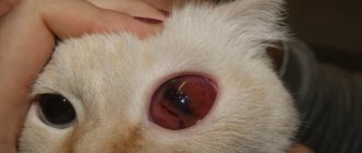
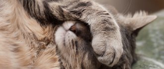
Glaucoma in cats is an increase in intraocular pressure associated with a malfunction of the eye. This condition is accompanied by an increase in eye volume, pain, squinting, clouding of the cornea, swelling of the eyelids, and increased injection of blood vessels into the sclera.
Glaucoma can significantly worsen your pet's condition and lead to refusal to eat, general weakness and sometimes even aggression.
Most often, the problem manifests itself as unilateral damage to the eye, although cases of bilateral damage are also possible, which can lead to symmetrical squinting and tearing.
Glaucoma can occur due to increased production of intraocular fluid and disruption of its outflow from the eye.
Glaucoma in cats occurs:
- open-angle, when there is no narrowing of the iridocorneal angle;
- closed-angle, when there is a violation of the outflow associated with a narrowing of the system that drains fluid from the eye.
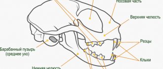
The structure of the cat's visual apparatus
Vision in cats performs all the same functions as in humans: it allows them to navigate in the surrounding space and on the basis of information received through the eyes:
- move;
- contact with people and other animals;
- avoid obstacles;
- find food, etc.
The structure of the visual apparatus in humans and cats is extremely similar. Its components are:
- cornea;
- lens;
- front camera;
- pupil.
Diagram of the structure of the cat's eye apparatus
At their core, the eyes of living creatures included in this class are represented by lenses created by nature, which provide:
- obtaining information by refracting light rays;
- send neurosignals to the brain center.
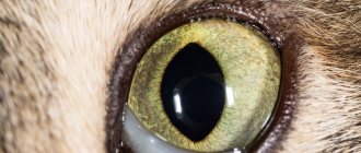
However, there are still some differences between the eyes of humans and our purring smaller brothers: the cat’s visual apparatus is equipped with unique nerve cells:
- chopsticks;
- cones.
These photoreceptors are responsible for refracting light and converting it into nerve impulses.
This is what photoreceptor nerve cells, rods and cones, look like
Please note: humans also have these unique nerve cells in their organs of vision, but their ratio will be 4/1, while in cats it is 25/1.
The unique ability of cats to see perfectly at dusk and in the dark is also due to the structure of the eye apparatus. So, inside their eyeballs there are formations called “tapetum”. This element determines:
- night vision of domestic predators;
- the glow of the eyes, which many owners are afraid of in the dark.
An interesting point: the color perception of representatives of the cat family, unlike humans, is mainly based on distinguishing thousands of shades of gray. The palette we are accustomed to is not known to cats; they will not be able to distinguish red from blue, even if they try. That is why, when choosing, for example, toys for pets, you should not think about the color of this or that item: this point is absolutely not important to your pet.
This is how cats see in the dark
You can read more about the structure of cat eyes in a special article on our portal.
Arterial hypertension in cats: clinical symptoms from the visual apparatus
Bernard Clerc, Sabine Chaori
With the development of blindness and/or hemorrhage in the eye area, the possibility of arterial hypertension should be considered. Treatment with amlodipine may provide beneficial results in some cases.
Hemorrhages that occur at the level of the visual apparatus, as well as sudden blindness in older cats, are quite often a reason for consultation with a veterinary specialist. According to Littman MP (1999), one of the main causes of these disorders is blood pressure (BP). Since the first publications in veterinary ophthalmology (Boldy K., 1983), more and more people are learning about this pathology.
Blood pressure (BP) measurement has not yet become part of routine veterinary practice. This is often due to lack of diagnosis of blood pressure (Littman MP, 1999). It is likely that the percentage of cats with elevated BP is much higher than expected, so systematic determination of BP in older cats over ten years of age is required.
Indirect methods of measuring blood pressure are carried out both using a special Doppler apparatus and using an oscillometric study. The first method allows you to estimate systolic pressure and is used more often. The average for a healthy cat is 118 mm (Littman MP, 1999). Systolic pressure exceeding 170 mmHg is considered abnormal (Littman MP, 1994).
Changes in the visual apparatus are often an alarming symptom for owners. External ophthalmological examination is not difficult, but the most characteristic damage is localized in the fundus (Glaze MB, Gelatt KN, 1998; Hayreh SS, 1996; Morgan RV, 1986). Therefore, determining blood pressure requires good practical skills in direct and indirect ophthalmic examination. This approach is based on knowledge of the semiology of the visual apparatus (Appendix 6).
Appendix 6. Reminder on semiology
• Significance of fundus examination The fundus is the back part of the eye and is examined using an ophthalmoscope. Morphologically, it is represented by three membranes, two of which (the retina and the choroid) are partially transparent (in most cases, the vessels of the choroid cannot be visualized). The shells are penetrated by many identifiable vessels, which are reference points for recording the location of damage. Blood extravasation can be localized under, above, or inside different membranes. The hemorrhagic picture corresponds to the location of the pathology. Consequently, an ophthalmological examination allows one to identify the hemorrhagic area and thus establish its nature. Fluorescein angiography can provide additional information.
Identification of the structure of vessels that acquire a red color is carried out by illuminating this area with white light. Hemorrhage has a natural red color, but this color can be modified through the interposition of incompletely transparent structures, such as the pigment epithelium or the retina itself.
• Detachment and elevation of tissue layers. The vessels of the retina are always visualized in the upper position of its transparent part. They are a good control of retinal elevation because their displacement is accompanied by a change in its position. If the hemorrhage is located in front of two layers of tissue, then they are not visualized; if it forms under the retina, then they rise.
If the hemorrhage is localized under the choroid, then the two raised inner membranes do not distort the visual picture of the fundus of the eye. Therefore, it is quite possible to distinguish between preretinal, subretinal or choroidal hemorrhages. Their color varies from red in the first case to dark red for the remaining hemorrhages. The connection between the vitreous and the retina is very weakened, and therefore blood in this area can cause a pocket-shaped detachment with a horizontal level, which is explained by the sedimentation of red blood cells (picture of the underwater part of a ship). Due to the transparency of the retina, hemorrhages are controlled and can be localized quite accurately.
Blood penetrates between retinal cells that are subject to dissociation. Hemorrhages in the superficial layers of the retina spread diffusely between glandular cells, which form a fibrillar “felt” in a horizontal position. They are directed towards the optic nerve.
In this case, hemorrhages have the shape of a brush or flame. Bipolar cells and photoreceptor cells in the form of columns located perpendicular to the surface of the retina limit the diffusion of blood. Red blood cells become trapped between these columns and form dot-shaped hemorrhagic spots. Hemorrhagic perivascular images, shaped like a sleeve, are easily identified by their location and appearance in the form of a semicircular ridge. Sometimes they are monoliform (bead-shaped) in nature (Fig. 2).
| Figure 2. Localization of hemorrhages at the bottom of the eye. |
• Changes in vascular morphology - Diffuse dilatation A diffuse increase in the size of retinal vessels is caused by the difficulty of outflow of contents from the vessels (blood hyperviscosity syndrome and polycythemia). Hypertension mainly leads to local vascular dilatation.
— Local dilatation of blood vessels: aneurysms Arterial aneurysms are local dilation of arteries detected by visual examination. They are predominantly localized on capillaries and in dogs usually have a subclinical course, and also appear in diabetes mellitus after several years of its development.
The first stages of the development of hypertension provoke a change in the diameter of blood vessels with dilation of the veins at the bottom of the eye. Hemorrhages are combined with changes in the fundus. If blood does not circulate in the vessels, they lose color. The picture of the vessels may be camouflaged by the resulting perivascular “sleeve,” which gives rise to a false assumption about a change in the diameter of the vessel itself.
• Hemorrhages of the fundus of the eye Localization of hemorrhages of the fundus of the eye depends on damage to the vessels, which take the form of a geographical map or puddle.
— Map- or puddle-shaped hemorrhages Hemorrhages are map- or puddle-shaped when they form between layers of tissue. They qualify as follows:
- preretinal, when effusion forms between the vitreous body and the retina of the eye; have a red living color if widespread. When the animal is standing, a horizontal level is visible. The retinal vessels are not visualized;
- subretinal, formed as a result of effusion that forms between the neurogenic part of the retina and the pigment epithelium, or under the pigment epithelium, where they have a peculiar tint (red “shaded” due to the absorption of light by the retina and its vessels, which become visible).
— Local hemorrhages Hemorrhages localized inside the retina, if they are small in size and penetrate into the structures of its tissues:
- perivascular: superficial, running along the vessels;
- in the shape of a flame or fan: superficial, located in the layer of glandular cells;
- in the form of points: have an intermediate position, delayed by the forming columns of bipolar cells.
With pronounced tissue dissociation, the form of hemorrhages varies. The photographs in this article illustrate all of the listed vascular modifications.
• Perivascular modifications Perivascular modifications (called “cobs of cotton”) occur as a result of reactions around disease-prone vessels. They are provoked by microvascular ischemia due to hypertension, diabetes mellitus and systemic lupus erythematosus. The artery is also engulfed in tissue reaction.
Reasons for consulting cats if their general health is compromised are blindness or hemorrhages inside the eye. An ophthalmological examination with the detection of hypertension makes it possible to make a diagnosis. In any case, it is necessary to identify other causes of hemorrhage or vascular pathology. In relation to prognosis and treatment, the article takes into account the experience that we have acquired in relation to representatives of this species of animal.
Reason for consultation
Arterial hypertension in cats is a disorder that, judging by numerous publications, is currently diagnosed quite often (Crispin M., Mould RB, 2001; Glaze MB, Gelatt KN, 1998; Hayreh SS, 1992; Littman MP, 1994). Animals susceptible to this disease come to the ophthalmologist for two reasons:
- when the owner discovers they have a “red eye” (eye hemorrhage, sometimes uveitis);
- if the owner notices a decrease or loss of vision (impairments in both eyes).
In these cases, damage to the visual apparatus leads to the need to identify hypertension. It is very important to conduct a fundus examination, which can most accurately guide the clinician in making a diagnosis. All the described disorders cannot always be found in one animal.
Methodology, symptoms and damage
It is impossible to separate the patient's ophthalmological examination from the clinical examination. Cats often have poor general condition, dull and brittle hair. Most of them, as it turns out during the consultation process, are blind. On average, vision loss occurs on the eighth day. Sometimes hemorrhages can be observed directly both under the mucous membranes or in the anterior chamber of the eye, and in the vitreous body. Visual function is often impaired or absent (personal observations).
The examination of the visual apparatus is carried out in a classical way. After functional assessment (photomotor reflex, cotton swab pain test, and danger blink reflex), examination is performed using a transelluminator and/or slit lamp. This allows us to note modifications in the anterior chamber of the eye, as well as in the iris, lens and vitreous body.
An ophthalmological examination is carried out after measuring intraocular pressure, which is usually normal. The iris is dilated with drops that cause mydriasis (tropicamide, Mydriaticum R). In this case, you can obtain the most complete information, because hypertension is often accompanied by hemorrhages in the fundus and/or retinal detachment (Appendix 2 and 4).
Appendix 2. Manifestations from the visual apparatus when hypertension occurs in a cat
- Frequently manifested manifestations at the level of the visual apparatus: - Hyperemia. - Hemorrhage. — Exudate determined using a cotton brush. - Limited peeling. - Complete detachment. — Periarterial hemorrhage in the form of a coupling. — Degenerative changes at the level of the choroid plexus.
- Rarely encountered changes in the visual apparatus in hypertension: - Glaucoma - Anterior uveitis - Chorioretinitis - Retinal degeneration - Papillary edema
Appendix 4. Retinal detachment and eye damage
In the vascular network of the eye, hypertension provokes a change in the pattern of the anterior and posterior segments.
• In the anterior segment, hemorrhages can be observed in both eyes, symmetrically or not (photo 1), or in only one eye (photo 2).
| Photo 1. Cat of European breed, female, 13 years old. Bilateral hemorrhages of the visual apparatus with periodic resorption and relapse. The blood coagulates; in the left eye, retraction of the clot leaves color in the peripheral part of the iris. | |
| Photo 2. Cat of the European breed, neutered, aged 11 years. There is hemorrhage in the anterior chamber of the eye, formed by one compressed clot, which partially masks the iris and pupil. The opening of the pupil is impaired due to the formation of synechiae, forming a picture of three leaves. The lens is susceptible to cataracts. The iris bears elements of neovascularization, which are explained by the chronic inflammatory process and reaction of the iris. | |
Hemorrhage in the anterior chamber of the eye is often organized; the cause of its origin may be associated with the iris or ciliary body. After therapeutic treatment, complications persist on the iris (Figure 3) with damage to its surface, indicating the consequences of uveitis.
| Photo 3. Cat of European breed, neutered male, age 13 years. There is deformation of the iris and a change in its color (dim, irregular, with modification). There is a complication due to successive inflammations caused by hypertension and hemorrhages. The iris on the surface bears elements of neovascularization. Intraocular pressure is normal. There is no Tyndall effect. | |
An organized nodule is present on the iris at six o'clock in relation to the open base of the pupil. Hemorrhagic lesions regress with therapy aimed at reducing hypertension.
• in the posterior segment of the eye, manifesting as hypertensive retinopathy (when compared with the normal fundus of the eye, photo 4).
| Photo 4. Cat of European breed, male, the fundus corresponds to the norm. Nerve fibers are not subject to myelination, which explains the gray color at the level of the papilla; hemocirculation is present. The carpet area has a green color and a homogeneous pattern. |
Vitreous hemorrhages are identified by examination through a transluminator after dilatation or by ophthalmoscopic examination. The most common and characteristic disorders are detected using an ophthalmoscope. An ophthalmoscopic examination can reveal retinopathy, choroidopathy or optic neuropathy.
• Retinal detachment - Numerous serous type retinal detachments (photos 5, 6, 7) were observed in the evolving stage of the disease. Bullous or so-called serous retinal detachments could be observed with inflammation of the fundus of the eye. The cause of detachment is determined by measuring blood pressure.
| Photo 5. Fundus of a cat prone to blindness and in a state of hypertension. Elongated gray spots indicate retinal edema, which is caused by an acute inflammatory reaction that has recently occurred. The process is reversible as a result of treatment with the prescription of an antihypertensive drug (amlodipine). | Photo 6. A pattern of leopard color visible on the bottom of a cat’s eye: the carpet area looks like butterfly wings or peacock feathers. Gray spots correspond to acute edematous areas of the retina, which partially absorb incoming light through lacunae and give a picture of dark gray spots. |
| Photo 7. An 11-year-old Persian cat in a state of hypertension. The fundus of the eye indicates modification of light in the carpet and equatorial zones. Bullous detachment is better felt with binocular visualization and lends itself well to examination. We are talking about the manifestation of hypertension, which can be comparable to the restoration of vision. | |
— Bullous inflammation is the only one that can be extensive (photo 8). In this case, it is similar to macular detachment of the serous type, which is described in humans. It is located in the area centralis of the retina, that is, in an area depleted of blood vessels, which is the main object of visualization.
| Photo 8. The right eye of a cat susceptible to subretinal (located under the retina) hemorrhage in the form of a geographical map, red and slightly shaded in color, where there are also elements of hemorrhage in the form of brushes, lines and dots. The green carpet area has a scattering of darker dots that are the result of hemorrhagic scars. |
— Detachment of the retina with ruptures and hemorrhages. Retinal detachment may spread, be accompanied by palpable hemorrhages immediately after iris dilation (Figure 9), and may be a consequence of choroid hemorrhages and edema that occur with choriocapillary disorders.
| Photo 9. The cat's left eye. Its bottom is subject to the formation of an infinitely red zone with a rectangular border (a), which corresponds to the site of retinal detachment, elevated due to the formation of serous-hemorrhagic effusion. In this cliche, the rupture zones are not controlled, but the vessels are sclerotic and have a white color (b). Other vessels are also observed in the 7 o'clock area. This indicates long-standing retinal detachment. |
• Hemorrhages - Hemorrhages, shaped like a geographic map, are located between two layers of tissue. They can be preretinal, subretinal, or localized under the pigment epithelium (photo 8, 9, 10). It is very difficult to differentiate hemorrhages in the form of a geographical map from those located inside the retina. However, their pronounced distribution and dark red color are additional elements indicating subretinal hemorrhages.
| Photo 10. Fundus with typical hemorrhage in a cat with high blood pressure. There are variations in the visual image of hemorrhage: in the form of a geographical zone on the upper border of the retina and below it (a), brushes (b), and perivascular points (c). |
In photo 10, the blood has a very clear location in the subepithelial layer. As for Photo 9, the hemorrhage is widespread under the detached retina, which is likely accompanied by a tear in the ora serrata. The retinal vessels undergo degeneration with blood loss, which materializes into white tracts passing through the visualized field.
- Hemorrhages inside the retina in the form of dots, flames, cotton brushes and perivascular. Retinal hemorrhages form around its structures. Superficial hemorrhages are located along the entire length of the ganglion cell fibers and around the vessels (photo 10).
Hemorrhages of the middle zone develop between bipolar cells. They have the shape of dots (photos 8, 11) and when they disappear, they leave traces in the form of black dots.
| Photo 11. Observed modifications in a cat in the area of the carpet zone of the retina. Small brown dots are visualized, which correspond to complications caused by pinpoint hemorrhages. Wider beach shapes with increased reflection are explained by reorganization in the carpet zone due to hemorrhages. |
• Choroid lesions have been well studied by various authors in the field of veterinary medicine (Crispin M., Mould RB, 2001) and may be responsible for damage to the retina or located under its layer.
Diagnostics
1. Preliminary diagnosis
Ocular damage associated with hypertension has been observed in older cats (usually 11 to 17 years of age) presented for consultation due to hemorrhage or visual dysfunction. This is the first element and guides the diagnosis of hypertension. At the ophthalmology center of the Alfort veterinary school, this method of preliminary diagnosis is systematically confirmed by measuring blood pressure using a Doppler apparatus (Fig. 1 and photos 13, 14).
| Figure 1. Installation of the Doppler apparatus 1. Air-filling cuff placed on the tail. 2. Doppler device placed on the tail artery. 3. Pressure gauge for reading pressure. | |
| Photo 13. Blood pressure measurement (Doppler method). A cuff inflated with air and an air insufflation (supply) system, which is fixed on the tail. | Photo 14. Doppler trap and recording system (Vettex R) of arterial systolic pressure. |
Measurements are carried out in a quiet room in the presence of the owner. An average blood pressure value of 265 mmHg was obtained in twenty-one clinical cases. Sometimes this figure exceeded 300 mmHg (seven cases out of twenty-one). Fundus hemorrhages indicate that hypertension is particularly severe, probably over many months. Auscultation was systematically performed in such animals. The unsatisfactory general condition of patients and accompanying heart murmurs were noted quite often. An echocardiographic study was also carried out (cardiology service). At the first consultation, a biochemical blood test was performed (urea, creatinine, glucose, thyroid hormone T4). Quantitative indicators of urea and creatinine often exceeded the physiological norm (Appendix 1).
Appendix 1. Results of a general examination of 28 cats susceptible to hypertension presented to the ophthalmology clinic of the Alfort veterinary school
- Average age of animals: 14.3 years
- Reason for consultation: - hemorrhage in the eye area: 7/28 - blindness: 17/28 - others: 4/28 (red eye, mydriasis, anisocoria, neurological causes).
- Clinical symptoms of the visual apparatus: - loss of vision: 25/28 - hemorrhage of the visual apparatus: 22/28 - retinal detachment: 19/28
- Urea and creatinine (21 cases): - uremia (g/l): < 0.7 (5/21) 0.7-1 (6/21) >1 (10/21) - creatininemia (mg/l): <20 (7/19) 20-25 (5/19) >25 (7/19) Similar studies have been carried out in other departments of veterinary medicine.
2. Differential diagnosis
Blood pressure measurement allows for a definitive diagnosis. Other vascular, blood or inflammatory processes can generate the development of hemorrhages, retinal detachment and alter vascularization. The pattern of vascular modifications is relatively specific and can facilitate etiotropic diagnosis (Appendix 3). In older cats, the likelihood of this type of hypertension-related damage is statistically 90%.
Appendix 3. Differential diagnosis of the causes of hemorrhages at the level of the fundus of the eye
| Cardiovascular disorders | Ophthalmic symptoms | Other clinical symptoms | Elements required in the diagnostic field |
| Polycythemia, polyglobulia. | Redness of the conjunctiva. Dilatation of small vessels. Dilatation of retinal vessels, mainly veins. Rarely occurring hemorrhages. Rarely occurring retinal detachment. | Redness and blood filling of all mucous membranes. Neurological symptoms. Difficulty performing physical activity. | Red blood cell counting. Hematocrit > 60%. |
| Hyperviscosity syndrome. | Permanent dilatation of retinal vessels. Evolving hemorrhages. | Epitaxis. Hemorrhages of the digestive tract. Symptoms of kidney failure. | Protein electrophoresis. Myelogram. |
| Thrombocytopenia. | Hemorrhagic effusion in the ocular or periocular area. | Petechiae, ecchymosis of the mucous or submucosal layer. | Clinical analysis and blood leukemia. Platelets. Increased bleeding time. |
| Anemia. | Paleness of the vessels of the retina. Hemorrhages. | Ictericity. Pallor of mucous membranes. Tachycardia. Polypnea. Heart murmurs. | Quantitative indicator and leukoformula of blood. |
| Impaired hemostasis. | Hemorrhages of the visual apparatus. Subconjunctival and orbital effusions. | Hemorrhages. Melena. ICE. | Bleeding time ↑. Time of cephalin-kaolin ↑. Temps de Quick ↑. Platelets N or ↓. |
| Arterial hypertension. | Hemorrhages in the fundus of the eye. Hemorrhages of the anterior segment (anterior chamber) of the eye. +/- retinal detachment. +/- swelling of the papilla. | Unsatisfactory general condition of the body due to a chronic disease. Emaciation. | Determination of blood pressure. Identifying the cause of hypertension |
| In cases where the cause of fundus damage is due to hypertension, both hemorrhages and scarring after hemorrhage are present, which may be associated with swelling in the papilla and/or at the level of retinal detachment. | |||
- Polyglobulia and myeloma increase blood viscosity and provoke an increase in the diameter of blood vessels, in particular veins (Hribernik TN, 1982). These vascular disorders are rarely complicated by hemorrhages. Dilatation is uniform throughout the entire length of the vessels. In cases of polyglobulia, it is moderately expressed.
- The development of arteritis and diabetic vasculitis at the microvascular level in diabetes mellitus has been well studied and has been reproduced experimentally (Herrtage ME, Barnett KC, Mac Dougall DF, 1985). Capillary aneurysms and sleeve-shaped perivascular hemorrhages are described as classic ophthalmic manifestations and may indicate hypertension with the manifestation of pinpoint hemorrhages in the retina in the form of inflammation. The spread of hemorrhages is often limited. Full-blown diabetes mellitus occurs more often in adult dogs than in cats.
- Hemostasis disorders are also often the cause of hemorrhages. They can appear everywhere, which proves the need for a more careful examination of all the patient’s mucous membranes. However, localization of hemorrhages to the eyeball is possible, which can cause confusion. A general clinical blood test supplements information about endogenous and exogenous pathways of hemostasis**, which in turn creates conditions for differential diagnosis.
- Acute uveitis is also included in the differential diagnosis of hypertension; they predominate in the pathology of the cat’s visual apparatus (Martin CL, 1982). Severe uveitis can occur with diseases of a parasitic, bacterial or viral nature (FeLV, FIV). Feline infectious peritonitis is also responsible for the development of a vascular reaction (vasculitis): it is likely responsible for most diagnostic errors. Exudates, as a rule, with this pathology are enriched with red blood cells (photos 15 and 16). The initial inflammatory process in the eyeball, the symptom of uveitis, are convincing elements in the diagnostic orientation. Examination of the obtained fluid sample indicates that the effusion is not a component of blood, but the exudate is enriched with red blood cells. Additional studies with counting of blood cells and leukoformula, as well as determining the titer of antibodies responsible for a specific disease, are a condition for differential diagnosis.
| Photo 15. A 10-year-old cat presented for consultation due to bilateral hemorrhage in the anterior chamber of the eye. These disorders, reminiscent of hypertension, are caused by the development of feline infectious peritonitis (serology, electrophoresis). | Photo 16. A six-year-old cat is susceptible to retinal detachment and uveitis. The iris has a fat pattern, suggesting anterior uveitis, while the detached retina is “overlaid” on the lens. The iris is subject to inflammation, which causes thickening of the tissue (white color). Vessel traces are visualized through the “perivascular coupling.” Pinpoint hemorrhages indicate intraretinal formation of some hemorrhages. |
Prognosis and treatment
Typically, cats presented for consultation because of vision loss due to hypertension have progressive vascular compromise (pressure generally 250 mmHg or greater). In this case, we are talking about forms of the disease with an unfavorable prognosis, because disorders, even when treated, leave serious consequences. In a limited percentage of cats (according to our studies, 10-20%), vision restoration occurs after administration of the calcium channel inhibitor amlodipine (amlodipine) and a diet depleted in table salt.
Modifications of the fundus in the form of bullous retinal detachment indicate a more favorable prognosis. In this form, the transudate formed under the retina, according to our research, is reversible.
Treatment, even with changes in pressure, requires the use of amlodipine (Amlor). Initially, it is usually used at a rate of 0.6 mg/day for cats weighing 4 kg (1/8 tablet) and 0.9 mg for larger individuals (Glaze MB, Gelatt KN, 1998; Henik RA, 1997; Herrtage ME, 1985 ;Littman MP, 1999; Synder PS, 1998). Such treatment, which must be carried out urgently, does not exclude a full cardiac examination and identification of the cause of hypertension. Six out of eleven cats responded excellently to treatment. If the response to therapy is not entirely sufficient, then the prescribed dose of the drug increases from 0.9 to 1.25 mg/day. Prescribing other medications for the cat, such as angiotensin converting enzyme (ACE) inhibitors, did not produce a positive result. Such drugs are used to eliminate some causes of hypertension, but together with amlodipine. According to some authors (Glaze MB, Gelatt KN, 1998; Henik RA, 1997; Herrtage ME, 1985; Littman MP, 1999; Synder PS, 1998), they did not observe any adverse effect when using amlodipine. Measuring blood pressure at the first stage of treatment is necessary, as it allows us to assess the effectiveness of manipulations. Treatment allows for the disappearance of effusions and hemorrhages; at the same time, owners constantly report an improvement in the general condition of patients. However, restoration of vision directly depends on the severity of complications caused by formed precipitates, as well as scarring. A 50% recovery of vision in animals during treatment has been reported (Crispin M., Mould RB, 2001). The percentage of vision restoration according to our research is being clarified. However, it is significantly lower (less than 20%). This indicator depends on timely diagnosis and treatment. It is very important to carry out early diagnosis, which eliminates serious complications in animals susceptible to hypertension.
Conclusion
A cat with visual damage or vision loss due to hypertension often exhibits pathognomonic symptoms. Examination of the fundus of the eye provides additional information and allows you to confirm the preliminary diagnosis by determining systolic blood pressure. Early administration of antihypertensive drugs at the level of the vessels of the system sometimes allows for restoration of vision, which proves the need for a constant search for hypertension in case of corresponding disorders of the visual apparatus.
Basic provisions
- In an elderly cat, arterial hypertension is the most common cause of hemorrhages in the visual analyzer (in the anterior and posterior chambers of the eye), as well as vision loss.
- Examination of the fundus is necessary in order to detect the most characteristic lesions for this category of diseases and assess the prognosis.
- Determining blood pressure is a test that allows you to make a final diagnosis.
- Bullous retinal detachment is probably the most favorable in terms of prognosis, in contrast to other damage to the fundus of the eye caused by arterial hypertension.
- For differential diagnosis, it is recommended to exclude: polyglobulia, myeloma, arteritis, diabetic vasculitis, as well as impaired hemostasis and acute uveitis.
SVM No. 5/2003
Appendix 5. Visual assessment of retinal detachment and damage to the visual apparatus due to hypertension in a cat.
• Detachment of the retina at the level of the serous membrane. Choroid disorders are identified by a change in the color of the carpet zone of the retina, which is represented by the choroid structure (photo 8, 12). The modification of this picture is simultaneously caused by a change in the choroid and a disruption of the retina due to a change in its trophism (more or less flickering character in the affected area).
| Photo 12. Cat, female, 13 years old with complete modification of the fundus. An isolated papilla is visualized, not subject to vascularization. The vessels are completely sclerotic (white area in the form of vascular covers). The fundus is reorganized into a gray color in the form of beaches outside the carpet area. |
• Optic neuropathy secondary to vascular hypertension has been described in experimental studies. Swelling of the papilla is noted due to vascular damage. This type of injury is rare or not identified on clinical examination due to retinal edema, detachment, or hemorrhage, which may mask papillary edema. Symptoms due to anatomical features (as opposed to a dog) are less likely. There are subtle angiographic findings that more accurately reflect the incidence and extent of papillary edema in a hypertensive cat. The final assessment is for optic atrophy (Figure 12). A twelve-year-old cat was observed for six months despite her owner's refusal to undergo antihypertensive treatment. Damage in the form of papillary atrophy was associated with degeneration of the entire retinal vascular system and the presence of organized exudate. This is the final stage of degenerative changes in the retina.
• Late modifications of the fundus in a cat with high blood pressure Retinochoroid atrophy accompanies the final stage of degenerative changes in the retina and provokes changes in pattern and color, mainly noticeable in the carpet area.
Tags Arterial hypertension Cardiology Ophthalmology
Eye diseases: classification
The similarities between human and cat eyes also mean that these animals often suffer from the same eye diseases as us:
- conjunctivitis;
- keratitis;
- glaucoma;
- cataracts;
- inflammation of the cornea;
- inversion of the upper eyelid;
- blepharitis, etc.
There is much more in common between the eyes of humans and cats than it seems
The occurrence of each of these diseases, including glaucoma, is directly dependent on many different factors, including belonging to a particular breed. So, the most likely lesions of glaucoma in the eyes of cats are:
- Siamese;
- Persian;
- Burmese , etc.
more about the symptoms, causes and treatment of cat eye diseases on our portal.
Forecast
In the secondary form of glaucoma, the prognosis for a cat is often unfavorable. With early treatment of uvitis, glaucoma can be reversible. The animal manages, although not fully, to retain its vision.
We invite you to read: Broiler ducks: description of breeds, maintenance and care
It all depends on the stage of glaucoma at the time of contacting the clinic. Permanent medical treatment or surgical treatment for glaucoma may be offered if visual function is preserved. However, today any treatment for glaucoma is not a treatment in the sense that we would like to understand this word. That is, we cannot undergo treatment and recover. You will need treatment for the rest of your life, perhaps with varying degrees of success.
In the same case, if at the time of admission the cat has no vision, the eye causes pain, and is enlarged in size, such a patient is offered removal of the eye as a radical but effective measure to relieve pain and the risk of eye perforation. Lifelong medical treatment or a series of surgical operations on the blind eye is unlikely to be a good plan for such a patient and its owner.
Glaucoma in cats: what does the owner need to know?
To move on to considering the scheme and stages of treating an animal for glaucoma, you should first consider the mechanism of its occurrence. Thus, in cats it is directly related to the impaired process of draining fluid from the internal structures of the organs of vision. Unremoved fluid accumulates inside the eye, stretching it like a balloon. However, the organs of vision in cats, despite their plasticity, sooner or later will not be able to resist the pressure of the liquid and will still collapse.
An enlarged eyeball is a characteristic symptom of glaucoma in cats.
The fluid that accumulates inside the eyeball is necessary for the normal functioning of the entire visual system. It is one of the systemic elements of the so-called ciliary body. In a healthy cat, this fluid is drained by the body in the required volume, and “water” retention does not occur. In the presence of various visual pathologies, moisture retention occurs, accumulating in the space separating:
- cornea;
- iris.
The more fluid there is in the eye, the greater the pressure inside it. The body of the eyeball begins to destroy the optic nerve, which as a result stops working, atrophies and ceases to be a conductor of impulses to the brain.
Information ceases to flow to the main center of the nervous system due to the destruction of the optic nerve, so the cat becomes blind, partial or complete
Symptoms of open angle glaucoma
Open-angle glaucoma does not develop very quickly in cats. Most often occurs in animals aged 10 years and older. Manifested by the following symptoms:
- Enlarged eyeballs. This happens due to the accumulation of fluid.
- Changes in eye shape. They become convex and sometimes may not close completely.
- Painful sensations.
- The pupil stops responding to light.
- Tear production increases.
- The vitreous fluid liquefies.
- Redness of the eyes is observed.
Causes of glaucoma in cats
Unfortunately, the risk group for the occurrence of the disease we are interested in can include many cats and cats that seem to have excellent health.
Glaucoma can develop in older cats
The fact is that the appearance of glaucoma is influenced by many factors. For example:
- age-related changes that occur in all body systems, including visual;
- long-term treatment for any ailment with anti-inflammatory or hormonal drugs;
- chronic diseases of the eye apparatus;
- diabetes;
- arterial type hypertension;
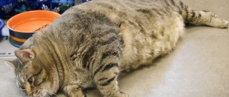
- overweight and obesity in your pet;
- dislocation of the lens of the visual organ;
- tumors in the eye;
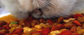
- improperly selected diet;
- low quality feed or natural food;
- any eye injuries;
- poor heredity of the animal.
How common is diabetes in cats?
So, quite often glaucoma becomes a consequence of the development in your cat of a disease such as uveitis, in which the anterior section of the eyeball is affected by inflammatory processes, which ultimately leads to clouding of the lens.
This animal has both eyes affected by the disease.
Is it possible to completely cure glaucoma?
Despite the high effectiveness of treatment methods, glaucoma remains an incurable pathology.
It is important to remember that timely contact with a qualified specialist can ensure:
- significant alleviation of symptoms;
- reduction of pain syndrome;
- elimination of most clinical manifestations of the disease.
For this purpose, veterinarians prescribe pilocarpine drops: this drug is intended to provide the sick animal with the most comfortable standard of living. Drops are prescribed strictly by a veterinarian based on the results of a comprehensive examination of the pet.
Types of glaucoma
According to the existing medical classification, there are several types of glaucoma in cats. You can see how they differ from each other in the table.
Table. Types of glaucoma in cats
| Type of disease | Causes and clinical manifestations |
| Genetic glaucoma | This disease develops due to the animal’s initial predisposition to the appearance of this disease. The predisposition is written into the animal’s genetic code, so most often it affects both eyes of the cat at once, and its treatment is very difficult. The main cause of genetic glaucoma is the gradual manifestation of congenital disorders in the drainage system of the animal’s visual apparatus. Provided that your cat had an initial hereditary predisposition to this disease, the probability that the disease will occur is at least 98%. You can determine whether you should be wary of illness by looking at the health of your pet’s parents. |
| Secondary glaucoma | This form of the disease in question is the most common among all possible ones. It occurs against the background of an existing disease of the visual system of various etiologies. In this form, the disease affects only one eye and goes away quickly after the cause is discovered. |
| Open angle type | During this disease, the development of dystrophic processes in the optic nerve is noted. The affected element of the system gradually atrophies and can no longer perform the tasks assigned to it by the body. |
| Closed angle type | This type of disease occurs against the background of the development of oncological processes in the eye, less often - against the background of inflammatory processes. |
Adult cats aged 9 years or more are at risk
The age mark at which many cats experience glaucoma is around 8-9 years of age. However, it is characterized only by a more frequent occurrence of the disease - under the influence of many negative factors, it can develop much earlier.
Classification of the disease
There are several types of the disease, differing in origin, involvement of one or both eyes in the process, degree of damage, severity of the process, and others.
By origin
Based on their origin, primary and secondary glaucoma are distinguished:
- The primary variant of the disease is an independent pathology of the ocular mechanisms that ensure the balance of the intraocular fluid (ciliary body, drainage system and optic nerve head). It can be either congenital or acquired.
- The secondary option is a complication of injuries and a symptom of inflammatory or other ophthalmological diseases. It can only be purchased.
By involvement in the process
Based on the involvement in the process, unilateral or bilateral glaucoma is distinguished (depending on whether one or both eyes are affected).
According to the state of the iridocorneal angle
The walls of the anterior chamber (iris and cornea), where intraocular fluid accumulates, together form the iridocorneal angle. How freely moisture can circulate in the thickness of the eye depends on its condition. Depending on the degree of its damage, open-angle and closed-angle glaucoma are distinguished:
- Open-angle glaucoma is not accompanied by blocking of the iridocorneal angle. It is often chronic in nature.
- In the angle-closure form, the drainage system of the eye and the iridocorneal angle of the anterior chamber are blocked, which further disrupts the balance of the intraocular fluid. This type of disease is often acute.
According to severity
Depending on the severity of the course, there are four stages of glaucoma:
- Initial. It is reversible. The structures of the eye are not damaged, the fields of vision are almost not narrowed.
- Advanced glaucoma. The field of view is narrowed slightly. There is a slight disturbance (excavation) of the optic nerve head.
- Advanced glaucoma. Concentric narrowing of visual fields. Excavation of the optic nerve disc (OND) is defined as subtotal.
- Terminal. It's practically blindness. The stage is characterized by irreversible damage to the optic nerve and total excavation of the optic disc.
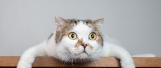
Symptoms of glaucoma in animals
The danger of the disease lies in the fact that in the initial stages almost any of its manifestations are absent. This is due not only to its “quiet” flow, but also to the fact that cats, like any other animals:
- can endure, without showing it, unbearable pain;
- have excellent orientation in space even if the visual apparatus is poorly functioning.
There are legends about how our pets endure any, even very severe, discomfort. Sometimes they calmly behave with such injuries that would probably cause a painful shock in a person. In addition, nature has endowed these animals with a large number of different devices for orientation in space. So, in addition to eyes, cats have ears and whiskers. This is why it is difficult to track the disease in a cat whose condition has not changed much.
A partially or completely blind cat can navigate in space using its whiskers and ears.
At later stages of the disease, various kinds of symptoms begin to manifest themselves. This is about:
- swelling of the eyeball;
- redness of the white of the eye;
- incomplete contact of the eyelids when closing the eyes;
- the pupil is constantly dilated;
- clouding of the cornea;
- a change in the general condition of the animal, for example, aggression, apathy, an excessive reaction to an attempt to examine the eye, or lack of appetite appears;
- tearfulness, discharge of purulent secretion from the eye;
- fear of light, etc.
Watery eyes are one of the symptoms of glaucoma.
The further the disease progresses, the larger the affected eyeball becomes. Moreover, it increases in size so much that it seems to be protruding, as if it were planted on a leg. At the same time, severe redness of the eye develops due to an increase in the size of the blood pathways passing through the organ. Due to the enlargement of the eye, it ceases to close completely, which, in turn, leads to drying out and the appearance of ulcers.
The animal's eye stops closing and gradually dries out, becoming covered with ulcers.
Please note: above we described the symptoms for glaucoma, which develops sequentially, from mild to severe stages. However, sometimes it happens that the work of an organ is immediately disrupted by the disease in the acute stage. In this case, symptoms such as:
- vomit;
- apathy and pronounced weakness of the animal;
- immunity to humans, food, water;
- other manifestations.
Provided that you are seeing such a clinic in front of you, and on top of everything else, the animal’s eye is inflamed or inflamed, you need to urgently go to the doctor. Only in this case will you be able to preserve the animal’s health, and possibly life.
A cat that is sick or suffering from glaucoma will most likely behave in such a way that only the most loving owner would suspect the disease. And, nevertheless, there are some behavioral signs of the animal that you immediately need to concentrate on. Contrary to popular belief, it is necessary to pay attention to them, and not to changes in the shape, color of the eye or the presence of cloudiness in it.
Cat with glaucoma avoids sunlight
We are talking about fleeting inconsistencies with the animal’s usual behavior. So, for example, a calm cat:
- becomes too shy;
- looks for secluded dark places to hide there;
- avoids loud noises;
- stops choosing cabinets, shelves and other heights as a place to rest;
- the animal’s movements become even more accurate, sometimes it seems that it is rearranging its paws, as if it is walking through a minefield.
Symptoms of the disease
In the early stages, it is difficult to notice that a cat's vision has deteriorated, since she continues to navigate well thanks to her keen sense of hearing and smell. Vibrissae also help her with this. But, from a certain point, the peculiarities of the animal’s behavior become noticeable to the owner.
Symptoms of angle-closure glaucoma
The acute (angle-closure) form of the disease is characterized by the following manifestations:
- sore eyes (the cat won’t let you touch them);
- clouding of the lens, which can be seen with the naked eye;
- rapid loss of vision, which affects the cat’s behavior. She begins to feel insecure, lethargy or aggression appears. The animal refuses to play. It feels discomfort because its fields of vision are narrowed, vision becomes, as it were, “tunnel”;
- Nausea and vomiting may occur.
If at this moment you show the cat to a veterinarian, then during the examination, the initial stage of atrophy of the optic nerve and retina will most likely be detected, which will confirm the preliminary diagnosis.
Symptoms of open angle glaucoma
The chronic (open-angle) nature of the disease is more characteristic of older animals. Its symptoms develop gradually:
- an increase in the size of the eye due to the accumulation of fluid due to poor functioning of the drainage system;
- protrusion of the eye, and, as a result, non-closure of the eyelids;
- gradually increasing soreness in the cat’s eyes;
- lack of pupil response to light;
- redness of the eye and increased tearing;
- liquefaction of the vitreous.
Symptoms of congenital glaucoma
Symptoms of congenital glaucoma can be noticed by careful observation of the cat:
- some blurred eyes due to insufficient transparency of the lens;
- avoidance of light, desire to be in the shadows or darkness;
- increased lacrimation;
- congenital strabismus may also indicate congenital glaucoma.
Diagnosis of glaucoma in cats
If you suspect glaucoma, before going to see a veterinarian, you can independently test the animal for the presence of this disease.
The first test is carried out as follows: place your animal on the edge of a table and let it jump off it. Exactly how the cat copes with the jump will be an indicator of the presence of the disease:
- if the jump occurred as naturally as possible, without loss of balance or coordination, then everything is fine with the animal;
- provided that the cat was confused, meowed, and asked you for help, and eventually jumped off extremely clumsily, then, most likely, she simply did not see where and how to move, which means that glaucoma (or another visual disorder) is present.
If the cat did not lose its balance during the jump, then everything is fine with it.
The second test you can do to diagnose this disease in your own pet is to have her play with a laser pointer. If your animal does not react in any way to the laser, most likely he has problems not only with vision, but also with his general condition, changes in which may also indicate glaucoma.
If your cat doesn't play with the laser, it most likely has vision problems.
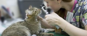
Of course, self-diagnosis is a useful thing, but only a veterinarian can correctly diagnose a pet and, moreover, prescribe an effective drug that fights this disease. A specialist in the field of diseases of our little brothers will examine your pet, prescribe an examination by an ophthalmologist, or conduct it himself. So, to identify the first stages of the disease, the following is carried out:
- slit lamp examination;
- measuring the pressure inside the animal's eye;
- ophthalmoscopy.
Only a veterinarian can accurately and reliably diagnose glaucoma.
When measuring, the doctor will focus on normal pressure values, which in a healthy animal are about 15-25 millimeters of mercury for each organ of the visual system. Provided that there is a difference between them of approximately 10 millimeters or more, the animal probably has glaucoma.
If the above examinations are not informative enough, a so-called gonioscopy can be performed. This procedure is an examination of the affected organ of vision using special instruments - medical mirror ophthalmoscopes. With their help, the doctor will be able to examine the following segments of the eye:
- areas of the retina located along the periphery;
- the front part of the organ;
- ciliary body.
To prevent the animal from experiencing discomfort, it receives preliminary local anesthesia
Provided that the doctor suspects the presence of pathologies that could also affect the development of glaucoma, an ultrasound examination is performed. Usually it helps to detect:
- tumors;
- detached retina;
- other diseases.
Since the clinical picture of the disease we are interested in is similar to the manifestations of other pathological processes affecting the organs of the visual system, it is also necessary to exclude at the examination stage:
- keratitis;
- degeneration of the eye elements;
- conjunctivitis;
- uveitis, etc.
To understand that your cat has glaucoma, you need to rule out other diseases with similar symptoms.
Only after a full examination of the animal can a veterinarian make a conclusion about the presence or absence of glaucoma and prescribe the appropriate treatment.
Please note: although we have taught you how to do several simple tests to check if your cat has glaucoma, we urge you to refrain from self-diagnosis and arbitrarily prescribing treatment for your pet. The fact is that medications for one disease can seriously accelerate and aggravate the course of another. By taking on such responsibility, you can cause suffering to your pet.
Glaucoma is incurable
Glaucoma, unfortunately, is an incurable disease. Most often, its occurrence leads to the animal losing its vision completely or partially. At advanced stages of the disease, obvious deformation of the organs of vision occurs:
- they increase;
- seriously blush, etc.
The affected organ can no longer be restored, however, if the disease is detected in the early stages, its symptoms can be stopped and blindness can be stopped.
If glaucoma is a consequence of any disease, then it can be quickly cured by eliminating the root cause
In addition, if glaucoma begins to develop as a secondary disease, by eliminating the root cause, it is possible to stop the development of the disease altogether.
Main symptoms
Glaucoma is a disease that affects vision. Therefore, the main symptom is its violation. However, it is quite problematic for the owner to determine in the early stages that the animal’s vision has become worse. The fact is that cats have very well developed other senses. We are talking about smell and hearing. At first, this helps the pet to navigate well in space, not paying attention to vision. But at a certain point the situation worsens and, accordingly, the animal’s behavior changes.
Treatment of the disease
As we mentioned above, the principles by which the animal will be treated will vary depending on what particular form of the disease the cat suffers from. So, provided that an open-angle variation of this disease is detected, which occurs rapidly and acutely, the animal must be taken to a veterinarian immediately - otherwise death is also possible.
If the disease is in its early stages, therapy will be conservative, using only medications. If the stage is advanced, then surgical intervention may be required.
Sometimes surgery is unavoidable
Conservative treatment
Conservative therapy has several main objectives:
- elimination of disease symptoms;
- improvement of the condition of the affected organ of the visual system.
Comprehensive treatment of glaucoma
To carry them out, drugs are used that have the following effects:
- triggering or enhancing the drainage of fluid accumulated in the eye;
- reducing the production of “water” in the eye;
- constricting pupil;
- diuretic;
- anti-inflammatory (usually hormones);
- pain reliever;
- improving nerve conduction, etc.
Thus, the most popular remedies for combating glaucoma in the field of veterinary medicine are presented in the list below.
Firstly, this is “Pilocarpine” - a drug that:
- constricts the pupil of the eye;
- relieves pain caused by the disease;
- slows down the progression of the disease.
This remedy is given once a day, sold in the form of drops. Each eye of the animal (including the healthy one) must be instilled once a day when the disease progresses slowly; in case of acute progression, the product is used every hour.
Pilocarpine constricts cat pupils
Secondly, Amlodipine is a drug that helps remove moisture accumulated in the eye, seriously improving the condition of the sick animal. This medication is given to the pet once a day, the approximate dosage is approximately 0.6 milligrams. An important nuance: taking the medicine should coincide with meals.
"Amlodipine" removes excess fluid from the eye
The third drug, Mannitol, showed excellent results in the early stages of the disease. The drug is administered inside the animal's body through an injection into a vein. The dosage is calculated as follows: one gram of medicine per kilogram of live weight.
"Mannitol" helps defeat glaucoma in the initial stages
Fourthly, Betoxolol is a drug that helps normalize pressure inside the organ of vision. The medication is administered every 12 hours, it is dripped directly into the animal’s eye.
Betoxolol normalizes intraocular pressure
A mandatory part of drug treatment will also be a limited drinking regime for your pet. In addition, during therapy, food containing various preservatives and additives is excluded from consumption (by the way, this food waste is not suitable for feeding even a healthy animal), and the pet is also given fortified complex preparations that improve its general condition.
Every living organism needs vitamins in the right ratio
Please note the following important fact: each drug indicated in the list above has a number of contraindications. You cannot give it to your pet on your own, since only a veterinarian can judge the potential side effects that may occur.
Here are the most common examples:
- A diuretic should not be given if the cat has a history of urolithiasis;
- Some eye drops can worsen retinal detachment. about the side effects of Ciprovet eye drops in our article.
Surgical intervention
If it is too late to take medications or their use has no effect, veterinarians will recommend surgery to save the animal’s life. Exactly how this intervention will be carried out will depend on how:
- the disease progresses;
- at what stage is the disease;
- whether the animal retained vision in the affected eye.
If medications do not help, the animal will require surgery.
Thus, the operation in which a laser is used as a surgical instrument is considered the most acceptable and has demonstrated a high level of effectiveness. When used, the symptoms of the disease are quickly eliminated, and rehabilitation occurs as quickly as possible. The few disadvantages of this procedure are as follows:
- Not every veterinary clinic has such expensive equipment;
- the operation costs a lot of money, although a loving owner will not consider this circumstance an obstacle to the treatment of pets.
Russian veterinary clinics, as a rule, do not provide laser surgery for glaucoma, but use techniques such as:
- cauterization of the ciliary body at low temperatures;
- implantation of the Ahmed valve;
- cyclodialysis.
Some veterinary clinics offer a laser solution to the problem.
The interventions listed above are aimed at reducing the pressure inside the visual organ affected by the disease. Unfortunately, despite their high effectiveness, they do not protect against:
- postoperative complications;
- re-development of the disease.
If the disease reoccurs, the pet will need surgery again. That is why, if possible, contact a clinic that works with laser methods for treating glaucoma. Thus, you will not only cure your pet, but also save on repeated surgery.
If atrophy of the optic nerve has occurred and it is not possible to restore its functioning, surgical intervention will be performed to eliminate the symptoms of the disease and its consequences, as well as to restore the organ and preserve the appearance of the cat.
The animal is fitted with a prosthesis or undergoes resection of the eyeball
Treatment with folk remedies: reviews from owners
In the comments, cat owners often ask about folk remedies to combat glaucoma. The fact is that this disease cannot be cured in this way. However, reviews say that with the help of traditional medicine it is possible to enhance the effect of drug therapy. So, let's look at what the owners advise.
- Wash your eyes with aloe decoction several times a day.
- Infuse strong tea with honey and drop a few drops into each eye.
This, of course, will not help get rid of glaucoma, but it will reduce inflammation and pain. The owners claim that the animals are becoming calmer.
Methods for preventing glaucoma in cats
Unfortunately, we cannot recommend any specific measures that would help prevent glaucoma in cats, since they simply do not exist. However, to prevent your pet's intraocular pressure from increasing, protect his visual apparatus from:
- injuries;
- mechanical damage;
- burns, etc.
In addition, monitor the general health of the animal, starting the fight in time against:
- overweight;
- diabetes mellitus;
- other diseases that could potentially provoke pathology.
Prevention is about maintaining a high level of cat health
Among other things, you should monitor such aspects of the animal’s life as:
- feeding with high-quality feed or natural food, and the food should also be sufficiently fortified;
- living conditions that meet the requirements for temperature conditions, environmental humidity levels and other nuances.
Prevention
Cat owners should systematically pay attention to eye hygiene of their furry pet. Wash your cat's eyes only with special preventive solutions.
Animals that have a genetic predisposition to the development of glaucoma should not be allowed to undergo planned mating.
It is equally important to strengthen your pet’s immunity and promptly treat eye diseases at an early stage of their development. Take your cat to the clinic two or three times a year for a routine check-up. Most diseases can be cured only at an early stage of development.
There are no preventive methods to prevent the formation of glaucoma in cats. The disease is easily transmitted from one damaged eye to another. Timely treatment can delay the progression of the disease to a more severe form.
READ How to understand that a cat is giving birth - main signs
Attention: the above is for educational purposes only and does not constitute professional medical advice or scientific material.
Since the onset of glaucoma is difficult to diagnose, your cat should undergo periodic ophthalmological examinations at a veterinary clinic. This primarily applies to breeds that are at risk for glaucoma, as well as elderly animals.
To prevent glaucoma, animals must receive high-quality care, which includes a balanced diet in accordance with age and breed, quality eye, ear and coat care, and an active lifestyle.
Let's sum it up
Glaucoma is a serious pathology that affects the visual system of cats and other animals. In the best case, you can get rid of it, in the worst case, the animal will die from the disease and the associated infection. Most often, owners who are slow to respond to changes in their pet's behavior have to deal with the fact that the animal completely loses its vision.
Cat health is the responsibility of the owner
When seeking help from a veterinarian, listen carefully to all the specialist’s instructions and follow the recommendations prescribed by him. Correctly selected treatment will minimize the damage from the disease or cure it altogether.
The most common diseases and treatments
Although there are a large number of pathologies that affect the eyes of cats, some of them are much more common than others.
Below is a list of the most common diseases and symptoms of eye diseases in cats, as well as methods of treating them.
How to treat?
Therapy depends on the cause of the disease. The goal of the therapeutic course is to stop the development of glaucoma and restore normal fundus pressure. Treatment of glaucoma in cats is carried out on both organs at once, even if only one of them is affected. To remove inflammatory processes, diuretics (Mannitol) are prescribed. Bimatoprost or Amlodipine help with uveitis and stagnation of ocular fluid. To reduce blood pressure, beta blockers are used: Temolol or Betoxol.
Vitamin complexes will help compensate for the lack of useful microelements.
In addition, the following groups of drugs are used:
- vitamins;
- anti-inflammatory;
- antibiotics;
- antispasmodics.
Drug treatments help stop the disease only in the first stages. At the last stages, surgical intervention is used: drainage of the anterior chambers of the visual apparatus or removal of the ciliary body. It is also possible to treat glaucoma with a laser. This method is painless and effectively eliminates pathology. If therapy is not possible, then the diseased organ is completely excised. This helps the cat get rid of constant spasms.
Prevention to prevent diseases
Treating an already developing pathology is much more difficult than taking measures to minimize the chance of an animal becoming ill. In order for your cat to be healthy and the risk of disease to be minimal, it is enough to follow a few simple rules:
- Conduct a preventive examination at the veterinary clinic and receive vaccinations recommended by the veterinarian on time;
- avoid contact with street animals; after a walk on the street, examine the cat for eye injuries and the presence of foreign objects;
- add vitamins and healthy supplements to your cat’s food;
- Regularly trim nails, comb and examine eyes for discharge.
When faced with an eye disease in a cat, the first thing you need to do is take the animal to a veterinarian.
Many owners, when their pet becomes ill, begin to look for information and methods of treatment based on photos of eye diseases in their cats, which should not be done under any circumstances, since improper treatment may lead to consequences in the form of complications or the development of more serious diseases.
Reasons for appearance
There are congenital and acquired forms of glaucoma. The first type occurs due to disturbances in the outflow of intrauterine fluid. Typically, such a pathology is preceded by a serious illness in the mother cat. The acquired form is a secondary disease and develops against the background of other disorders. Quite often, uveitis, purulent diseases of the visual organs, injuries of the cornea and lens become the source of pathology. The cat's hereditary predisposition to cataracts is of great importance. The disease usually affects breeds with short, flat faces, such as Siamese, British or Persian breeds.
Excess body weight can be a symptom of diabetes.
Causes of glaucoma:
- diabetes;
- excessive weight;
- stress;
- taking hormonal and steroid medications;
- neoplasms in the visual organ;
- pathological diseases of the cornea;
- chemical burn.