What diseases require eye removal?
Surgery to remove an eye in cats, as well as dogs, may be required in the presence of a serious pathological process. But first you need to carry out a series of therapeutic procedures that can save at least part of your vision. If the situation is very serious, the veterinarian often recommends removing the cat’s eye.
Surgery to remove a cat's eye may be required for the following pathological processes:
- the presence of internal infectious lesions that can spread to other organs;
- damage to the back of the eyeball, but within the orbit;
- congenital deformations of the visual organs;
- oncological tumors;
- infectious and inflammatory processes of the eyeballs (superficial and internal), which can only be cured using surgery;
- glaucoma with a severe course;
- the presence of severe physical injuries from which it is impossible to recover independently.
Many cat owners often have a question: why remove the eyeball from an animal? Enucleation is required to reduce pain in animals, as well as to eliminate all suffering. In addition, the damage can often affect other internal organs in addition to the eye.
The cat's eye was removed - how to care for it?
Cats are naturally inquisitive and also need their own territory. At the intersection of these two personality traits, pets often experience injuries, minor or serious, at the wrong time. One of the most dangerous injuries is eye injury. If the cat's eye is infected with a dangerous infection that can spread further throughout the body, or the organ of vision is damaged beyond repair, the veterinarian may decide to enucleate. Enucleation is the removal of the eyeball followed by suturing the resulting hole. Caring for a cat after eye removal will require the owner to carefully follow the doctor's instructions and constant attention to the pet. So, how should you care for your cat after eye removal?
For what diseases does a cat's eye need to be removed?
- Internal eye infections that can spread to the rest of the body.
- Involvement of the space behind the eyeball, but within the orbit.
- Congenital eye deformities.
- Eye cancer.
- Infections or inflammations of the eyeball (both superficial and internal) that cannot be treated without surgery.
- Severe glaucoma.
- Severe physical trauma to the eyeball, from which recovery is impossible.
Some pet owners ask why a blind cat's eye should be removed. Veterinarians perform enucliation on blind animals to reduce pain, to remove their suffering from an organ that is unable to see and ceases to serve a useful function for the animal. If a cat is diagnosed with eye cancer, and the owners cannot provide the pet with the proper medications and treatment for any reason, a decision may be made to remove the eye with the consent of the cat's owners.
We recommend reading: Vomiting in a Cat After Eating
Features of caring for a cat after eye removal
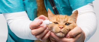
Please follow your veterinarian's instructions responsibly. It is worth paying special attention to the dosage of prescribed medications, changes in the cat’s diet, and the purchase of additional equipment (bandages, collars, etc.). Your cat's reaction to the anesthesia and the surgery performed will also affect the management of subsequent care.
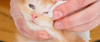
Periodically inspect the sutured eye socket in order to notice tissue swelling or seam divergence in time. Do not be alarmed if minor bleeding and slight swelling appear within a few days (2-4) after surgery. Sometimes an animal can sneeze with blood, but this is also not a pathology (this is how the cat gets rid of residual blood after surgery). Sutures are usually removed after about 7-10 days, depending on many factors. Within 6-8 weeks, fur and whiskers grow back around the removed eye. A mustache will be especially necessary to help the “blind” side navigate and avoid bumping into obstacles.
You will also have to purchase an “Elizabethan” collar to prevent the cat from scratching the stitched eye socket, which can lead to deep scratches and seams coming apart. When putting on the collar, make sure it fits firmly and securely around your pet's neck. However, make sure that the collar straps do not put pressure on your cat's skin or cause discomfort.
Pay attention to the cat's behavior. After anesthesia, animals are usually lethargic and have little initiative, but after some time (no more than a day), the cats’ condition returns to normal, they willingly accept food and water. If your pet refuses to eat and has been in apathy for a long time, experiencing weakness, contact your veterinarian about this issue.
Antibiotics, anti-infectives, and some other medications are usually prescribed for post-enucleation care. Follow dosage and other instructions from your veterinarian. Compliance with them helps prevent the development of an inflammatory process (infection due to illness or introduced during surgery), which can greatly complicate a cat’s life.
Sometimes both eyes are removed in case of particularly severe diseases. However, 99% of enucliations involve one eye. This allows the cat to partially retain vision, and with it activity and capacity.
Please follow your veterinarian's instructions responsibly. It is worth paying special attention to the dosage of prescribed medications, changes in the cat’s diet, and the purchase of additional equipment (bandages, collars, etc.). Your cat's reaction to the anesthesia and the surgery performed will also affect the management of subsequent care.
Rules for eye removal surgery
When performing an operation to remove an eye from a cat or dog, veterinarians must follow certain rules that can prevent dangerous consequences for the health of the animal.
When performing this procedure in a clinic, specialists must fulfill the following requirements::
- During the operation, the doctor must cause minimal harm to the cat’s health;
- The doctor must perform the operation correctly in compliance with all necessary measures, this will avoid complications after surgery;
- The procedure should be done using anesthesia so that the pet cannot feel all the unpleasant feelings. In addition, so that the cat does not feel pain after removal of the eye, it is necessary to inject special painkillers;
- The anesthetic during surgery should be used in small quantities so as not to cause serious harm to the health of the animal.
When is removal necessary?
Indications for surgical intervention are pathologies that can cause serious harm to internal organs, or cause severe pain to a kitten or an older animal. These conditions include:
- Panophthalmitis. It is an inflammation of the cavity of the eyeball, which is purulent in nature. The organ cavity is filled with pus, which cannot be eliminated using conservative methods of therapy. If surgery to remove the eye is not performed, inflammation affects adjacent tissues, sometimes even the brain, resulting in death.
- Glaucoma that cannot be cured with medication. When conservative therapy does not bring the required effect and the cat’s eyeball falls out of the socket, the veterinarian decides to remove the cat’s eye to prevent damage to the optic nerve and brain.
- Severe traumatic injuries. If medications do not help tissue scarring, the cavity of the visual organs fills with pus, resulting in panophthalmitis.
- Eye loss in a cat.
- Tumors of the visual organs of a malignant nature.
- Scleral rupture.
- Congenital deformation of the eye.
We recommend reading: Are Worms Transmitted From Scratches From Cats?
Return to contents
Types of operations
If the doctor detects symptoms of a serious illness in a cat, he can undertake various medical treatments, including surgery.
Operations can be carried out using several methods:
- enucleation _ During this procedure, the pet’s eyeball is removed, but the muscle fibers and ligaments in the visual organ apparatus are fully preserved;
- evisceration . With this method of treatment, the apple and intraocular structures are completely excised. After completion of the operation, the size of the eye decreases throughout the entire recovery period; this process takes about a month. But after this procedure, postoperative inversion and inversion of the lower eyelid may appear;
- exenteration _ During this procedure, both the eyeball and other important parts of the visual organ are removed - extraocular muscle fibers, lacrimal glands, orbital fatty tissue, and the third eyelid. After this, the edges of the eyelids are sutured. This method of surgical intervention is performed only for severe forms of purulent lesions with an inflammatory nature, as well as for oncological formations.
In order for the cat to recover quickly, the doctor must follow all the necessary rules during the operation. It is also important to provide adequate care for the cat after eye removal, which can speed up the recovery process. The veterinarian at the clinic will be able to tell you how to care for it and what actions need to be taken after the operation.
Surgery to remove a cat's eye may be required for the following pathological processes:
Exenteration
This is an operation that is a radical way to eliminate pathological processes in the eye. This method is an enucleation combined with the removal of extraocular muscles, fatty tissue of the orbit, upper, lower and third eyelids, lacrimal gland, as well as all conjunctival tissues. The cut edges of the eyelids are sewn together. This surgical intervention technique is indicated for the treatment of severe purulent pathologies affecting the eyeball and orbit, as well as for neoplastic processes.
In some cases, the subsequent placement of an intraocular prosthesis can eliminate the unattractive cosmetic effect after surgery. Such an event is characterized by low trauma. However, a thorough assessment of the safety of the procedure is required. Thus, contraindications to prosthetics include panophthalmitis, uveitis, panuveitis, and suspected intraocular neoplasm.
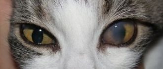
Symptoms of eye disease
The best way to preserve your pet's vision is to have regular eye exams. You can do this yourself at home or in a veterinary clinic. If problems are detected in the early stages, they can be eliminated with medication without resorting to surgery. When examining the animal, be sure to wash your hands and treat them with alcohol. Gently open the eyelid without pressing on it.
If you notice the following symptoms of eye disease in cats, contact your veterinarian immediately:
- Redness is the most common problem, which can be caused by diseases of varying severity, which can lead to complete loss of vision.
- Pus is released - a sure sign of conjunctivitis. This inflammation occurs quite often and is accompanied by purulent discharge and the appearance of a crust on the eyelid.
- An injury occurred and due to severe pain, the cat does not allow him to approach him.
- Blepharospasm is the inability to open the eyelid or constant squinting.
- Increased tearfulness, fear of light.
Prognosis and prevention
In most cases, the prognosis of the disease is favorable. If temporary sutures were applied, entropion may recur. In this case, either re-stitching or eyelid surgery is performed.
A complication after plastic surgery is eyelid inversion, which goes away on its own and does not require treatment.
The main preventive measures include:
- Protecting your cat's eyes from injury and burns.
- Timely diagnosis and treatment of eye diseases.
- Increased attention to the visual organs in breeds predisposed to the disease.
- Annual vaccination against viral infections.
Operations on the eyes of a cat
Surgeries on the eyes of cats are performed for the following pathologies:
We recommend reading: Cause of Bad Odor from Dog's Mouth
- Corneal ulcer is the most difficult disease that requires immediate treatment. Extensive and deep injuries must be operated on urgently to prevent blindness.
- Entropion of the eyelid is less common than in dogs, but such cases still occur. This is a pathology when the skin fold turns inward. Because of this, eyelashes and fur touch the cornea, irritating the mucous membrane. If the problem is not corrected in time, this will lead to loss of vision and even removal of the eyeball.
- Cataract - characterized by clouding of the lens, deterioration of vision to complete blindness.
- Enucleation - removal of the eyeball. It is required in cases where the visual organ cannot be saved, its functions are lost or it threatens the life of the animal.
Benefits of treatment at the Komondor veterinary clinic
The feline ophthalmology department at the veterinary office accepts patients with eye problems. The clinic is equipped with European ophthalmological equipment for complex diagnostics and the required conservative or surgical treatment.
Our veterinary center employs experienced, highly qualified ophthalmologists who will provide professional medical care to your pet.
If you notice symptoms of eye disease in cats or you see that your pet has been injured, consult a doctor immediately. Timely detection of pathology and prescription of treatment will avoid eye surgery and maintain the health of the visual organ.
- Corneal ulcer is the most difficult disease that requires immediate treatment. Extensive and deep injuries must be operated on urgently to prevent blindness.
- Entropion of the eyelid is less common than in dogs, but such cases still occur. This is a pathology when the skin fold turns inward. Because of this, eyelashes and fur touch the cornea, irritating the mucous membrane. If the problem is not corrected in time, this will lead to loss of vision and even removal of the eyeball.
- Cataract - characterized by clouding of the lens, deterioration of vision to complete blindness.
- Enucleation - removal of the eyeball. It is required in cases where the visual organ cannot be saved, its functions are lost or it threatens the life of the animal.
Treatment
Entropion of the eyelid requires mandatory surgical intervention. Prolonged mechanical irritation can lead to serious complications including blindness and loss of the eye.
There are 2 types of surgery - eyelid surgery and temporary suture for correction. In the second case, the method is used for young animals up to 7 months.
Using a special mattress suture, the skin of the eyelids is simply sutured to the lower edge for fixation. This design ensures that entropion does not recur and helps the cat improve his life until the age when full eyelid surgery can be performed.
Most often, due to young age and the final formation of the skull, such a suture is sufficient. As the kitten's head grows, the inversion of the eyelid resolves on its own.
This intervention is performed without anesthesia.
If the animal is older than 7 months, then eyelid surgery is used - in this case, a small piece of excess cat is cut off, and the remaining flaps are sewn together. In this way, the physiological position of the eyelashes and eyelid is achieved.
Features of preparation for surgery and postoperative care
12 hours before surgery, the animal is prohibited from giving food; water is allowed, but in small quantities. It is also advisable to conduct a full examination of the cat to determine the tolerability of general anesthesia and the operation itself.
Cost of eye surgery for cats
Video of an ophthalmological examination of a cat:
Read reviews about our veterinary center. Call the number and schedule a consultation right now or request a call back. (c) Veterinary center for the treatment and rehabilitation of animals “Zoostatus”. Varshavskoe highway, 125 building 1.
(c) Veterinary center for the treatment and rehabilitation of animals “Zoostatus”.
Varshavskoe highway, 125 building 1. tel.
Types of the most common eye surgeries in cats:
For what diseases does a cat need eye removal?
The main indications for surgical removal of the eyeball are diseases that can cause irreparable harm to internal organs and the brain, and also cause unbearable pain to cats.
- Panophthalmitis is a purulent inflammation of the cavity of the eyeball. The entire cavity is filled with purulent exudate, and, unfortunately, the inflammation cannot be treated therapeutically. Without removing the affected gas, inflammation can spread to surrounding tissues and even to the brain, which leads to the death of the pet.
- Glaucoma with increased intraocular pressure without therapeutic effect - increased intraocular pressure has a negative effect on the optic nerve and causes blindness. The eyeball is bursting and it “falls out” a little from the orbit. If the animal does not respond to therapeutic treatment, it is recommended to remove the organ to prevent functional and structural damage to the optic nerve and brain.
- Severe injuries to the eyeball - these can be various wounds that cannot be healed with medication. Without surgery, the cavity of the eyeball becomes clogged with pus and panophthalmitis develops.
- Loss of the visual organ with changes in the tissue structure - changes in the color of the retina, rupture of blood vessels and the optic nerve.
- Malignant tumors of the eye
- Scleral rupture
To what extent does evisceration and enucleation spoil the appearance of an animal?
It all depends on the perception of others and on the extent of the owner’s love for his four-legged pet. Of course, one cannot expect the same appearance, but in this case the question arises not about a cosmetic effect, but about saving the life of the animal.
ATTENTION!!!
Unfortunately, it is not always possible to save an animal’s eyeball, especially in cases of late contact with an ophthalmologist or in cases of severe eye injury, tumors and complicated cases of eyeball prolapse. The eye is removed only when vision is irretrievably lost and when it causes suffering to the animal, as well as in cases of risk of involvement in the pathological process of organs and tissues adjacent to the orbit or the neighboring eye.
Unfortunately, removal of the eyeball
– the most common orbital surgery in animals. This situation arises for many reasons. The main reason is that even the most attentive owner cannot notice in time a developing disease inside the eye of his animal and turns to a specialist, as a rule, if the pet has obvious signs of the disease. By this point, the eyeball may already have irreversible changes. Removal of the eyeball is indicated if its functions are irreversibly lost, if it is painful or poses a threat to the life of the animal. Surgery to remove the eyeball should not be performed in place of diagnostic procedures to make a correct diagnosis. It can only be recommended after a preliminary or final diagnosis has been made.
Examples:
Suspicion of an intraocular neoplasm in an animal. Additional studies include ophthalmoscopy, examination of the anterior segment of the eyeball using a slit lamp, and ultrasound of the eyeball. In the case where the detected neoplasm is inoperable, a preliminary diagnosis is made: inoperable intraocular neoplasm. Removal of the eyeball is recommended. The final diagnosis is made based on histological examination of the material. (Fig. 1, 2).
Young dog with an enlarged eyeball. The owner's history indicates that some time ago the dog got into a fight with a cat. (Fig.3). During the examination, a study of the pupil's reaction to light, reaction to glare, measurement of intraocular pressure, and ultrasound of the eyeball were performed. In the absence of pupillary-motor reactions in combination with an enlarged eyeball and a changed lens, a final diagnosis is made: end-stage glaucoma due to traumatic cataract and placement of an intraocular prosthesis or removal of the eyeball is recommended. (Fig. 4)
Direct indications for eyeball removal:
- Panophthalmitis (Fig. 5,6.).
- Severe panuveitis (Fig. 7).
- End-stage glaucoma with increased intraocular pressure, which is not controlled by therapeutic methods (Fig. 8).
- Buphthalmos with associated complications (Fig. 9, 10).
- Blunt trauma to the eyeball with rupture of the sclera in the fundus (Fig. 11, 12).
- Severe penetrating injury to the eyeball with damage to intraocular structures.
- Some cases of eyeball prolapse (Fig. 13).
- Inoperable intraocular neoplasms (Fig. 14).
- Aphthalmia, microphthalmia with accompanying pathologies (Fig. 15).
All of the above pathologies lead to the fact that the eyeball becomes a source of suffering for the animal, and the process developing in it can threaten not only the health, but also the life of the animal. We are talking about conditions when the intraocular structures are severely damaged, and visual functions cannot be restored. Once the veterinarian has determined that the eyeball has lost visual function and requires removal, a method for removing the eyeball must be selected.
Rules for operations to remove the eyeball
- Use of general anesthesia
- Compliance with the rules of asepsis and antiseptics . The operation is performed in a sterile operating room. Before the procedure, the hair around the eye is shaved and disinfected with available isoseptic agents.
- Using local anesthesia to prevent pain in the cat.
Before the operation, owners are prescribed antibiotic therapy and the use of hemostatic agents 5 days before the intended operation. Convenient if it was planned. During unscheduled surgical interventions during eye surgery, drugs are used in the postoperative period.
Types of operations
There are three types of surgical intervention. Each of them is prescribed depending on the indications.
- Enucleation is the removal of the eye with all its contents, without affecting the structural components of the orbit - muscles and ligaments. Possible consequences include cosmetic deformation, chronic entropion of the eyelids and conjunctivitis.
- Evisceration – removal of the eyeball and ligaments.
- Exenteration is the removal of the eyeball along with muscles and ligaments. Recommended in case of panophthalmitis or cancerous tumors affecting surrounding tissues. Gives a good cosmetic result without complications.
During the operation, it is possible to install an implant that will replace the removed eye in the orbit. This procedure is carried out at the owner’s request, regardless of the type of surgical intervention.
Postoperative care for animals
- Antibiotic therapy for 5-7 days
- Treatment of the seam 2 times a day for two weeks
- Use of non-steroidal anti-inflammatory drugs for a week.
- Use a protective collar if the cat is bothered by a stitch in the eye area.
- Limit the cat from contact with the street and other animals.
- Provide her with a source of heat during the recovery period.
We recommend reading: How Long Does Childbirth Last in Large Breed Dogs?
The diet should include a large amount of protein, which will be the foundation for the restoration and healing of damaged tissue.
Vitamin complexes with ascorbic acid accelerate regeneration. They can be given through intramuscular injections or along with food.
Removing the eyeball is a simple operation that does not pose a danger to the animal’s life. The absence of one eye does not affect the pet's quality of life.
In this article I will talk about one of the most common types of surgical interventions in pets – eye removal. I will consider for what diseases of cats the procedure is performed, the rules of surgical intervention, types of operations and postoperative care for the animal.
Evisceration
- Antibiotic therapy for 5-7 days
- Treatment of the seam 2 times a day for two weeks
- Use of non-steroidal anti-inflammatory drugs for a week.
- Use a protective collar if the cat is bothered by a stitch in the eye area.
- Limit the cat from contact with the street and other animals.
- Provide her with a source of heat during the recovery period.
Properly selected diet therapy plays a special role in a cat’s recovery.
The diet should include a large amount of protein, which will be the foundation for the restoration and healing of damaged tissue.
In the postoperative period, it is important to follow all the veterinarian’s instructions to prevent the development of secondary infections in the cavity of the eye orbit.
Removing the eyeball is a simple operation that does not pose a danger to the animal’s life. The absence of one eye does not affect the pet's quality of life.
Eye damage in cats is a serious and dangerous traumatic injury. During examination, the veterinarian can provide assistance, but it is not always possible to save this organ. In these cases, it may be necessary to remove the cat's eye. But in order for this procedure to be successful, you need to know the main features of its implementation.
This is a surgical procedure in which the eyeball itself and part of its auxiliary apparatus are removed while preserving the muscular-ligamentous apparatus of the organ. In this case, there are several approaches to performing the operation - transconjunctival and transpalpebral. The latter is recognized as the cleanest method.
The advantage of this method is the extraction of the orbicularis eyelid muscle and a significant reduction in skin prominence deep into the orbit in the postoperative period. However, the intervention is accompanied by significant bleeding, so in some cases there is a need for ligation of blood vessels. Enucleation gives a stable result and a low percentage of postoperative complications.
The operation assumes that it is necessary to completely remove the contents of the eyeball and all intraocular structures: the retina, lens, vitreous body, choroid. Despite the scale of the work, this method is low-traumatic and does not require the use of special equipment. After the blood clot dissolves, a fibrin framework remains in the cavity. After a few years, it resolves and the eye completely atrophies.
The disadvantages of evisceration are an unsatisfactory aesthetic result and the possibility of developing complications in the postoperative period: inversion and eversion of the upper and lower eyelids, which provoke injury to the conjunctiva, an increase in lacrimal and mucous secretions, and the appearance of chronic conjunctivitis in the dog. Prolapse of the third eyelid gland may occur.
Bleeding occurs after ophthalmic surgery. Among the complications that can develop in the postoperative period in dogs, it is worth noting emphysema, fistula, purulent inflammation of the orbital cavity, damage to the chiasm as a result of tension on the optic nerve, etc. Therefore, it is so important to provide the pet with proper care and not neglect regular examinations by a veterinarian, who will control the healing process.
After surgery, the four-legged patient is prescribed a course of painkillers and anti-inflammatory drugs. To avoid the risk of wound infection, systemic antibacterial therapy is carried out. Local treatment of the seam is required once or twice a day. If necessary, a special protective collar is placed around the dog's neck. After 10–14 weeks, the stitches are usually removed. After this, the doctor will prescribe a histological examination of the eyeball.
We suggest you read: Treatment of cat colds with antibiotics
Make an appointment for your dog to see the doctor. To do this, fill out the online application form. If you have questions, you can ask them to a TIM clinic specialist by phone.
Severe injuries with destruction of the eye;
Purulent inflammatory processes (panophthalmitis, endophthalmitis);
Painful absolute glaucoma;
Malignant tumor processes in the eye area;
Threat of sympathetic ophthalmia;
Long-term inflammation in the blind eye;
Atrophy and subatrophy of the eyeball;
Removal of an eye for cosmetic purposes.
Enucleation of the eye is a very aggressive procedure that requires deep anesthesia. Very often the operation is accompanied by significant blood loss, and changes the appearance of the animal far from for the better. This circumstance is extremely painful for animal owners, especially those with an exhibition animal.
Enucleation, execution technique
First of all, the conjunctiva is separated from the limbus, then the eye muscles are grabbed with a hook, stitched (except for the oblique muscles) and cut off. Special scissors are carefully placed behind the eye and used to cut off the optic nerve (after moving the eye anteriorly). Stopping bleeding is an important point; it is carried out using tamponing with a solution of 3% hydrogen peroxide.
Enucleation of the eyeball in dogs, as well as in cats, takes place under conditions of absolute sterility using the most modern methods, instruments and drugs used in veterinary medicine.
In recent years, a method of cosmetically restoring the appearance of animals has been developed and successfully implemented so that owners do not perceive the visual defect of a pet too much.
Ocular prosthetics are used after enucleation and are produced to restore the cosmetic appearance of dogs and cats.
Maintains normal shape and size of the eyeball;
Ensures the anatomically correct location of the eyeball in the orbit and adherence of the eyelids (and third eyelid) to the eyeball;
Preserves the normal functioning of the extraocular muscles;
Provides an excellent cosmetic effect, the animal looks almost the same as in normal condition;
The operation to install the implant does not last longer than half an hour and is performed under general anesthesia. But, it is contraindicated for intraorbital and intraocular neoplasia, purulent inflammatory processes in the eye, ulcerative and degenerative changes and lesions of the cornea, which are accompanied by its thinning.
After enucleation, the animal must wear a special protective collar for at least a month to prevent damage to the suture. Local use of antibiotics in solution and keratoprotectors is indicated (2-3 times a day, for a month). Systemic therapy, nonsteroidal anti-inflammatory drugs, and antibiotics are used. The use of absorbable suture material allows the sutures not to be removed; they gradually dissolve on their own.
Evisceration of the eye in animals
Evisceration - consists of the complete removal of all intraocular structures, after which only the fibrous membrane remains.
Technique of the operation
After the conjunctiva is separated from the limbus and sclera, the cornea is removed with special scissors, capturing a small strip of the sclera. The contents of the eye are removed with a surgical spoon, leaving only the fibrous membrane. The cavity is washed using hemostasis lavage and treated with antiseptic solutions. Evisceration of the eye is performed using 4 incisions on the sclera, and the conjunctiva is sutured with a continuous suture, and a drainage is inserted into the eye cavity.
There is evisceration of the eyeball with excision of the posterior pole and neurotomy.
Frequently asked questions to the doctor.
How difficult is the recovery period for a dog after eye enucleation?
It all depends on the severity of the pathological process and the traumatic mechanism that preceded the operation. As a rule, the recovery period takes place in at least a month.
Is it necessary for the animal to wear a surgical collar after surgery?
Yes, it is desirable, since the animal will definitely “try” to wipe the newly operated eye with its paw. As a result, the sutures will be removed, and pathogenic microorganisms will inevitably enter the fresh wound.
To what extent does evisceration and enucleation spoil the appearance of an animal?
It all depends on the perception of others and on the extent of the owner’s love for his four-legged pet. Of course, one cannot expect the same appearance, but in this case the question arises not about a cosmetic effect, but about saving the life of the animal.
Veterinary
Eye injury in cats is a common problem. The injury can be blunt, the eye injury can be penetrating or non-penetrating, but in any case, this condition requires the help of an ophthalmologist to assess the damage and preserve vision.
Blunt eye injury in cats occurs when falling from a height, car injury, or fights (Fig. 1). The following clinical signs are noted: blepharospasm, miosis (constriction of the pupil), fibrin in the anterior chamber of the eye, hyphema (hemorrhage in the anterior chamber of the eye), hyperemia of the vessels of the iris, hemorrhage under the conjunctiva of the eyeball. These signs may be mild and may be partially present in cases of minor injury.
After an ophthalmological examination, a veterinary ophthalmologist assesses the severity of the eye condition and prescribes symptomatic treatment (anti-inflammatory drugs); in case of minor injury, the eye condition stabilizes after a few days, the eyeball can perform its functions normally.
In cases of severe blunt trauma to the eye in cats, proptosis (prolapse of the eyeball), rupture of the sclera with severe intraocular hemorrhage may occur. Proptosis of the eyeball in cats is a rare pathology because the cat's orbit securely anchors the eye, unlike some breeds of dogs that are predisposed to proptosis, which means that trauma must be very serious to cause this condition in a cat.