Polyps that form in the area of the mucous membranes lining the nasal passages, the area of the Eustachian tube, and the middle part of the ear are called nasopharyngeal. They are benign neoplasms of a non-tumor type. Typically, nasopharyngeal polyps form in domestic cats and appear as a pedunculated mass.
Pathological growths take second place after lymphomas in the frequency of diagnosis in pets. In other pets - dogs, respiratory tract polyps are diagnosed in isolated cases; more often they grow into the middle ear area.
Nasopharyngeal polyps can be unilateral or bilateral. In the literature, respiratory tract polyps are referred to as otopharyngeal or inflammatory.
What are polyps
A polyp is a benign neoplasm that is localized in the nasal passages, nasopharynx of pets, other parts of the body or internal organs.
The formations have a stalk to which they are attached to the tissues, predominantly round in shape, different in size, fairly dense in consistency, gray-pink, light brown, red-gray in color.
Nasal polyps are characterized by a cystic structure, are located in the nasal passages, and are not associated with the tympanic cavity or the Eustachian tube. Moreover, in advanced cases, if the cat does not receive medical attention, it can spread into the nasopharynx.
What complications can occur during and after the operation?
1. Intraoperative bleeding
accompanies every operation for total resection of the external auditory canal, this is facilitated by the extreme vascularization of the operated area.
The branches of the external carotid, maxillary arteries and jugular vein “entangle” the external auditory canal from the oral, aboral and ventral sides, so conventional methods of hemostasis for this operation are not effective. To stop bleeding, it is advisable to use electrothermosurgical equipment. The use of this technique minimizes blood loss and significantly improves the conditions for the intervention. In addition, in the preoperative period, it is appropriate to take a course of medications that improve blood clotting. 2. Paresis of the facial nerve
occurred in all operated patients.
This is an inevitable complication of this operation, and it is associated with the anatomical features of the location of the facial nerve. The facial nerve emerges from the opening of the facial canal of the temporal bone and bends around the auditory canal on the ventral side. Therefore, nerve damage is inevitable during such a traumatic operation, and this entails nerve paresis. However, in the vast majority of cases, the phenomena of paresis disappear 14-20 days after surgery. Despite the inevitability of paresis, the most careful operating technique and protection of the facial nerve from mechanical and thermal influences should be used. 3. Failure of sutures, periwound phlegmon, and arrosive bleeding
occur in many patients on days 5–7 after surgery. The cause of these complications is the initial purulent, often months-long and even long-term inflammation of the external auditory canal and middle ear. As a rule, microorganisms found in the external auditory canal (Staphylococcus; Pseudomonas; Proteus; Escherichia coli; Streptococcus, etc.) are resistant to most antibacterial agents. Resistance is due to the use of multiple local and general courses of antibiotic therapy during the period of conservative treatment. The most important measure to prevent these complications is daily sanitation of the surgical wound cavity with a 1% solution of dioxidine through a tubular perforated drainage, which is installed during surgery and removed on the 10th - 14th day of the postoperative period. Equally important is creating peace in the operated area; For this purpose, special bandages and protective collars are used to prevent scratching and excessive shaking of the ears.
ATTENTION!!!
Failure to comply with postoperative treatment regulations in most cases leads to the rapid development of purulent surgical infection. Dear owners, strictly follow the doctor’s recommendations during the postoperative period.
The need for this kind of surgery arises when a tumor or tissue hyperplasia of the external auditory canal is suspected. Quite often, these two problems arise after a long, chronic illness. Neoplasms of the auditory canal
and are more often malignant and appear after 7 years of age, and
hyperplasia
, which is benign initially, can appear at 2 years of age.
The treatment of these two diseases is basically surgical. For malignant tumors, it is necessary to remove the entire external auditory canal with excision of the submandibular lymph node (total resection of the external auditory canal with lymphodenectomy). This is the so-called ablasticity, when the tumor is removed, including healthy tissue and a nearby lymph node. The part of the ear that is not visible to the eye is removed and is felt as if inside, below the auricle. After such an operation, the animal does not hear in one ear, but the quality of life improves and, in small stages of the tumor process, a complete recovery occurs. Veterinary doctors recommend that you treat your animals as carefully as possible, since the earlier the owners notice the tumor, the greater the chance of curing the animal.
After the operation, the animal remains in the veterinary clinic for at least a day, because... the operation is considered complex and requires close supervision by trained personnel. At home, the animal needs antibiotic therapy, rinsing of the postoperative internal cavity through drains and suture care. A collar is recommended, removal of drains on days 5-7 and sutures on days 12-14.
How to notice a tumor at the initial stage? It's not that difficult. As we have already said, the tumor often grows in animals with chronic otitis, which means the ears are periodically examined and treated. Plus, the tumor often bleeds even at a small stage of the process. The tumor appears as a bright red growth inside the ear canal. But visualization is not always possible without a special instrument (otoscope). It is better to entrust the diagnosis to an experienced doctor (dermatologist, oncologist). If your animal is suspected of having a tumor of the external auditory canal, show up, he will easily diagnose you. It is worth remembering that oncology can be treated and even cured, but in the early stages of the tumor process.
At the third stage of the process, when the tumor has already grown into the surrounding tissues and the submandibular lymph node is affected, it is no longer possible to cure the animal; you can only try to reduce the volume of the tumor using radiation therapy. This will prolong the life of your animal and improve the quality of life, but will not save it from the generalization of the process. Metastases in such animals appear within several months and are most often localized in the lungs. They are diagnosed using.
When an animal with a tumor of the external auditory canal is diagnosed with metastases on an x-ray of the chest cavity at the initial appointment, we are talking about the fourth stage. Unfortunately, this stage cannot be treated.
For hyperplasia of the external auditory canal, that is, benign tissue growth, depending on the degree of neglect of the process, two types of surgery are used. When the ear canal is almost completely closed, a total excision is performed, as in a tumor process. Otherwise, severe inflammation may develop and subsequently spread to the membranes of the brain. When there is moderate growth of connective tissue in the ear canal, plastic surgery is performed. Those. a window is made in the lower part of the vertical ear canal so that the ear can “breathe”, so that air can enter and ventilate the passage, preventing inflammation from developing.
Only a doctor can decide which operation is indicated for your animal. And only early diagnosis of the disease will give us a chance to completely cure your animal. Remember this, get checked by a doctor on time and don’t get sick!
Tumors of the ceruminous glands are degeneration into a neoplasm of ordinary sulfur glands of the external auditory canal. The ceruminous glands themselves are present in large numbers on the skin of the ear canal, they are modified sweat (apocrine) glands, their secretions create optimal conditions on the surface. There is both a benign transformation, in the form of adenoma and cystic degeneration, and a malignant one, in the form of adenocarcinoma of the ceruminous glands (the latter type is observed somewhat more often in cats). Other types of neoplasms can also develop in the external auditory canal of cats, but most often it is the ceruminous glands that degenerate.
The exact causes of the development of tumors of the ceruminous glands in cats have not been established, as well as the factors predisposing to their development.
Clinical signs
For tumors of the ceruminous glands of cats, the main reason for contacting a veterinary clinic is inflammation in the external auditory canal. In some cases, the owner of the animal may notice the appearance of protruding masses visible to the naked eye. In some cases, partial deafness may occur. The severity of the symptoms depends on the volume of the newly grown masses. In rare cases, when the tumor spreads (metastasis), various neurological disorders may occur.
Adenoma and cysts of the ceruminous glands, as mentioned above, are benign tumors, externally presented in the form of separate round formations with a smooth surface, but they can ulcerate and bleed. The average age of development of these tumors in cats is 9 years. Ceruminous gland cysts typically appear as black, round, multiple dots with a smooth surface and less than 5 mm in diameter. Adenoma of the ceruminous glands, compared to adenocarcinoma, is observed at a younger age, and the age of the animal can be used to predict the nature of the tumor. Adenoma and cysts of the ceruminous glands tend to grow locally, develop secondary inflammation of the ear, bulge from the ear, and metastasis is not observed in them.
Adenocarcinoma of the ceruminous glands (malignant form) in cats, occurs somewhat more often than adenoma (benign form), is observed at an older age, the average age of development of the disease in an animal of this species is 12 years. The malignant version of the tumor is characterized by greater growth into the underlying tissue (usually into the cartilage of the auricle), rather than occlusion (blocking) of the external auditory canal through bulging. With adenocarcinoma of the ceruminous glands, distant metastases quite often form - the development of a tumor focus outside the primary localization zone, often affecting the tympanic bladder, submandibular and cervical lymph nodes, and somewhat less frequently the lungs.
When examining adenocarcinoma through an otoscope (a special device for visualizing the outer ear), outwardly they look like ulcerated pink masses, very fragile in nature, and bleed easily. Most cats have concomitant purulent inflammation of the outer ear (suppurative otitis media).
It should be remembered that definitively distinguishing adenoma of the ceruminous glands from adenocarcinoma is only possible by examining a small piece of the tumor under a microscope. This type of diagnosis is called a pathomorphological examination and is available only in specialized laboratories. The appearance of the tumor only suggests the nature of the disease.
Diagnostics
When diagnosing tumors of the ceruminous glands, a clinical examination of the sick animal is first carried out; if masses are identified in the external auditory canal, otoscopy under anesthesia may be necessary to more fully determine the spread of the formation. If adenocarcinoma of the ceruminous glands is suspected, the submandibular and superficial cervical lymph nodes are palpated, as well as a radiographic assessment of the tympanic bladder and lungs; these methods make it possible to determine further spread or metastasis. To accurately determine the extent of the process, visual diagnostic tools such as computed tomography and magnetic resonance imaging (CT and MRI, respectively) can be used.
The final diagnosis of the type of tumor requires a competent pathomorphological examination of a piece of the removed tumor. This type of research is available only in third-party laboratories.
Treatment and prognosis
Regardless of the benignity or malignancy of tumors of the ceruminous glands, the basis of treatment is surgical excision of the primary foci of tumor growth.
For benign tumors of the ceruminous glands, types of operations such as resection (removal) of the lateral wall of the external auditory canal, amputation of the outer part of the auditory canal, or total amputation may be required. Only with aggressive surgical tactics with excision of a large mass of affected tissue is further complete recovery (a state free from disease) possible. For benign tumors, temporary treatment is possible in the form of removing part or all of the tumor without affecting the cartilage of the ear canal - but, as a rule, this only leads to temporary relief.
For adenocarcinoma of the ceruminous glands, the basis of treatment is also surgical excision; the best results are achieved with complete amputation of the external auditory canal and osteotomy of the tympanic bulla. This is a very difficult type of surgery, but with this approach, re-development of tumors is observed much less frequently. With complete amputation of the external auditory canal in cats, the average period free from the disease is 42 months. When only part of the ear canal is removed, the average disease-free life is only 10 months, and the recurrence rate is 66.7% of cases.
If adenocarcinoma of the ceruminous glands grows beyond the external auditory canal (usually into the surrounding soft tissue), as well as when tumor metastases form (tympanic bladder, lymph nodes, lungs), radiation therapy is indicated for further control of the disease. High-quality radiation therapy is currently poorly available in Russia.
Photo.
Tumors of the ceruminous glands in a 12-year-old Persian cat.
Veterinary clinic of Dr. Shubin, Balakovo
Due to their curiosity and mobility, furry pets are often subject to injuries to various organs, including the ears, which result in hematomas. This pathological formation is a hemorrhage of varying degrees into the space between the cartilage and the skin of the ear.
The danger of a hematoma lies in the risk of developing unpleasant complications in the form of inflammatory processes, abscesses and even blood poisoning. After a hematoma, deformation of the auricle often develops.
Read in this article
Causes of nasal polyps in cats
Until now, the causes of formation in the nose, nasopharynx, and other parts of the body of cats and other animals have not been fully established. Polyps are formed due to the intensive proliferation of epithelial cellular structures and hyperplasia of mucous tissue.
Possible causes of polyps in a cat's nose:
- chronic respiratory infections;
- inflammatory processes in the nasal cavities, upper respiratory tract (chronic post-infectious, allergic rhinitis);
- breed, genetic predisposition;
- tendency to cancer, neoplasia;
- viral, bacterial, fungal infections, diseases;
- asthma, allergic rhinitis;
- congenital anomalies of the structure of the nasal passages;
- nasal injuries;
- diseases of the gums and teeth;
- facial deformities;
- stenosis (narrowing) of the nasopharynx, nostrils;
- autoimmune, immunosuppressive diseases;
- problems in the functioning of the tear ducts;
- chronic otitis, conjunctivitis.
Important! Often, polyps are diagnosed in cats with herpesvirus infection, in animals that suffered from calcivirus or chlamydia at an early age.
REMEMBER! POSTOPERATIVE CARE CAN BE PERFORMED IN A HOSPITAL CONDITION.
For malignant tumors, it is necessary to remove the entire external auditory canal with excision of the submandibular lymph node (total resection of the external auditory canal with lymphodenectomy). This is the so-called ablasticity, when the tumor is removed, including healthy tissue and a nearby lymph node. The part of the ear that is not visible to the eye is removed and is felt as if inside, below the auricle. The operation to remove the external auditory canal consists of excision of tissue by gradually separating the skin inside the animal’s ear, sometimes with the removal of part of the cartilage. After such an operation, the animal does not hear in one ear, but the quality of life improves and, in small stages of the tumor process, a complete recovery occurs. With a slight growth of connective tissue, plastic surgery will be required, the formation of a “window” in the lower part of the vertical auditory canal, the ear begins to “breathe” and inflammation does not develop. Since the tissues of the external auditory canal are abundantly supplied with blood vessels, bleeding control will be required during the entire operation. Towards the horizontal part of the ear canal, the operation becomes more difficult, the tissue becomes denser, and the ear cartilage becomes thicker. At the end of the operation, a drainage system is installed to carry out preventive douching of the cavity and drainage of exudate.
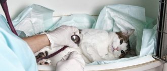
View of the ear canal before surgery
Installation of drainage into the cavity of the surgical wound
View of the ear after surgery
ATTENTION!!!
It is necessary to treat your animals as carefully as possible, since the earlier the owners notice the tumor, the greater the chance of curing the animal.
How to tell if your cat has nasal polyps
The first manifestations of pathology can be noticed only after the polypous formations have reached a significant size.
Symptoms of nasal polyps in cats:
- labored breathing;
- wheezing, grunting, wheezing when breathing, snoring;
- decreased sense of smell;
- dyspnea;
- change in behavior (lethargy, apathy, inactivity);
- nasal discharge from both, one nostril (mucous, purulent, bloody);
- fatigue after short periods of activity;
- frequent sneezing, coughing fits, choking;
- nausea, gagging, vomiting, dysphagia;
- decreased appetite;
- neurological disorders;
- mucous discharge from the eyes, tearfulness;
- pallor, cyanosis of mucous membranes.
The cat may feel anxious, be reluctant to make contact, rub its nose with its paws, shake its head, tilt its muzzle to the side, breathe with its mouth open, especially in stuffy rooms during the hot season. If, in addition to nasal polyps, the cat has nasopharyngeal polyps, drooling, difficulty eating, and weight loss are noted.
In severe cases of polypous formations, especially if polyps are detected in two nostrils, deformation of the cat’s nose is possible.
Treatment of the auricle
The choice of treatment tactics for the disease largely depends on the size of the hemorrhage and the stage of the pathological process. For small hematomas, cold is indicated in the first hours after injury. Frozen foods from the freezer wrapped in cloth are suitable for this purpose. You can cool the damaged ear for 10 - 15 minutes.
If there is an open wound, it is treated with disinfectants. To reduce blood leakage, a pressure bandage is applied to the ear.
For extensive hematomas, due to the fact that there is a high risk of infection, the most effective method of treatment is surgery. In addition, when the pathology progresses spontaneously in the final stage, fibrinous tissue grows at the site of damage. This leads to deformation of the ear cartilage, which is undesirable for show animals.
During the operation, the veterinarian opens the hematoma using an incision at the base of the ear. Blood clots and fibrin are removed. At the end of the manipulation, a fixing suture and bandage are applied. Surgery is usually performed under local anesthesia. After the operation, the animal must be in a special collar that prevents the paw from scratching the ear.
Pumping out blood with a syringe
The positive aspect of the surgical treatment method is the minimal risk of relapse, as well as the preservation of the natural shape of the ear cartilage. The disadvantages of surgery include a small stitch in the cat's ear.
In addition to surgical treatment, a veterinarian can use special dressings for show animals. A special feature of this method of treating hematoma is the absence of postoperative scars. The disadvantage is that the owner cannot apply the bandage himself; constant visits to the veterinary clinic are required.
A common way to treat small, fresh hematomas is to drain the blood with a syringe. The method is often used when the blood that has escaped from the blood vessels has not yet clotted.
Ointments for the treatment of ear hematoma in cats
Anti-inflammatory and antibacterial ointments are widely used in the treatment of the disease: levomycin, syntomycin, levomycin. For suppuration, ichthyol and Vishnevsky ointment are used. Additionally, if the hematoma becomes infected, the animal is prescribed a course of antibacterial therapy. For this purpose, penicillin antibiotics, tetracyclines, and cephalosporins are used.
Diagnostics
A veterinarian will help determine the presence of polyps, their size, location, quantity, as well as the possible root cause after conducting a number of diagnostic tests.
When making a diagnosis, medical history, breed, age, general condition, and clinical signs are taken into account. Differential diagnostics, biopsy, MRI, CT, fluoroscopy, endoscopic studies (rhinoscopy) are performed, which allow a complete assessment of the condition of the nasal mucosa.