Types of cysts in cats and places of their occurrence
In veterinary medicine, the following classification of cysts in domestic animals is accepted, based on the nature of the contents of the blisters.
Atheromas are widespread in the cat population.
Based on location, veterinary specialists distinguish the following types of neoplasms:
- Epidermal tumors. This form of cyst-like cavities is characteristic of the outer layers of the animal's skin. Cysts are soft swellings with clear boundaries that do not cause concern to the pet. Most often, epidermal neoplasms are observed on the back and side surface of the body. Often the cyst breaks out on its own, releasing a paste-like content of gray-yellow color.
- Hair cyst. It is usually formed when the sebaceous glands of the skin become clogged and inflamed and is diagnosed in animals after 5 years of age. Such a cavity is most often filled with keratin contents.
- Dermoid cystic tumor. It occurs most often in young individuals and is localized in the auricle. It is innate in nature. The cavity of the dermoid formation is filled with a sebum-like secretion. The cyst has a spherical shape, is soft to the touch, and does not cause pain to the animal.
- Internal cysts. Neoplasms can affect many organs: ovaries, kidneys, uterus, liver. Internal tumors are not as harmless as external ones. Polycystic kidney disease is dangerous due to the development of secondary bacterial infection and the development of sepsis.
- Mucocele . A separate type of tumor that occurs when the salivary ducts are blocked. The disease is divided by localization into sublingual, pharyngeal and zygomatic mucocele. This type of neoplasm often degenerates into an oncological tumor.
- Breast cyst . The most common formation in domestic cats. The main reason for the formation of this type of pathological cavities is hormonal imbalances in the female’s body.
The variety of cystic tumors often frightens owners. Most cysts are benign. However, some formations can degenerate into malignant tumors and threaten the health and life of cats.
Types of inflammation of the salivary glands
In veterinary practice, only two types of inflammation of the salivary glands most often occur.
Sialadenitis
The classic type of inflammation is sialadenitis. This is a nonspecific damage to the tissues of the salivary glands due to trauma, general systemic infections, etc. Large glands are most often affected. An acute course of the disease is characteristic, accompanied by swelling of the affected areas, an increase in local and even general body temperature. In slightly more rare cases, purulent inflammation develops, which, if left untreated, “mutates” into necrosis of the gland tissue.
Mumps
This is also a classic inflammation of the salivary glands, but mumps in veterinary medicine is considered a viral type of pathology. The symptoms are the same as in the previous case. This type of disease is considered more dangerous, since pathogens from the paramyxovirus family can affect not only the salivary glands, but also other types of glands in the cat’s body.
Animals at risk
The reasons for the formation of cystic cavities are not fully understood. It is believed that some types of cysts are caused by a hereditary predisposition, while others appear due to a violation of the hormonal status of the body. The conditions of keeping and feeding the pet are also important.
Veterinary specialists and experienced breeders believe that the following pets are at risk:
- Unsterilized cats. Estrus, pregnancy, childbirth, and feeding offspring are often accompanied by disruptions in the female’s hormonal system.
It is the disruption of hormonal status that is the main cause of the development of polycystic ovary syndrome and cystic tumors of the mammary gland.
- The use of hormonal drugs to control estrus and sexual behavior is a serious blow to the cat’s endocrine system. Veterinary experts consider the use of tablets, drops, and injections based on sex hormones to be the main cause of the development of ovarian and mammary cysts in domestic cats.
- Injuries and mechanical damage. If the integrity of the skin is damaged, a keratin-like substance can penetrate into the layers of the skin. Epidermal cysts often develop through this mechanism.
- Breeds such as the Persian, Himalayan, British Shorthair and Exotic Shorthair are at risk for developing polycystic kidney disease.
- Keeping pets in poor hygienic conditions and the presence of thorny indoor plants provokes the development of hair and epidermal tumors.
- Violation of feeding rules. Eating bones is a risk factor for the development of mucocele in the domestic cat.
Causes
The occurrence of a cyst depends on its type.
Epidermal ones occur when a keratin-like substance enters the thickness of the skin traumatically or when the follicular canal is blocked.
A dermoid cyst is most often a congenital disease. Changes occur during ebryogenesis, when part of the epithelium is formed in the thickness of the dermis. Because of this, a cyst forms in its place, which is filled with skin-like epithelium, sebaceous and sweat glands, and in rare cases, underdeveloped teeth.
Ovarian cysts can be caused by the frequent use of hormonal agents to regulate sexual heat in cats; the most dangerous is the use of long-acting hormonal injections. It is also possible to have a hormonal imbalance as a complication after childbirth.
Polycystic formations in the organs of the genitourinary system are considered hereditary diseases, and Persian breeds are most often affected by them.
With a mucocele, veterinarians often cannot figure out the root cause, but believe that in most cases the cyst occurs due to traumatic injury.
Breast cysts occur due to hormonal imbalances.
Clinical symptoms of a cyst in a cat
Identifying an external neoplasm in an animal is not difficult for an attentive owner. Typically, the disease has the following symptoms:
- A spherical or spherical compaction is visible above the surface of the skin.
- On palpation, the tumor is often soft and elastic. In the case of a keratinized formation, the cavity is dense when palpated.
- The animal does not experience pain on palpation.
- The general condition of the animal is usually satisfactory. The cat does not lose its appetite and physical activity.
- When opening the cavity, a complication in the form of wound infection is possible. In this case, the symptoms will depend on the nature of the inflammatory process.
Mucocele has characteristic features. The owner observes excessive salivation, poor appetite, and loss of body weight in the cat. With the development of the pathological process, saliva contains blood impurities.
It is not difficult to detect mammary cysts in a domestic cat. At the first stages of development, the pathological formation does not cause concern to the pet. However, breast tumors are prone to rupture and infection.
Damage to internal organs is much more difficult for the owner to determine. So, polycystic kidney disease has no obvious symptoms. Clinical signs of the disease are similar to those of renal failure. The animal becomes lethargic and urination is delayed.
Polycystic kidney disease in a cat
Videos and illustrations
Gallbladder cyst on x-ray
Gallbladder cyst
Breast cyst
Salivary gland cyst or mucocelle
Multiple follicular cysts
Polycystic uterus
Polycystic
Diagnosis of cyst formation in an animal
In a specialized institution, your pet will not only be diagnosed, but also the type of cystic formation will be determined. Modern veterinary medicine has the latest techniques. The sick animal will first undergo a biopsy. Using a puncture, the veterinarian will puncture the pathological bladder and take its contents for examination.
The procedure is important for the differential diagnosis of a cyst from an abscess, inflammatory infiltrates, and various types of malignant formations.
Histological examination of biological material allows for a correct diagnosis.
If the development of cyst-like cavities in the internal organs is suspected, the veterinarian uses the following methods for diagnosis:
- Ultrasound examination of the kidneys, ovaries and other internal organs. Diagnostics allows the animal to painlessly determine the presence of pathological blisters in the ovaries, kidneys, uterus, liver, estimate their number, and determine their exact location.
Ultrasound of a cat: polycystic kidney disease
- An X-ray examination of the abdominal organs allows one to identify pathological structures in the kidneys and uterus. An X-ray of the cat's head will allow us to determine the exact location of the mucocele.
For a differential diagnosis, methods such as general and biochemical blood tests, determination of hormone levels, and urinalysis can be used.
For information on diagnosing polycystic kidney disease, watch this video:
What is it, what types of cysts are there?
This is the name of a capsule with a dense or soft wall, which is most often filled with liquid contents. It can be located anywhere, even on the head. Contrary to popular belief, not all cysts pose an immediate danger to the life and health of the animal, and in some cases they even resolve spontaneously.
Cats often have cysts caused by echinococcosis or alveococcus. If this is so, then the bubble in the tissues is extremely dangerous for humans, since it is filled with parasite larvae. Thus, in particular, a kidney cyst can develop (including in humans).
Scientists have long developed a general classification of cysts, according to which the following types are distinguished:
- Atheromas (including the epidermal variety). This is the most common type of cyst in cats. These formations can occur anywhere. Cysts are usually small, up to a maximum of 2.5 cm in diameter, most often dense to the touch, filled with sebum-like contents. They are (most often) benign, showing no tendency to grow or increase.
- Keratinized cysts contain grayish caseous (curdled) contents.
- Follicular cyst. Another common type is the classic bubble filled with liquid.
- Hairy cysts appear as a result of clogging of the sebaceous glands and inflammation in the hair follicles. Sometimes they appear with demodicosis.
- Dermoid cyst. It develops from skin tissue; its thickness contains numerous sebaceous glands. The dermoid cavity is filled with sebaceous contents. Quite often found in the ear.
- Apocrine cysts. A very dangerous variety, as they are found exclusively in the form of clusters. If they appear in internal organs, the functionality of the latter is seriously affected.
We invite you to read: Herd horse breeding key points
Treatment of cysts in cats depending on location
If a formation of any size is detected, the most important thing is to consult a doctor. Only he can know exactly how to act in a given situation, which examination will more accurately show the problem, and also prescribe the most effective treatment.
Should I always delete
Surgical treatment does not always have indications. If the animal is elderly or weakened by disease, the operation can be dangerous.
Contraindications to tumor removal are often concomitant diseases or conditions in which anesthesia is undesirable.
If surgical intervention is not possible, fine-needle puncture is performed. The procedure makes life easier for the pet with large tumors and reduces the risk of spontaneous opening and infection.
Surgery as an option to get rid of a cyst
The most effective way to cure a sick animal is surgical removal of pathological cavities. Excision of the tumor occurs within healthy tissue. The capsule must be excised.
Prognosis for an animal with a cyst and after removal
Veterinary specialists do not classify cystic formations as life-threatening pathologies for pets. However, this is true for cavities that are not complicated by infection. Spontaneous opening of tumors is often accompanied by the development of a secondary bacterial infection. This phenomenon leads to a deterioration in the general condition of the animal, an increase in temperature, and the development of pain. The risk of developing sepsis increases.
After removal of the cyst, veterinarians usually give a favorable prognosis if the tissue does not degenerate into oncological formations.
Measures to prevent inflammation of the salivary glands
There are the following measures to prevent inflammation of the glandular organs:
- The cat must be vaccinated on time, reducing the risk of the pet contracting dangerous infectious diseases.
- At least once a week, your pet’s mouth should be examined for heavy plaque deposits. If any are found, clean the cat’s teeth using a gauze swab and a weak aqueous solution of soda.
- It is necessary to carefully monitor the quality of your pet's diet. You should never give cats bones!
- In addition, once a quarter the cat is taken to the veterinarian for a preventive examination. This measure will help prevent a lot of trouble.
Preventing the appearance of cysts in cats
Preventive measures depend primarily on the type of cystic pathology:
- If a pet is diagnosed with polycystic kidney disease, the owner must decide to remove the cat from breeding, since the pathology is hereditary.
- It is necessary to protect the animal from injury and mechanical damage to the skin.
- Timely sterilization of the cat before the start of the first heat allows to minimize the formation of pathological cavities in the ovaries and uterus.
- Do not use hormonal drugs that affect sexual heat in reproductive animals.
- Keeping your pet clean.
- Exclusion of bones from the diet, follow the diet.
- Carrying out regular deworming and vaccination.
The formation of cysts in furry pets has a different etiology. The variety of forms and types of neoplasms requires a qualified approach in diagnosing pathology. The most effective treatment method is surgical removal of the cyst. The prognosis for uncomplicated forms is usually favorable.
How dangerous is hypertrophic cardiomyopathy in cats and how is it treated? Types of cysts in cats, methods of diagnosis and treatment.
The difference in the forms and manifestations of cysts in cats is associated with a variety of reasons leading to the development of pathology.
It is worth noting that salivary gland cysts in cats are often prone to recurrence, therefore, even after successful treatment.
Polycystic ovary syndrome - what is it?
This is a dishormonal disease accompanied by multiple formation of cysts. On average, their diameter ranges from 0.5 to 5 cm. However, large cysts with a diameter exceeding 10 cm can also occur.
In cats there are:
Functional cysts . They occur against the background of minor hormonal disorders, in particular they can be caused by long-term use of hormonal drugs. However, functional cysts resolve on their own and do not require special treatment.
Organic cysts . Their main feature is their rapid increase in size. Organic formations can grow to large sizes, and therefore often lead to negative consequences for the entire body. Requires veterinary treatment.
Skin cysts
Svetlana Belova, Estonian University of Life Sciences, www.vetderm.eu Photos of the author are used in the article
A cyst is a pathological cavity (in our case in the skin) with a wall lined with epithelium. The contents of the cyst depend on the secretory activity of this epithelium. Cysts can be acquired (for example, blockage of the excretory duct of the skin gland) or congenital.
In the skin, there are cysts of sweat and sebaceous glands, follicular cysts and dermoid cysts/sinuses.
A sweat gland cyst is a small (several millimeters) sized cavity with a bluish tint (photo 1). It is filled with clear liquid (sweat) and protrudes above the skin level. It is relatively rare in both dogs and cats. Single sweat cysts, as a rule, do not cause discomfort and do not require treatment.
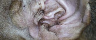
Multiple cerumen/ceruminal (modified sweat) gland cysts in the external auditory canal and pinna of cats are a relatively common occurrence and are called ceruminal cystomatosis . While the cysts are small in size (they look like dark gray, almost black smooth elevations a few millimeters in diameter), they do not cause any discomfort (photo 2). However, when they reach a certain size and quantity and block the lumen of the ear canal, they cause external otitis media with secondary infections, soreness and itching (photo 3). Cysts can degenerate into adenomas and adenocarcinomas of the sulfur glands. Treatment consists of surgical excision (preferably laser excision).
Sebaceous gland cysts are small (1–3 mm) elevations of a whitish or yellowish hue (photo 4). They are relatively common, especially in dogs. They do not cause discomfort and do not require treatment.
Follicular cysts (FC) are most often formed by the epithelium of the mouth of the hair follicle and are a cavity filled with dead keratinized epithelium - keratin. FC occurs quite often in dogs (predisposed breeds are German Shepherds, Pekingese, Shih Tzu and Boxers) and rarely in cats. Clinically, FC appears as a rounded raised area of grayish skin ranging from a few mm to 6 cm, often with a hairless and comedonal surface (Figure 5). FC is often mistakenly called a sebaceous gland blockage. They can be single or multiple and have a progressive course. Follicular cysts that have reached significant sizes often have a “pore” on the surface of the skin through which a curdled content (keratin) of a grayish tint is released. When drying, keratin forms a kind of cutaneous horn on the surface of the skin (photo 6). Secondary bacterial infection of the FC is also not uncommon (especially after an attempt to squeeze it out!), in which erythema is noticeable and pain and itching appear (photo 7). Main differential diagnosis: abscesses, tumors. Diagnosis is made based on history, clinical presentation, cytological examination of cyst aspirate (keratin, cholesterol crystals +/- signs of infection) or histology. Treatment consists of surgical excision. Retinoids (isotretinoin 1.5–3 mg/kg/day) may be effective in inhibiting the growth of old cysts in dogs and preventing the formation of new ones.
A dermoid cyst (DC) is a congenital anatomical anomaly in which a fully functioning piece of skin with all appendages (hair follicles, sweat and sebaceous glands) is enclosed in a closed cavity. DC becomes noticeable already in puppyhood, most often on the midline of the head (photo 8). It looks like one or more soft, round or oval-shaped bumps that can be infected and have a “pore” on the surface, from which a tuft of hair often sticks out. Treatment is surgical excision. A dermoid cyst may be connected by a tubular tubule to the spine (dermoid sinus), which naturally creates a risk of bacterial infection of the nervous system. Rhodesian Ridgebacks are especially predisposed to this anomaly, in which the presence of a crest is predetermined by a mutation of certain genes inherited as an autosomal dominant trait and is associated with the formation of dermoid sinuses (photo 9). The diagnosis is made on the basis of anamnesis, examination, contrast radiography, magnetic resonance imaging. Treatment is surgical excision (photo 10).
Lumps under the skin in cats
The appearance of strange formations under the skin of a pet is not that uncommon. They can be malignant or benign, become inflamed, cause discomfort to the cat, ooze exudate (serous or purulent) or be absolutely painless and not annoy the pet in any way. But any subcutaneous formations in a pet deserve the owner’s attention.
Under the skin, between the skin and the muscle layer, nodules or capsules with liquid contents may form in your pet. They are “dough-like”, mobile and do not cause trouble for your pet. The lumps may be hard and painful.
Subcutaneous formations consist of degenerated tissues, or are capsules filled with contents (dense or serous). They have normal body temperature or are dense and hot to the touch. At the same time, the animal’s general body temperature may increase, and other signs of general intoxication may appear (weakness, refusal to eat, irritability or lethargy, dyspepsia). Formations prone to rapid growth deserve special attention.
Causes of bumps
Lipoma, also known as fat, is a benign neoplasm that can appear on any part of the body. Prone to growth, can reach large sizes, but does not metastasize. The formation is soft, limited, painless. General symptoms are related to the location, that is, the mechanical pressure of the tumor on surrounding tissues. The formation of lipomas on the limbs can lead to lameness of the animal, and in the area of the carotid artery to death from suffocation. The general condition of the animal is usually normal. Unfortunately, lipomas often occur in association with malignant tumors (fibrosarcomas or sarcomas). More often this happens when the tumor appears on the back of the animal. Lipomas are more common in older animals.
Abscesses in animals can be post-injection, develop as a result of wounds or after insect bites, as a consequence of notoedrosis.
Post-injection abscesses form after an unsuccessful injection, if a bacterial infection was introduced during the injection process or the animal “got a cold” at the injection site. Thin deep punctures often fester. Because anaerobic microflora enters the narrow, deep wound channel and actively multiplies, since it is difficult for oxygen to enter such a wound. Abscess tumors are painful, they lead to dysfunction, and are accompanied by fever and local temperature.
Lymphadenitis or an inflammatory process in the tissues of the lymph nodes, can occur with “feline distemper”, leukemia, tularemia, or fungal infections. This disease is caused by some species of Bartonella, Pasteurella, and Fusobacterium. In this case, swelling appears under the jaw, in the groin and axillary area. Swollen lymph nodes are very painful, even if the swelling is minimal, and they are barely palpable. Lymphadenitis is usually accompanied by general intoxication and a disturbance in the condition of the animal.
Nodules under the skin can form as a result of encapsulation of a foreign body trapped under the skin of an animal or a source of bacterial infection. In this case, the formation is dense and painless. Discomfort in the animal can be caused by pressure on the nodule if it presses on the underlying tissue. The general condition of the cat is not disturbed, the nodule is not prone to growth.
A mite embedded under the skin and feeding is very similar to a small skin formation. You need to carefully examine such a “growth” and remove it. A tick, even one that has eaten, is small, usually no larger than the nail plate of a person on the little finger of his hand. There is no disturbance in the animal's condition if a tick attaches itself to it.
Polycystic kidney disease in cats: how long can they live and can it be cured?
Polycystic disease is a pathology in which many cavities (cysts) filled with fluid form in the kidneys. Part of the organ becomes a vesicular formation. The disease affects both kidneys at the same time. Since a significant part of the parenchyma is lost, the function of filtering biological waste is not fully performed. Metabolites accumulate, causing intoxication.
Polycystic disease is a hereditary disease. Man created many breeds of cats according to his own will, endowing them, in addition to the given qualities, with unexpected deformities. Among cats suffering from polycystic kidney disease, the most common are Persian and British breeds.
The pathology does not threaten the pet with instant death, but makes his life a series of painful sufferings. It must be treated immediately after diagnosis. In addition to constant intoxication, the contents of the cyst provide a breeding ground for conditionally pathogenic microflora.
The cavities tend to grow and replace the parenchyma of the excretory organ. Symptoms of polycystic disease become noticeable in cats over 7 years of age, as the enlargement of the cavities occurs slowly.
The cat adapts to life with defective kidneys and for a long time the disease proceeds without revealing itself. The disease cannot be cured. The veterinarian’s task is to stop the development of pathology.
Treatment of any disease is effective if it is detected in the initial phase. This has nothing to do with polycystic disease. Until the cavities occupy a significant part of the kidney area, signs of the disease cannot be detected. If upon palpation you notice pain in the abdominal cavity, the prognosis is poor.
Specific signs of polycystic kidney disease include:
- Loss of appetite.
- Oppression.
- Hematuria.
- Thirst.
- Emaciation.
- Vomit.
- Pollakiuria.
- Nervous phenomena.
Diagnostics
A presumptive diagnosis is established by clinical signs, urine analysis, and in-depth palpation of the abdominal chamber. To confirm the assumption, a biopsy is performed. This study allows us to establish the nature of the cavities and determine the degree of their danger to the life of the cat. Cultures of pathological material reveal the presence or absence of secondary infection of the kidneys and excretory channels.
Ultrasound allows you to see the tumor and determine the ratio of intact and modified tissue in the organ. Modern clinics have mastered the method of gene testing. If both methods indicate polycystic disease, it becomes possible to carry out preventive treatment and prophylactic measures that can slow down the development of cysts. Radiography reveals the disease in its final stage.
Polycystic kidney disease cannot be cured non-surgically. If the pathology is detected in the early phase, and the affected area is small, then there is a chance to get rid of the disease. But such a development of events is possible when the cat owner himself insists on conducting an examination. In most situations, the veterinarian has to deal with the second or even third phase of the disease. The goal of treatment is to ensure satisfactory well-being and prolong life.
If the size of the cystic blisters is large, periodic removal of fluid is carried out with a puncture needle. Drug treatment is aimed at minimizing developing chronic renal failure.
The following methods are used in the treatment of polycystic kidney disease:
- Diet therapy.
- Rehydration detoxification.
- Normalization of blood pressure.
- Gastroprotectors.
- Prevention of anemia.
- Antibiotic therapy.
Diet therapy
They use specialized wet food for cats with kidney disease, with reduced levels of Phosphorus and protein, which provoke the development of symptoms of chronic renal failure. To normalize phosphorus-calcium metabolism, vitamin D preparations are used.
Rehydration detoxification
To remove toxins, parenteral administration of rehydration solutions is necessary.
Normalization of blood pressure
The medical drug Amlodilin is in demand for cats. To maintain the dosage, you will need a tablet knife.
Prevention of anemia
Use of Erythropoietin, as well as drugs for the treatment of anemia.
Gastroprotectors
The main purpose is to relieve attacks of nausea and prevent the formation of stomach ulcers. They use drugs that reduce the production of gastric juice - antacids.
Antibiotic therapy
If bacterial culture reveals the presence of secondary microflora, antibiotic therapy is prescribed. First, universal drugs are used, and when the results of a test for the sensitivity of microflora to antibiotics become available, treatment correction is applied.
When carrying out maintenance therapy at the first stage of the disease, the prognosis is favorable, in the opposite situation it is doubtful or negative. The lifespan of sick cats can be several years or two months.
Prevention
Care should be taken when breeding polycystic-prone Britons and Persians. Pets with this diagnosis are not allowed to be bred and are recommended to be sterilized. For breeds predisposed to the disease, it is necessary to use specialized feed.
Sterilization of cats
Sterilizing a cat is a surgical operation that veterinarians around the world have been successfully performing for many years. The methods used by different doctors may differ, but the essence is the same: during the operation, the reproductive organs are removed, which helps to reduce hormonal levels and stop the reproductive functions of the body.
What is the purpose of sterilization?
The question of sterilizing a domestic cat sooner or later arises before every owner. With the onset of puberty, a cat can turn a quiet house into a real hell, and drive the owner into a white heat with her endless purring and loud concerts at night. Such active vocalization is not due to the fact that your pet’s character has deteriorated, and not to her desire to not let you rest. This is simply how she informs her relatives that she is ready for mating and calls the cat “on a date.” That's the nature of a cat. Her wild ancestors have shaped this type of behavior for centuries, and she is simply obeying ancient instincts.
But what to do if there is no cat nearby, and the owner has no desire to breed his pet and then deal with the adoption of kittens? A mutually beneficial solution is to sterilize the cat. After the operation, the production of female sex hormones (estrogens) stops within about a month, the hormonal levels decrease, and the cat again turns into a cute, harmless pet. With a high degree of probability, sterilization will also help rid the cat of excessive aggressiveness. As the level of hormones in the blood decreases, the character improves, the animal becomes calmer, and aggression towards others and the owner decreases or disappears altogether.
Sterilization and castration of cats
The term “sterilization” is usually applied to cats (females), and the term “castration” is applied to cats (males). It is believed that cats are spayed and male cats are neutered. For ease of communication with clients, veterinarians also use the same terminology. However, female animals undergo both spaying and sterilization. Let's look at the differences.
Surgery to sterilize a cat involves removing only the ovaries, without removing the uterus (oophorectomy). Castration is the complete surgical removal of all reproductive organs (ovariohysterectomy).
What is better - castration or sterilization? Let's figure it out. During an oophorectomy, only the ovaries are removed, while the uterus lives its own life in the abdominal cavity and does not perform any useful function. It will not atrophy or die, since its blood supply is not impaired. She will never fulfill her destiny and will not be able to bear fruit. But since the organ is completely alive, the risk of developing uterine diseases remains fully intact. These can be neoplasms and inflammatory diseases (endometritis, pyometra, etc.). So why preserve an organ that will act as a time bomb, with the risk of exploding at the most inopportune moment? That is why, at present, almost no one performs ovariectomy (classical sterilization); veterinarians prefer castration as the only reliable method. But for ease of communication, they still say “sterilization.”
Where is it better to perform the operation - in a clinic or at home?
It is better and safer to do any abdominal surgery in a veterinary clinic. In an operating room there are more opportunities to take emergency measures if something suddenly goes wrong.
It’s even better if the clinic provides a postoperative hospital service. Then your pet will be under the supervision of specialists throughout the crisis period. Of course, emergency measures are not always required.
In the photo: day hospital in our veterinary clinic. The animal remains under the supervision of a doctor until it completely recovers from anesthesia.
Typically, young and healthy cats tolerate ovariohysterectomy well and, with minimal care recommendations, recover well at home. You will only need one more visit to the clinic to remove the stitches.
The advantages of sterilization in the clinic : all the necessary conditions for the operation are provided, the risk of emergency situations is reduced, and there is a high probability of overcoming them.
Disadvantages of sterilization in the clinic : the owner spends time traveling and waiting for the end of the operation.
Despite the obvious advantages of surgery in a veterinary clinic, sterilization of cats at home is also practiced by most veterinarians and, with proper training and responsibility of the specialist, is technically no different from surgery in a clinic.
In the photo: preparing to sterilize a cat at home.
The advantage of sterilization at home is the absence of stress for the cat from traveling and being forced to stay in a place that is foreign to it (some cats are terrified of leaving the apartment), as well as the absence of risks of contracting infectious diseases (if the animal is not vaccinated). In addition, the doctor can be invited at a time convenient for the owner, which is also very important for people who are constantly busy at work.
Disadvantage of surgery at home : it is difficult to ensure high-quality sterility.
Age of sterilization of cats
We recommend spaying cats after they reach 7-8 months of age. Earlier sterilization is not advisable, because The animal’s body is not yet fully formed and there is a high risk of postoperative complications. In addition, there have been cases of retardation in growth and development of those cats that were sterilized at 4-5 months of age. The operation is allowed at 6 months of age, provided that the cat is large and weighs at least 2.5-3 kilograms.
In a later period (over 8 months), cats, of course, are also sterilized. But it should be remembered that the risks of postoperative and post-anesthesia complications increase in proportion to the age of the animal. Do not delay the operation - every year your cat gets older, the risk of negative consequences increases, and the effectiveness of the operation decreases.
Our long-term practice of sterilizing animals shows that cats aged 7 months to 10 years tolerate the operation well and without problems. Animals over 10 years of age require additional examination. For such cats, we recommend donating blood for a biochemical analysis, diagnosing the functioning of the heart and consulting with a cardiologist.
Can a cat be spayed while in heat?
During the hunting period, the operation can be performed, but recovery from anesthesia and healing of the sutures may be somewhat more difficult. We generally recommend surgery two weeks before or two weeks after the heat. However, it often happens that a cat’s heat lasts for a very long time, with minor breaks (from 1 to 5 days). In this case, it is not possible to maintain a two-week interval, and we recommend sterilizing the cat to avoid exhaustion and deterioration in the quality of life.
Should a cat be allowed to give birth before being spayed?
No! If you do not plan to breed cats, then it is better to carry out the operation as early as possible, without allowing the cat to be bred. Moreover, you should not specially knit it, knowing that you will sterilize it later. Many cat owners are mistaken, believing that a cat needs to first give birth, “feel like a woman,” “let it know the joy of motherhood,” etc. (quotes from real conversations with cat owners at the reception). I will try to explain why this should not be done. At the hormonal level, the process of cat “purring” and night cries is carried out through the production of estrogens, which cause typical manifestations of sexual heat. Initially, these hormones are produced only in the ovaries and after sterilization, all unwanted effects for the owner disappear. If the cat has already kitted, then other endocrine glands begin to produce female sex hormones, and sexual behavior can persist for a long time, if not forever. Thus, the owner’s stereotypical thinking can delay the achievement of the main goal of cat sterilization - to avoid sexual estrus in the pet.
Is it possible to sterilize a pregnant cat?
Yes, you can. The procedure is called “extirpation of the pregnant uterus” and is usually performed for medical reasons to save the life of the female. We usually do not recommend sterilizing a healthy pregnant cat, since during pregnancy the animal’s body undergoes significant changes at both the hormonal and physiological levels. There is a possibility of high blood loss and longer recovery of the animal after sterilization. In addition, the suture after sterilization of a pregnant cat is much larger in size.
However, situations are different and, if the owner is aware of the risks associated with such an operation and insists on it, we perform extirpation of the uterus and fetuses.
Does a cat need to be vaccinated before sterilization?
It is highly desirable that the cat be vaccinated at least a month before sterilization. In this case, there is no risk of getting a viral infection when visiting a veterinary clinic, because animals with various diseases, including viral ones, are brought there. Anesthesia given to a cat during surgery can reduce the body's immune response, and an unvaccinated animal will become ill. Many veterinarians offer to protect the cat by injecting her with a special hyperimmune serum (globulin), which provides protection against infections for several weeks. Unfortunately, the introduction of globulins does not provide 100% protection. Therefore, it is advisable to vaccinate your pet in advance and plan surgery in a month.
Methods for sterilizing cats
We will describe the most common surgical methods used throughout the world.
1. Classic method . A skin incision 1 to 3 cm long is made in the center of the abdomen, 2-3 cm below the navel. The abdominal wall is incised along the linea alba. The uterus is removed through the incision, a ligature of absorbable suture material (catgut, caproag, PHA, etc.) is applied to the vessels or a coagulator is used. After this, the uterus along with the ovaries is removed, and sutures are placed on the peritoneum and skin. The skin suture can be either removable or non-removable. The first one must be removed after at least 7, maximum after 10 days. There is no need to remove the second one.
In the photo: removal of the uterus and ovaries from a cat using the classical method 2. Method of sterilizing a cat through a side incision . It differs from the previous one only in the place of tissue separation. A skin incision is made on the side, the muscles are separated in a blunt manner. The method is considered less traumatic than the classic one, but there are several nuances. First, a muscle tear does not guarantee that there will be no bleeding. Secondly, sterilization rather than castration is often performed through such a wound, since it is not always possible to extract and remove the uterus completely. The method is recommended for sterilization of stray animals, as it does not require suture care. If a special intradermal suture is applied, the cat can be released into the wild immediately after the operation.
In the photo: applying a ligature to the ovarian ligament during sterilization of a cat using a side incision. Photo courtesy of colleagues. As you can see in the picture, the bleeding is no less than with classical sterilization. 3. Method of sterilization through an ultra-small incision using a surgical hook . Some veterinary clinics position this method as “laparoscopic sterilization,” but it has nothing to do with laparoscopy. The skin and peritoneum are dissected in the same way as in the first case, only the incision is made less than a centimeter long. The surgeon inserts a sterile hook into the wound, picks up the ligament and removes the ovary. After applying the ligatures, either only the ovary is removed along with part of the ligament, or the uterus is pulled into the wound and removed completely. As in the previous case, it is not always possible to completely remove the uterus.
In the photo: oophorectomy using a surgical hook through a very small incision 4. Laparoscopic method of sterilization of cats . Using endoscopic technologies, complete removal of the uterus and ovaries is performed through one or more punctures of the skin and abdominal wall. The operation is technically complex and requires expensive equipment and special personnel skills. In Russia it is not used everywhere and is much more expensive than conventional sterilization.
In the photo: capture of the uterine horn during the laparoscopic method of sterilization of cats.
I would also like to note that the best method of performing an operation is one that the surgeon is fluent in . Any technique mastered by a doctor to its intricacies will lead to a successful outcome in cat sterilization. You should not force the doctor to do what you read on the Internet. He may do it your way, but he will do it better and safer in his own way.
Another method of sterilizing cats should also be mentioned: medication. This method is based on the introduction of a special implant with medicine under the skin. Slowly released and entering the blood, the active substances cause a reversible castration (sterilization) effect in cats, lasting up to 3 years. In this case, you will not have to expose the animal to anesthetic risks. Using a special device, the veterinarian will insert a small implant under the skin in just a few seconds. Read more about the medical method of sterilizing cats in our article.
Preparing a cat for sterilization
There is no need to take any special measures to prepare the animal for sterilization. A prerequisite is to ensure a 12-hour fasting diet. If the operation is planned, say, at 10 o’clock in the morning, then the cat can be fed for the last time at 10 o’clock in the evening, and then the food can be removed altogether.
If several animals live in the house, everyone will have to go hungry. Or the cat will need to be placed in a separate room, without access to food. On the day of surgery, in the morning, it is recommended to remove the water.
This requirement is due to the side effects of substances used for anesthesia in cats (most veterinary clinics use a combination of xylazine and tiletamine), which manifests itself in the urge to vomit when there is food or water in the stomach. Aspiration of vomit can cause a serious complication - aspiration pneumonia. Therefore, we recommend that you take a responsible approach to the recommendation of fasting before surgery.
Caring for a cat after sterilization
Typically, post-operative care is not difficult for the pet owner. The main thing is to have some free time to watch your pet.
It is advisable for the cat to have a horizontal, soft place in a warm place, preferably without access to direct sunlight (bright light irritates the cornea of the eye when recovering from anesthesia and the animal becomes more anxious).
When you bring your cat from the veterinary clinic, place it in a pre-prepared area and leave it to rest. It is advisable to limit the access of children and other animals to the recovering cat during the first 24 hours after sterilization so that they do not disturb it.
A small nuance that frightens many pet owners - cats do not close their eyes while under anesthesia. To ensure that the animal, when it begins to wake up, does not experience discomfort from the drying out of the cornea, it is recommended to periodically instill contact lens solution (artificial tears) or regular saline solution (0.9% NaCl). If none of this is available, you can simply close your eyelids every 5-10 minutes with a few light movements. The tear will spread over the surface of the eyeball, wetting the cornea. Following this recommendation will make recovery from anesthesia more comfortable for the animal.
In the photo: a cat in deep anesthesia. The eyes don't close. In order not to provoke the development of postoperative conjunctivitis, it is recommended to periodically moisten the cornea.
Over the course of 7-10 days, it is also necessary to check the condition of the skin sutures (if any). The seam should remain dry and clean after sterilization. The seam should not become soaked or rotted.
Treatment of seams usually involves wiping with antiseptic solutions (chlorhexidine, dioxidine, hydrogen peroxide). Some veterinarians prescribe additional treatment with wound healing ointments.
Sutures placed during sterilization in our veterinary clinic do not require additional processing at all.
In the photo: treating the seam with a spray that prevents infection from entering the wound.
In the photo: a suture in a cat on the 8th day after sterilization. After treatment with a special spray, the seam does not require special care.
To prevent contamination of the wound, the cat is given a special bandage (blanket), in which it must remain for the entire postoperative period, until the sutures are removed. The cat’s discomfort from the blanket usually disappears within the first 24 hours after sterilization, and then she calmly wears this forced “clothing.”
In the photo: the cat is wearing a special blanket that prevents damage to the seams
If the wound was sutured with an intradermal suture, it is simply wiped with a 0.05% chlorhexidine solution.
It is acceptable to have slight swelling around the wound due to tissue swelling during healing. Usually the swelling subsides within 2-3 days, no additional therapy is required.
Veterinarians prescribe postoperative antibiotic therapy during the postoperative period. As a rule, 2 injections of a long-acting antibiotic, given at intervals of 48 hours, are sufficient. The first injection is carried out immediately in the veterinary clinic after the end of the operation, the second is prescribed two days later. The owner can give the cat a subcutaneous injection himself (we give the drug in a syringe) or bring the cat to the veterinary clinic.
Sutures are scheduled to be removed 7, maximum 10 days after surgery.
Cat after sterilization. Answers to frequently asked questions
In conclusion, I would like to answer questions that our clients often ask when calling or after surgery.
How long does it take to sterilize a cat? The “net time” spent by our specialists on the operation itself is 10-15 minutes. But before the operation, it is necessary to examine the animal, talk with the owner about his health, give anesthesia and prepare the surgical field. And after sterilization, it still takes some time to put on the blanket and consult the owner. As a result, the average time a client spends in a veterinary clinic is 40-60 minutes.
When can a cat that has given birth and is nursing kittens be spayed? First you need to wait until the kittens are weaned. Then, after stopping feeding, it is advisable to maintain a time interval of 2-3 weeks so that the mammary glands completely return to normal. After this, sterilization can be carried out.
But during this time she will become pregnant again! It's OK. Sterilizing a cat in early pregnancy will not cause any serious complications.
How will a cat's behavior change after sterilization? Will she become fat and lazy? Changes in the body's hormonal background can stimulate physical inactivity, but obesity, as a rule, is stimulated by the owner himself. If your pet always has dry food in her bowl and the cat has unlimited access to it, yes, she will most likely develop obesity. If you follow the food manufacturer's recommendations printed on each package and feed food in portions according to the animal's weight, significant weight gain will not occur. In addition, many manufacturers offer a line of food for sterilized cats. These diets are specifically low in calories to prevent obesity.
Will my cat catch mice after surgery? Yes, it will. Sterilization does not affect the hunting qualities of the animal in any way. It's a delusion. The same as the fact that a cat must first be allowed to give birth.
Yesterday they did sterilization, but today the cat doesn’t eat anything. This is fine? Yes, it is normal. Depending on the physiological state of the animal, the cat may refuse to eat for up to 3 days after surgery. But, as a rule, appetite appears the very next day after surgery. In any case, if something bothers you, it is better to show the animal to a veterinarian.
Will sterilization help if a cat shits? There is no clear answer to this question. If the word “shits” means marking the territory, then after sterilization this will stop. If we mean lack of upbringing or if the cat takes revenge on the owner for something, then consultation with an animal psychologist and long-term responsible work on behavior correction will be required.
Dmitry Golovachev, chief doctor of the Agata veterinary clinic.
Have you read the article? Don't rush to leave! We are confident that we can convince you to entrust your kitty to our specialists!
Check out our special offer on cat sterilization
Read reviews about our veterinary clinic
Book your surgery online. We will call you back within an hour from 9:00 to 21:00 or the next morning.


