Diagnosing diseases in pets is often difficult even for experienced veterinarians. Unfortunately, pets are not able to communicate the nature of their illness, and their behavior and symptoms are often differentiated, that is, they have signs of several ailments at once. To refute or confirm the diagnosis, as well as to identify hidden pathological processes in the animal’s body, the doctor needs not only to examine it, but also to obtain the results of laboratory tests. A blood test in cats is the most accurate laboratory method for studying the state of general health and specific organs. Today we will talk about general (clinical) and biochemical tests: how the procedure is carried out, what is the difference between the two types of studies, what indicators are considered normal.
Blood test in cats
How to properly prepare an animal for blood donation
- Before taking blood for analysis, the cat is not fed for 10-12 hours, and there must be free access to water.
- We limit the animal’s physical activity (refusal of games, etc.).
- Taking any medications is prohibited. If your cat regularly takes any medications as prescribed by your veterinarian, consult about stopping them.
- Before taking blood, therapeutic procedures, ultrasound, x-rays, and massage are unacceptable.
When are red blood cells detected in stool?
Many diseases are accompanied by the appearance of blood in the stool. These are ulcerative lesions of the walls of the large intestine, polyps, hemorrhoids and other ailments can cause increased red blood cells in the feces. With the development of hemorrhoids and the appearance of anal fissures, blood, as a rule, does not mix with feces and is detected in the form of bright red clots on their surface. Traces of it can also be found on toilet paper or directly on underwear.
If red blood cells are found in stool, is it dangerous?
Of course, such analysis results do not bode well. If there are 1-1 red blood cells in the stool in the field of view, doctors must prescribe additional studies to find out the reason for the excess of the norm. Let us repeat that ideally there should be no blood in feces at all, but the reference values are 0-1 red blood cells in the field of view. You should also be aware that a positive reaction to hidden components may occur in the presence of bleeding from the nose and gums.
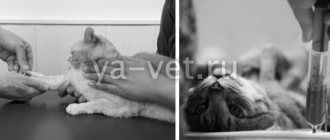
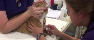
The procedure for taking blood for analysis
Blood is taken from the vein of the animal. To do this, the cat is placed on its side, and if the cat resists, it is secured in a special veterinary bag. Blood is taken from the front paw by shaving off a small area of fur. The injection site is treated with a disinfectant solution, a needle from a disposable syringe is inserted into the vein, blood in the amount of 2 ml is drawn into a test tube with heparin or sodium citrate.
What blood tests are performed in veterinary clinics?
In modern veterinary clinics, two laboratory blood tests are performed:
- General or clinical.
- Biochemical.
General blood test for a cat
A general blood test based on the number and condition of formed blood elements shows the health status of the cat’s body. When performing a general blood test, parasites such as dirofilaria (dirofilariasis), hemobartenella can be detected in a cat’s blood.
What indicators does the clinic’s veterinary specialist receive when conducting a general blood test:
- Hematocrit
- Hemoglobin.
- Average content and concentration of hemoglobin in an erythrocyte.
- Color indicator.
- ESR (erythrocyte sedimentation rate).
- Red blood cells.
- Leukocytes.
- Neutrophils.
- Lymphocytes.
- Eosinophils.
- Monocytes.
- Platelets.
- Basophils.
- Myelocytes.
Biochemical blood test for a cat
Biochemical blood tests allow veterinary specialists to identify subclinical (hidden) cat diseases. A biochemical blood test allows you to determine the functioning of the body’s enzymatic system and provide information about damage to a particular organ in a cat.
A biochemical blood test for a cat includes enzymatic, electrolyte, fat and substrate indicators.
Basic biochemical parameters:
- Glucose.
- Protein and albumin.
- Cholesterol.
- Bilirubin is direct and total.
- Alanine aminotransferase (ALT).
- Aspartate aminotransferase (AST).
- Lactate dehydrogenase.
- Gamma glutamyl transferase.
- Alkaline phosphatase.
- a –Amylase.
- Urea.
- Creatinine.
- Calcium.
- Magnesium.
- Creatine phosphokinase.
- Triglycerides.
- Phosphorus, inorganic.
- Electrolytes (potassium, calcium, sodium, iron, chlorine, phosphorus).
Indications: when is research necessary?
Coprogram in cats is carried out for the purpose of diagnosing and identifying gastrointestinal diseases. This allows you to evaluate the functioning of the digestive organs, the secretion of enzymes, and detect pathogenic microflora or hidden internal bleeding. Analysis is required if there is a suspicion of colitis, enteritis, foreign body entry into the gastrointestinal tract, hepatitis, infection with worms or bacteria of the E. coli group, clostridia or from the cocci family (streptococci, staphylococci), accumulated wool. The main indications for the study are the following changes in the pet:
- vomiting or diarrhea mixed with blood or mucus;
- constipation;
- yellowness of the mucous membranes and eye sclera;
- anemic pallor of the skin and mucous membranes;
- decreased or lack of appetite;
- false urge to defecate;
- abdominal pain;
- sudden weight loss;
- the appearance of dermatitis or rashes;
- general deterioration of condition.
Indicators of blood tests obtained and their characteristics
Each indicator of a blood test shows the functioning of individual organs or entire systems, while the veterinarian takes into account not only each data separately, but also the relationship to each other.
Hematocrit is a conditional indicator showing the ratio of all formed elements of blood to its volume, i.e. determines the thickness of blood. Shows how much blood can carry oxygen.
Hemoglobin is a protein contained in red blood cells that ensures the movement of oxygen and carbon dioxide throughout the animal’s body.
The average concentration of hemoglobin in an erythrocyte shows in percentage terms how much erythrocytes are saturated with hemoglobin.
The color (color) blood index shows how much hemoglobin is contained in red blood cells in relation to the normal value.
ESR is an indicator that determines the presence of an inflammatory process in the body.
Erythrocytes are red blood cells that take part in tissue gas exchange and maintaining acid-base balance. It’s bad when test results go beyond the norm, not only in the direction of decrease, but also of increase.
Leukocytes (white blood cells) - indicate the state of the animal’s immune system. Leukocytes include lymphocytes, neutrophils, monocytes, basophils and eosinophils. For a veterinarian, the relationship between these cells is of diagnostic importance.
- Neutrophils are responsible for destroying bacteria in the blood.
- lymphocytes - speak of a general indicator of immunity.
- monocytes - perform the function of destroying foreign substances in the blood,
- caught in the blood.
- eosinophils - perform the function of fighting allergens.
- basophils - together with other leukocytes, help the body recognize and identify foreign particles that have entered the blood.
Platelets are blood cells responsible for blood clotting. In addition to this function, they are responsible for the integrity of blood vessels. Both high and low levels of them are dangerous for the body.
Myelocytes are located in the bone marrow and normally should not be in the blood.
Indications for use
A general blood test is almost always prescribed for cats if there are alarming symptoms and suspicions of some kind of dysfunction. The procedure is considered ordinary and is carried out for all animals. Any veterinary clinic has all the necessary equipment for it. The analysis determines the number of blood cells and also identifies blood parasites, if any.
Specialists get an idea of the general condition of the body and can make predictions about the presence of certain diseases. Often this information is enough to diagnose the cat. In more complex cases, additional research is required. However, diagnosis is almost never complete without a general analysis.
Blood chemistry
Glucose is an informative indicator indicating the functioning of a complex enzymatic system in the body, including individual organs (liver, pancreas, kidneys). Glucose metabolism in the body involves 8 different hormones and 4 complex enzymatic processes. A disorder is considered to be either high or low blood sugar levels in a cat.
Total protein in the blood reflects the correctness of amino acid metabolism in the body. Shows the total amount of all protein fractions - globulins and albumins. Proteins in an animal’s body take part in almost all life processes of the body. For specialists, both their increased and decreased amounts are important.
Albumin is the most basic blood protein produced by the liver. Albumin in a cat’s body performs a large number of functions (transporting nutrients, maintaining reserve reserves of amino acids for the body, maintaining osmotic blood pressure, etc.).
Cholesterol is a structural component that ensures the strength of cellular structures and takes part in the synthesis of many vital hormones. Veterinary experts judge lipid metabolism in a cat’s body based on cholesterol levels.
Bilirubin is a bile pigment found in the body in two forms - direct and indirect. Indirect bilirubin is formed in the blood as a result of the breakdown of red blood cells, and bound (direct) bilirubin is converted from indirect bilirubin in the liver. Bilirubin shows the functioning of the hepabiliary system (biliary and hepatic). Refers to “color” indicators i.e. when its content in the body is increased, the tissues turn yellow (jaundice).
Alanine aminotransferase (ALT, ALaT) and aspartate aminotransferase (AST, ACaT) are enzymes produced by liver cells, heart cells, red blood cells and skeletal muscles. It is an indicator of the functions of these organs or departments.
Lactate dehydrogenase (LDH) is an enzyme involved in the final step in the breakdown of glucose. Veterinary LDH specialists monitor the functioning of the cardiac and hepatic systems, and also judge the risks of tumor formation.
ɤ-glutamyltransferase (Gamma-GT) – in combination with other liver enzymes, provides insight into the functioning of the hepabilary system, pancreas and thyroid glands.
Alkaline phosphatase is determined to monitor liver function.
ɑ-Amylase – produced by the pancreas and parotid salivary gland. Their work is judged by its level, but always in conjunction with other indicators.
Urea is the result of protein processing, which is excreted by the kidneys. Some remains circulating in the blood. Using this indicator, you can check your kidney function.
Creatinine is a muscle byproduct excreted from the body by the renal system. The level fluctuates depending on the condition of the urinary excretory system.
Potassium, calcium, phosphorus and magnesium are always assessed in complex and in relation to each other.
Calcium is involved in the conduction of nerve impulses, especially through the heart muscle. By its level, you can determine problems in the functioning of the heart, muscle contractility and blood clotting.
Creatine phosphokinase is an enzyme that is found in large quantities in the skeletal muscle group. By its presence in the blood, one can judge the work of the heart muscle, as well as internal muscle injuries.
Triglycerides in the blood characterize the functioning of the cardiovascular system, as well as energy metabolism. Usually analyzed in conjunction with cholesterol levels.
Electrolytes are responsible for membrane electrical properties.
Thanks to the electrical potential difference, cells pick up and execute commands from the brain. In pathologies, cells are literally “thrown out” from the nerve impulse conduction system.
What types of tests are there?
Stool examinations are as follows:
Biochemistry will reveal hidden blood in the animal’s feces.
- Macroscopic. Color, smell, consistency are taken into account. The norm is a moderately dense, well-formed brown fecal lump. A deviation from the norm is a liquid, foamy, too hard or mushy consistency, a black, greenish or light tint, a rotten or sour smell.
- Microscopic. Shows the presence of helminths and their eggs, as well as fats, leukocytes, neoplasm cells, fiber, tripelphosphates, cholesterol.
- Biochemical. Hidden blood in the stool, protein, stercobilin and bilirubin are detected.
Laboratory technicians use 2 methods:
- Native. A sodium chloride solution is applied to a glass slide and a piece of stool is ground. It is covered with another piece of glass and examined under magnification.
- Fulleborn method. The biomaterial is mixed with a sodium chloride solution, filtered and left for about 1.5 hours. Helminth eggs are attached to the resulting film, which are collected with a special loop, placed on a glass slide and studied.
Norms of blood tests in cats
- General (clinical) blood test
- Blood chemistry
| The name of indicators | Units | Norm |
| Ø hematocrit | % (l/l) | 26-48 (0,26-0,48) |
| Ø hemoglobin | g/l | 80-150 |
| Ø average hemoglobin concentration in an erythrocyte | % | 31-36 |
| Ø average amount of hemoglobin in a red blood cell | pg | 14-19 |
| Ø color indicator; | 0,65-0,9 | |
| Ø ESR | mm/hour | 0-13 |
| Ø red blood cells | million/µl | 5-10 |
| Ø leukocytes | thousand/µl | 5,5-18,5 |
| Ø segmented neutrophils | % | 35-75 |
| Ø band neutrophils | % | 0-3 |
| Ø lymphocytes | % | 25-55 |
| Ø monocytes | % | 1-4 |
| Ø eosinophils | % | 0-4 |
| Ø platelets | million/l | 300-630 |
| Ø basophils | % | — |
| Ø myelocytes | % | — |
Norms of blood tests in cats
General (clinical) blood test
Blood chemistry
| The name of indicators | Units | Norm |
| Ø glucose | mmol/l | 3,2-6,4 |
| Ø protein | g/l | 54-77 |
| Ø albumin | g/l | 23-37 |
| Ø cholesterol | mmol/l | 1,3-3,7 |
| Ø direct bilirubin | µmol/l | 0-5,5 |
| Ø total bilirubin | µmol/l | 3-12 |
| Ø alanine aminotransferase (ALT) | U/l | 17 (19) -79 |
| Ø aspartate aminotransferase (AST) | U/l | 9-29 |
| Ø lactate dehydrogenase | U/l | 55-155 |
| Ø ɤ-glutamyltransferase | U/l | 5-50 |
| Ø alkaline phosphatase | U/l | 39-55 |
| Ø ɑ-Amylase | U/l | 780-1720 |
| Ø urea | mmol/l | 2-8 |
| Ø creatinine | mmol/l | 70-165 |
| Ø calcium | mmol/l | 2-2,7 |
| Ø magnesium | mmol/l | 0,72-1,2 |
| Ø creatine phosphokinase | U/l | 150-798 |
| Ø triglycerides | mmol/l | 0,38-1,1 |
| Ø inorganic phosphorus | mmol/l | 0,7-1,8 |
| Ø Electrolytes | ||
| Ø potassium (K+) | mmol/l | 3,8-5,4 |
| Ø calcium | mmol/l | 2-2,7 |
| Ø sodium (Na+) | mmol/l | 143-165 |
| Ø iron | mmol/l | 20-30 |
| Ø chlorine | mmol/l | 107-123 |
| Ø phosphorus | mmol/l | 1,1-2,3 |
Leukocytes in infants
It was already noted above that their norm in children and adults is significantly different. So, in breastfed babies, there may be 10-12 leukocytes in the feces in the field of view, and in artificially fed babies - 10. However, even with a leukocyte count in feces of 15 in the field of view, this is not yet a pathology. It’s just that the functioning of the gastrointestinal tract has not yet been established in infants. Let us once again draw your attention to the fact that there should be no red blood cells in a baby’s stool, regardless of what kind of feeding he is on. Otherwise, inflammation in the intestines or the occurrence of infection cannot be ruled out.
Blood tests in cats (transcript)
All deviations in indicators are considered in complex and in relation to one data to another within the same results from the study of one blood sample. Only an experienced veterinarian should interpret blood tests (results).
General (clinical) blood test
| The name of indicators | Promotion | Decline |
| 1. Hematocrit |
|
|
| 2. Hemoglobin |
|
|
| 3. ESR |
|
|
| 4. Red blood cells |
|
|
| 5. Leukocytes |
|
|
| 6. Segmented neutrophils (mature) |
|
|
| 7. Band neutrophils (immature) |
| |
| 8. Lymphocytes |
|
|
| 9. Monocytes |
|
|
| 10. Eosinophils |
| |
| 11. Platelets |
|
- incoagulability of blood. |
| 12. Basophils | hemoblastoses | Normally absent |
| 13. Myelocytes |
| Normally none. |
Blood chemistry
| The name of indicators | Promotion | Decline |
| 1. Glucose |
|
|
| 2. Protein |
|
|
| 3. Albumin |
|
|
| 4. Cholesterol |
|
|
| 5. Direct bilirubin |
| |
| 6. Total bilirubin |
|
|
| 7. Alanine amino-transferase (ALT, ALaT) |
| |
| 8. Aspartate aminotransferase (AST, ASat) |
|
|
| 9. Lactate dehydrogenase |
| |
| 10. ɤ-glutamyl transferase |
| |
| 11. Alkaline phosphatase |
|
|
| 12. ɑ-Amylase |
|
|
| 13. Urea |
|
|
| 14. Creatinine |
|
|
| 15. Calcium |
|
|
| 16. Magnesium |
|
|
| 17. Creatine phosphokinase |
| |
| 18. Triglycerides |
|
|
| 19. Inorganic phosphorus |
|
|
| 20. Electrolytes | ||
| · potassium |
|
|
| · sodium |
|
|
| · iron |
|
|
| · chlorine |
|
|
| · phosphorus |
|
|
Why do the levels of these cells change?
As already noted, most often red blood cells in feces can be detected with the development of ulcerative colitis of the colon, with the appearance of anal fissures, and also in the case of hemorrhoids. It is important to emphasize that in the coprogram, red cells are recorded only in cases where there is damage to the lower sectors of the intestine.
Even massive bleeding from the upper secateurs of the digestive system cannot be the cause of the analysis result “red blood cells in stool 1-1 in the field of view,” since these cells are destroyed under the influence of enzymes. Thus, bleeding from the stomach manifests itself by staining the biomaterial in question black.
Less rarely, macrophages can be found in stool analysis, which are an important criterion in the diagnosis of pathologies such as amoebiasis and bacillary dysentery. A serious symptom is the presence of atypical malignant cells, which are observed in the coprogram during the disintegration of the rectal tumor. The study of the cytological indicator of this study has important diagnostic value in identifying ulcerative and inflammatory processes in the lower regions of the digestive system.
Not only red blood cells, but also white blood cells can be found in the stool. The norm is considered to be a white blood cell count within 1-2 in the field of view. If there are more of them, this indicates inflammatory processes. The less the leukocyte is changed, the closer to the anus the source of inflammation is located. However, the above standards are only true for adults. In children, there may be much more leukocytes in the stool. This does not apply to red blood cells.
Which children are recommended for analysis?
Examination of stool is called a coprogram. It is performed in most modern laboratories located in almost any hospital. What are the most common indications for ordering a test for red blood cells in feces? Typically, these are the following factors:
- Abnormal stool (color, consistency, smell, presence of impurities).
- The appearance of blood in biomaterial.
- Violation of bowel movements (we are talking about constipation, frequent bowel movements).
- The appearance of abdominal pain and discomfort during bowel movements.
- There are suspicions of digestive problems in the child.
- The occurrence of intestinal infections or suspicion of such.
- Pathological change detected during ultrasound examination of the abdominal area.
If red blood cells and mucus are present simultaneously in the stool, this is one of the symptoms of inflammatory processes in the intestines.