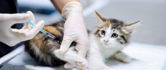
In cases of serious illness, all pets require medical attention. And often it has to be provided at home. If it is necessary to give an animal an IV or remove fluid from the bladder, many owners ask questions about how to place and remove a catheter from a cat.
Thanks to this device, it is possible to avoid constant injections. The catheter allows you to give your pet a drip or injection at any time necessary.
And in the postoperative period or in case of diseases of the urinary system, this device allows you to empty the animal’s bladder of excess fluid completely painlessly.
When is a catheter needed?
Typically, a healthcare professional should insert and remove the catheter from the cat's body. But sometimes you have to carry out this operation without leaving your home.
On the recommendation of a doctor, a catheter (branula) is inserted into the cat on its own in the following cases::
- when the pet is prescribed perenteral nutrition;
- the pet has hyperhydration or hydration of the body;
- the cat is prescribed regular multiple injections using medications;
- when the drug needs to be delivered into the body accurately and quickly and with a special concentration, as opposed to oral administration, when the drug may lose its properties.
After the device is correctly inserted into the vein, it will have to be removed after some time. And here the owners of purring pets always have a problem with how to remove an intravenous catheter from a cat.
Main indications for catheter placement
Basic Rules
Bladder catheterization sometimes becomes the only possible way to remove urine and save the animal’s life.
Urolithiasis in cats is the process of stone formation in the bladder and urethra. Clinically, when the disease occurs, urinary retention or urine leakage in droplets, and the appearance of blood in the urine are observed. The cat may sit in the litter box for a long time and try to urinate to no avail. In addition, the primary symptom of the disease is frequent, prolonged, painful urination. Urine is discolored and smells unpleasant. The cat has to undergo bladder catheterization to save his life.
Urolithiasis can be fatal. For example, if a cat fails to defecate within several days, the lethal end #8212; inevitable. To save the animal, urine should be drained in time using a catheter. For veterinarians, the problem of urolithiasis in cats is a daily occurrence. In this area, our specialists have extensive experience in catheterization and treatment of the disease. They can save even the most hopeless purr.
Treatment of urolithiasis in animals at home is carried out by professional veterinarians. You should check with your doctor over the phone about the cost of treatment. Unfortunately, without medical help you will not be able to help your pet.
- The therapeutic process begins with manual massage of the cat's bladder. This manipulation helps remove sand plugs. If urine does not pass on its own, there is a need for urgent catheter placement. Veterinarians perform this procedure with brilliant success, eliminating any complications. Bladder catheterization is a simple procedure, but the owner should not do it himself.
- Catheterization is performed at home under general anesthesia. You can put a catheter on a cat without anesthesia if there are contraindications: the animal is over 10 years old or is severely weakened. After the procedure, symptomatic treatment is prescribed using anti-inflammatory drugs, anesthetics, antispasmodics, homeopathic drugs, antibacterial, physiotherapeutic agents, and vitamins.
- Doctors may prescribe medications that help dissolve stones inside the bladder. While you are undergoing treatment, you will have to periodically catheterize your bladder.
- But one of the most important components, which is mandatory in the process of treatment and further prevention of urolithiasis in cats, is maintaining a proper diet. Mixtures of special feeds help speed up the treatment process.
- For drug treatment, intramuscular solutions of no-shpa and antispasmodic are used. In case of blockage of the urethra, a 0.5% solution of novocaine is administered through a catheter.
- Pain syndrome is eliminated using novocaine blockade with a 0.25% solution. Intramuscular administration of bicillin-3 helps relieve inflammation of the urinary tract.
- In addition, decoctions of corn silk, bearberry leaves, and biseptol can be used.
Catheter for urine excretion.
Symptoms of the disease in cats depend on the duration of urinary retention. A constant urge to urinate in an animal is considered a clear sign of urolithiasis. Observing the animal, you can notice how alarmed and exhausted it is, meows in pain during defecation, and then licks its genitals.
Palpation of the bladder allows you to determine that the organ is greatly distended. With prolonged urinary retention (more than 2 days), the bladder can rupture, causing the development of peritonitis and subsequent death of the animal. Therefore, catheterization of the cat’s bladder in such cases is extremely necessary!
In order to empty a full bladder, it is necessary to insert a catheter into the cat. This procedure is the only condition for saving the life of the pet. Owners should know that every fifth cat dies from untimely assistance for the formation of stones in the excretory system.
Catheterization for conservative treatment of urolithiasis
Catheterization is performed in the following situations:
- with conservative treatment of urolithiasis;
- urinary disorders of various etiologies;
- surgical intervention (for urine output during and after surgery);
- when therapeutic procedures are carried out (washing the urethra, bladder);
- to control urination, collect urine;
- for X-ray diagnostics using a contrast agent.
Catheters can be installed for a short period of time, for example, during a diagnostic examination, for collecting tests, or for a one-time excretion of urine. For these purposes, polypropylene models are most often used. The answer to the question of how long a cat’s catheter should last will depend on the severity of the animal’s disease and the characteristics of the pathology.
They often resort to permanent urethral catheters, for example, when the animal is in serious condition, with acute renal failure, or with bladder injuries. For permanent catheterization, models made of polyvinyl chloride are used, which are less traumatic for the animal compared to polypropylene.
Catheterization is a purely medical procedure that requires certain knowledge and skills. Therefore, it can only be carried out by qualified personnel. The owner must have an idea of how to place a catheter in a cat in order to understand the seriousness of subsequent care for the pet.
Catheterization also has contraindications. Manipulation cannot be carried out when:
- acute infectious diseases,
- damage to the urethra and bladder by tumors.
An animal may require catheterization not only due to urolithiasis, but also for a number of other indications. A cat is subject to bladder catheterization in the following cases:
- a conservative method of treating urolitasis - a urinary catheter is used, depending on the condition of the animal, once or continuously;
- traumatic injuries of the bladder;
- disturbances in the passage of urine in acute form due to any disorder when catheterization is required for emergency emptying of the organ, even before the cause of the phenomenon is determined;
- surgical interventions in which a catheter is inserted so that urine can easily pass both during the operation itself and after it, when the functioning of the bladder muscles may be impaired due to anesthesia;
- therapeutic lavage of the bladder and urethra, which is carried out so that medicinal substances can be delivered to the sore spot;
- determination of the volume of urination and the need to collect urine when catheterization is carried out for a short period of time, and after obtaining the material necessary for the study, the catheter is removed;
- X-ray of the bladder with contrast, in which catheterization is required to administer the contrast agent.
shutterstock
For one-time use for a short time, cheaper catheters made of polypropylene are used. If the presence of a catheter in the cat’s body is required for more than one day, then a device made of polyvinyl chloride is used, which is more comfortable for the animal and does not cause significant negative sensations. After catheterization, the cat must be completely isolated from the street to prevent bladder infection.
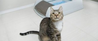
The main indication for installing a catheter is urolithiasis in a cat, in which there is impaired insufficient urine flow. This violation can be noticed by several signs.
- Small puddles outside the tray to the whole house. This happens due to the fact that the cat cannot suppress the acute urge to urinate, but due to impaired discharge, physiological fluid is released in excessively small quantities.
- The pet often goes to the litter tray. In this case, the cat sits for a long time in a position corresponding to the one it takes when urinating, but there is no urine output at all.
- A loud cry at a time when, in addition to physiological fluid, which is small, a significant amount of blood is also drained.
- A serious increase in the size of the bladder, which can be easily felt when palpating the abdomen even by a non-specialist. The manipulation must be carried out extremely carefully so that the maximally stretched walls of the overcrowded organ do not rupture.
If symptoms of urinary retention occur, after examining the animal, the veterinarian will determine whether bladder catheterization is necessary or not. If the cat is able to clear urine from the bladder, albeit incompletely, treatment without the use of a catheter can be done.
Before removing the catheter from a cat, you should check for kinks and distortions in the system, otherwise injury to the mucous membrane of the urinary tract is possible. Only a veterinarian should remove the catheter.
After the catheterization procedure, the animal is usually prescribed antispasmodic drugs to facilitate the process of urination. To prevent the development of a bacterial infection, antibacterial agents are prescribed. Since obstruction of the urinary tract is often a consequence of the disease, after the elimination of ischuria, treatment of the underlying pathology continues.
Owners often encounter various problems when catheterizing an animal. For example, a cat cannot pee after a catheter. This phenomenon is observed due to spasm of the urethral muscles. In this case, the animal is prescribed antispasmodic drugs. For this purpose, no-shpa, spazgan, and papaverine are used. Manual bladder massage is also effective.
If a cat pulls out a catheter, what should the owner do in such a situation? The animal, regardless of the reasons for which catheterization was performed, must be immediately taken to a veterinary institution.
If your pet has urological syndromes that indicate difficulty urinating, then catheterization is sometimes the only solution in this situation. Only a veterinarian should insert and remove a catheter. Independent manipulations are unacceptable, as they involve the risk of injury and rupture of the urethra and bladder.
How to remove an intravenous catheter
The cat owner has to carry out this procedure 5 days after installation, in case of mechanical damage to the catheter, or if the pet’s limb with branula is swollen.
The catheter is usually located on the cat's front legs. It is fixed with the turn of a regular adhesive plaster. In order to remove the intravenous catheter from the cat, it is enough to cut this dressing from bottom to top. Then the remaining patch must be carefully removed from the pet's hair. After this manipulation, you can remove the catheters from the cat’s paws by pulling out the plastic tube from the vein. Apply a thick bandage soaked in alcohol to the previous location of the scold, and bandage the paw for the next hour.
When carrying out this procedure, you should pay attention to the fact that:
- When the device is pulled out, the animal may try to escape. Therefore, removing a cat’s catheter at home is much easier with two people;
- when pulling out the tube, the movement should be careful, but as fast as possible;
- instead of alcohol, you can use a solution of furatsilin or hydrogen peroxide to wet the disinfectant swab;
- If suspicious symptoms appear after removing the catheter, it is better to seek help from a veterinarian. Such uncharacteristic signs when removing the device include: swelling of the extremities, discoloration of the skin, lameness or clenching of the paw, pain on palpation, development of hyperthermia, lack of appetite, weak and apathetic state;
- It is best to use nail scissors to cut the patch, as they will remove the bandage more accurately. If the animal twitches, then it is better to give preference to a device with rounded ends.
Technique
If pathological processes are detected in an animal, it may be necessary to rinse the bladder. The doctor will perform the procedure as follows:
- To relieve pain, the male needs sedation or general anesthesia. Clinicians use local aerosol analgesics (Lidocaine Asept (spray), Carmolis).
- Sterile lubricant is placed on the tip of the tube. An atraumatic tip lubricated with a synthetic lubricant is inserted into the lumen of the urethra. If the device cannot pass through the excretory duct, the veterinarian will drain the animal's bladder by placing a needle on the outside and releasing the urine using a syringe.
- Once the first 2 cm of the catheter is in the penile urethra, the non-dominant hand is used to pull the preputial opening caudally to continue insertion of the tube.
- When regulating water and electrolyte metabolism in an animal, parenteral fluid therapy is performed. The infusion volume is 45-68 ml of crystalloid per kilogram of body weight over 30 minutes.
- Removal of obstructive materials is carried out using solutions that destroy plugs and mucus clots.
- Surgery may be required to remove stones from the organ to prevent inflammation from recurring.
- When the device completely enters the excretory channel, liquid begins to be released from the tube. The clinician determines the condition of the organ by color, consistency and smell.
- Blood clots are eliminated using a solution of novocaine.
- Cats with life-threatening arrhythmias secondary to hyperkalemia are treated immediately with sodium bicarbonate (stimulation of intracellular potassium metabolism) or calcium gluconate (stabilization of the heart and changes in threshold potential).
If necessary, leave the instrument for several days. The veterinarian prescribes medication and diet therapy at home.
Catheter for diseases of the urinary system
If you have problems with urination (oncological diseases, prostate pathologies, urolithiasis, etc.) in a cat or dog, a catheterization procedure is necessary. This is the only way in some cases it is possible to save a pet’s life.
It may be necessary to insert and remove a catheter in the following cases::
- when you need to control your cat's urination;
- in the postoperative period;
- if there are injuries to the genitourinary system;
- to remove stones from the bladder;
- for therapeutic purposes (washing the urinary system);
- for one-time excretion of urine.
Why is a catheter placed?
Many diseases of the urinary system are accompanied by a symptom such as ischuria. Retention of urination when the bladder is full is a frequent companion to urolithiasis, tumor diseases, problems with the prostate gland in males, severe inflammatory processes, narrowing of the urethra of a traumatic nature, etc.
Most often, a catheter is placed in a cat for urolithiasis, since this pathology leads to obstruction of the urinary tract. Urinary retention can be acute or chronic. In the first case of ischuria, catheterization is a vital procedure. When a plug of urinary sand forms in the urethra, obstruction develops. Signs of urinary retention in a cat are as follows:
- urinating in the wrong places, outside the tray;
- frequent urges, the pet takes the appropriate position, but urine is not released;
- the animal is worried and screams when going to the toilet;
- urine is released in drops, often mixed with blood;
- palpation of the lower abdomen reveals a tense bladder. It becomes hard and increases in size to the size of a chicken egg.
Removing the urinary catheter
A Faley catheter is a thin tube that drains urine into a special bag. Its removal is necessary in cases where :
- the catheter stops functioning;
- the animal has suffered an injury to the urethra or bladder;
- The problem causing the device installation has been resolved.
The procedure for its removal is carried out only by a veterinarian. You should not try to carry out such an operation at home by watching videos from the Internet. Removing the catheter on your own can result in injury to the mucous membrane of the urinary tract.
How is a cat catheter placed?
The doctor must insert the device very carefully so as not to further injure the urinary tract.
Only a veterinarian can correctly place a tube to drain urine and carry out the necessary manipulation. The technique requires special care, since during the procedure there is a high risk of mechanical damage, which will lead to serious complications. A catheter equipped with a mandrel is inserted into the animal's urethra.
Before insertion, the tube is lubricated with a lubricant so that it can be inserted further and the walls of the organ are not damaged. If the catheter does not pass through during manipulation, then special solutions are injected into it to remove any blockages that have arisen. When urine comes out of the tube, this indicates that it has entered the bladder. During the procedure, a small amount of material is taken for laboratory testing. If urine is released with blood, damage to the walls of the organ is detected.
Long-term catheterization of the bladder in cats is often carried out, during which the catheter must remain in place for 2 days or more. In this case, the specialist stitches the tube to the red flesh. Thus, urine is removed from the bladder and collected in a special tank. With such a pathology, the cat needs to follow a diet and take medications. With the help of veterinary medications, it is possible to alleviate the symptoms that occur during catheterization.
Rules for drip infusion of drugs
There are several universal rules that must be followed when installing IVs for both humans and animals:
- First, you need to thoroughly wash your hands and disinfect the injection site to avoid infection. The area on the paw is shaved and treated with an antiseptic.
- The needle must be inserted with a quick but gentle movement, minimizing pain in the cat. It is advisable that a second person hold the pet at this moment so that it does not escape and cause harm to itself.
- The container with the medicine should be located on a raised surface to ensure free flow of liquid. The optimal height is 35–40 cm above the lying animal. You need to think in advance about how to secure the bag with the solution.
- The medicine should be at room temperature. Most often, the drug is kept in the refrigerator, so you need to hold it in your hands for some time to warm it up slightly.
- The drip system must remain sterile. You should not keep it unpacked; you should open it immediately before the procedure.
We have a difficult task - we need to give an intravenous injection to a dog!
We figured out how to assemble an IV, now we need to learn how to administer drugs intravenously to our dog. Don't be alarmed, you don't have to find the vein and puncture it yourself. To save you from the bloody procedure, they came up with a special catheter, which the doctor will install, but we will learn how to care for it in this article.
Valve for introducing substances, for example, heparin solution. In the photo it is blue, but it can be green, pink, gray or other - the different colors help determine the size of the device.
I noticed that when I have to install a catheter in the presence of the owner, he then worries about the huge needle that remains in his paw. Just so you don't worry, here's a photo.
Open the valve for administering the medicine (blue), you don’t need to turn it here, just pull it towards you. Connect a syringe with saline solution (without a needle) and press the plunger. Let's look at the photo.
If the syringe plunger moves easily, then we can connect the system and administer it intravenously to our dog. Close the valve with the blue cap.
We figured out how to give intravenous injection to a dog using a catheter, if everything goes well for us, but it doesn’t always work out so smoothly. Quite often a blood clot clogs the catheter, then you have to dance a little with a tambourine.
Again, we take a syringe with saline solution (or Ringer), connect it to the port with a blue cap and press the piston so that the liquid comes out of the cannula, that is, we repeat everything as during preparation.
Then we insert the syringe into the cannula (where the white cap was) and try to inject the solution, it doesn’t work, then we pull the piston towards ourselves - air will get into the syringe. We disconnect it, release the bubbles and reconnect it to the cannula and try to inject the solution.
Our task is to “loose” the blood clot. Let’s immediately dispel fears about blood clots -
Source
System installation features
Putting a drip or installing a catheter on a cat at home is much more difficult than giving a subcutaneous or intramuscular injection. This procedure requires skill, but owners do not always have the opportunity to take their pet to a veterinary clinic, so they have to install an IV themselves. By following simple rules, you can greatly simplify the procedure and make it almost painless for the cat.
For intravenous infusion: through a butterfly needle and through a catheter
A dropper with a butterfly needle is used for one-time or emergency infusion. Installation algorithm:
- An intake needle is inserted into the stopper of the container with the medicine, and next to it is a needle for air access. Some bags already have a system for oxygen supply, then an additional puncture is not needed.
- When the intake chamber is filled with liquid, the dispenser should be adjusted so as to completely stop the flow of the drug. Care must be taken to ensure that no air gets into the chamber.
- The animal is fixed in a lying position, the limb is shaved and treated with alcohol. Installing an IV is stressful for a cat, so it is better for the owner to be nearby, petting and calming his pet.
- Most often, the puncture is made on the front leg, but if it is convenient, you can stick the needle into the back leg. A medical tourniquet is applied above the shaved area. To find a vein, the limb is flexed and extended several times. When the veins swell so much that they can be felt with your fingers, insert the needle with a gentle but confident movement and secure it with the petals of the “butterfly”.
- The needle should be inserted parallel to the vein; it is prohibited to insert it perpendicularly. A sign of correct administration will be a few drops of blood in the tube. As the medicine arrives, the puncture site should be checked. If swelling appears around, it means that the IV is not installed correctly and the liquid is not getting into the vein, but under the skin.
About collecting and installing an IV for a dog
So, first you need to treat your hands with an antiseptic and put on sterile gloves. Next, you need to open the system and the bottle of medicine. There is no need to remove the entire metal cover from it - just its central part. Then you need to insert two intake needles into the rubber lid of the container with the medicine and fix the bottle with the bottom.
Next, move the dispenser lever all the way, make 2-3 smooth presses on the reservoir with the filter. He will draw the medicine into himself under pressure. Shake the chamber slightly to release excess oxygen. Lower the dispenser lever and watch the medicine go down. It is important that there are no air bubbles in the tube below the filter reservoir.
The dog must be prepared for the procedure as follows. The optimal place for infusion is the wide veins of the forelimbs, so you need to first shave or trim the hair between the pet’s wrist and elbow. Next, you need to pull the paw with a tourniquet above the puncture area. In dogs, the veins are clearly visible.
We insert a needle into the vein, disconnecting it from the dropper, parallel to the paw. This must be done carefully and slowly. The pressure should be moderate. Next, the needle is fixed on the paw with an adhesive plaster, and the tourniquet is removed. Only then does the system join. So, now the process of infusion of medicine has begun, and the owner’s task is to monitor the dog.
It is important that the solution for administration is warm. It often happens that a properly inserted needle comes out of the vein because the dog tenses the muscle. To prevent this from happening, you must constantly monitor the progress of the procedure.
After placing a drip, dogs may, in rare cases, experience allergic attacks. They are manifested by difficulty breathing, coughing, swelling of the lips and throat. If the above signs appear during the procedure, then it is necessary to stop it by removing the needle. In this case, the dog’s paw is pricked with a 0.1% solution of adrenaline. The same medicine must be administered intramuscularly. It is better to call a veterinarian if the animal’s condition does not improve after an injection of adrenaline.
Have you read it? Myocardial infarction in dogs ⋆ Heart Treatment
How to care for a catheter at home?
The catheter is usually installed for up to 5 days. All this time you need to look after it and keep it clean, otherwise the risk of infection increases. Catheter care rules:
- the cannula cover must be sterile and changed after each administration of the drug;
- the external part should be disinfected regularly - before and after the dropper, as well as during the day;
- the catheter should be regularly inspected for dirt, blockage and, if necessary, cleaned with saline;
- If the catheter is clogged, it needs to be changed at the veterinarian's office.
Catheter care rules
The main condition for caring for a urethral catheter in an animal should be compliance with the rules of asepsis and antisepsis. If a permanent urinary apparatus is installed, the owner should maintain its sterility. It is necessary to monitor the condition of the fur in the perineal area, keep it clean, and, if necessary, shave the hair to avoid getting it into the genitourinary tract.
The genital area should be regularly treated with antiseptic solutions of furatsilin or chlorhexidine.
This will prevent the development of a bacterial genitourinary infection.
With continuous catheterization for several days, it is necessary to flush the cat's urinary catheter. This procedure is performed to sanitize the urinary tract with a warmed saline or antiseptic solution twice a day. For rinsing, syringes without a needle are used. After removing urine from the catheter, about 60-80 ml of disinfectant liquid is injected with their help. Empty the bladder using empty syringes for this purpose. Having freed the organ from fluid, add another 60-80 ml of solution, close the catheter with a lid and leave for sanitation. After 20 minutes, the system is opened and the flushing liquid is removed using empty syringes. Carry out the procedure until the flowing solution becomes transparent. As a rule, 2–3 such manipulations are required.
If installation of a permanent urethral catheter is necessary, then experts, as a rule, recommend that owners leave the animal in the clinic’s hospital for bladder lavage.
How to remove a catheter from a paw?
The catheter is removed in several cases:
- the treatment period has ended;
- 5 days have passed since installation and a new one needs to be installed;
- the cat tried to tear off the adhesive plaster or damaged the puncture site.
How to remove a catheter from a cat? As with inserting a needle, removing the system must be done carefully but confidently. The animal is fixed in a motionless position, constantly calming. The puncture site is treated with alcohol or other antiseptic, and hands should be thoroughly washed.
Fixing adhesive plasters should be removed using scissors. There is no need to tear them off, this will cause pain to the cat, she will try to run away, and it will be much more difficult to remove the catheter. Place a sterile cotton ball over the skin, press lightly and slowly remove the tube from the blood vessel parallel to the paw, immediately pressing the wound with cotton wool. The cotton swab is fixed with a bandage or plaster for 1–2 hours.
How to place an IV correctly?
The dropper consists of a transparent plastic tube. One of its ends is connected to the catheter and vein. The other one needs to be placed in a bottle containing an infusion solution. The drip also consists of a device that regulates the rate of administration and a device that does not allow oxygen to enter the veins. True, modifications are also possible. But the sequence of actions is always the same.
- Turn off the drug administration speed regulator completely. To do this, turn the wheel on the regulator with your thumb all the way down. This way the wheel will squeeze the plastic tube through which the medicine flows. To understand when the tube is open and when it is closed, simply move the wheel down;
- Remove the aluminum protective cap from the medicine bottle. It cannot be removed completely. You only need to remove the round central part by prying it up and tearing it off. Under it you will find an elastic band. This is where the system needs to be installed;
- In order to remove all the air from the system, you need to remove the protective cap from the tip of the dropper. The plastic tip of the oxygen removal device must be placed in the stopper of the bottle. To do this, it is pierced. This should be done especially carefully with small bottles, because their stoppers may fall inside. In this case, the bottle must be replaced with a new one;
- We open the air duct and hang the bottle with the system upside down. Decide for yourself where to hang it. This could be a coat hook, a cabinet handle, a stepladder, or even a chandelier. A hanger for the bottle can be made from medical plaster (the kind that comes in rolls);
- With two fingers we squeeze the reservoir near the device that removes oxygen. Now we release and watch how the reservoir is filled with medicine;
- Open the injection speed regulator completely. Now the medicine is actively entering the reservoir, and the air is exiting through the air duct, which we previously opened. Now the medicine goes down the system and the remaining oxygen is removed from it. When the medicine begins to flow from the end of the tube and the stream is steady, the dropper is ready. The speed control can now be completely closed;
- With our left hand we hold the catheter on the paw of a dog or cat. It shouldn't move much. Unscrew the white cap on the catheter. If there is blood, it’s okay: it only means that there is no microthrombus in the catheter that interferes with the operation of the IV;
- We continue to hold the catheter on the cat's paw. We take the end of the dropper (the one made of plastic and near the rubber tube), insert it where you unscrewed the cap. There shouldn't be a needle at the end. To fix the dropper in the catheter even better, turn its plastic end slightly clockwise ¼ turn to the right. When disconnecting the dropper, unscrew the end to the left. There is no need for a needle; its role is played by a plastic catheter tube, which is inserted into the vein of a cat or dog;
- We fully open the drug administration speed regulator. The medicine now flows into the reservoir. We remind you that it should arrive drop by drop per second;
What difficulties may arise and how to resolve them?
Even experienced veterinarians can experience difficulties when placing an IV, not to mention a person who has never performed such manipulations before. The most common problem is fluid obstruction. What to do if the medicine does not drip?
If the needle was inserted directly into a blood vessel, you should check to see if the cat has pinched the vein with its paw. Due to stress, she could bend her limb and a muscle spasm could occur. The problem is solved by a light massage of the paw and soothing strokes.
The third reason for medication failure is improper use of the drip system. The air needle may not be installed. The problem is solved by introducing an additional needle into the bottle of liquid.
Some symptoms that appear after IV drips frighten pet owners. However, this is a normal reaction to a stressful situation and the administration of medication. Do not panic if your four-legged patient experiences:
- lethargic state;
- apathy;
- lack of appetite;
- one-time urinary incontinence;
- one-time attack of vomiting;
- slight swelling at the puncture site.
If your cat is vomiting repeatedly or has swelling of the face, which indicates an allergic reaction, you should immediately take her to the veterinarian. A dangerous condition can only be treated in a veterinary clinic.
If they are not given immediate help, they will die. And we, ordinary people, lack the skills and abilities to provide the necessary assistance.
Many diseases require serious attention that there is a need to give IVs, but it is not possible to visit a doctor.
But if you prick a dog or cat every day, then there will be no living space left on them. Therefore, an intravenous catheter becomes a great help, with the help of which you can make both a drip and an injection at any time.
When is the procedure performed?
Inserting a urethral catheter for a kitten and an adult is required not only for urolithiasis, but also for other pathological conditions in which the pet does not urinate. Veterinarians prescribe catheterization in case of mechanical damage to the cat’s bladder. Indications for which a urological catheter is required are the following situations:
- Conservative treatment for urolithiasis. This therapeutic technique can be used for pathology once or regularly to facilitate urine excretion.
- Impaired urine flow during acute illness. When abnormalities in the urinary system worsen, the cat is prescribed catheterization for emergency bladder emptying.
- Surgical intervention. After surgery in the area of the urinary system, it is difficult for the pet to walk when necessary, so a catheter is installed. When the muscles are restored, the cat will be able to urinate on its own.
- The need to rinse the cat's urethra and bladder. This measure is carried out during treatment, with its help it is easier to deliver medicines to the affected organs.
- Determination of urination volume and urine collection. Catheterization is performed in a short time, and after the study the catheter is removed.
- For diagnostic purposes. If a cat or kitten needs to take X-rays of the urinary system, then catheterization is performed, through which a contrast agent is injected.
When should an intravenous catheter be used in dogs and cats?
- Emergency conditions, resuscitation, operations that require quick access to the bloodstream (for example, if you need to administer drugs urgently and at high speed).
- Hospitalization of an animal with seizures.
- Prescribed parenteral nutrition.
- Overhydration or hydration of the body.
- Transfusion of blood products (whole blood, packed red blood cells).
- Repeated or continuous administration of drugs intravenously.
- The need for rapid and accurate administration of the drug in an effective concentration (especially when the drug can change its properties when taken orally).
Criteria for choosing a vein and catheter for dogs and cats
The lateral saphen vein is more accessible in dogs, whereas in cats it is easier to insert a central catheter through the medial saphen vein. Catheters inserted into the hind leg are more difficult to keep clean and are not recommended for patients with urinary incontinence or diarrhea.
With intravenous injections, the advantage remains with the peripheral veins. The veins should be soft and elastic, without compactions or knots. It is better to inject drugs into large veins, in a straight section corresponding to the length of the catheter.
- Vein diameter (the diameter of the catheter must be less than the diameter of the vein).
- Required speed of solution administration (the larger the catheter size, the higher the solution injection rate).
- Potential time the catheter remains in the vein (no more than 3 days).
System for drip administration or - dropper
On one side it is connected to a catheter in a vein, and on the other it is inserted into a bottle with a solution. There is a wide plastic needle at one end (with an air duct) 1
, filter for retaining dust and air bubbles,
2
– the dropper itself.
At the other end of the tube there is an orange soft rubber unit 5 ending with a cannula 4 and a needle inserted into the vein. The needles are in special caps. The tube has a device 3
(clamp-regulator) for adjusting the speed of solution administration.
Preparing dogs and cats for peripheral catheter placement
- Lay out a clean diaper or sheet (it is ideal if you iron or steam it with an iron).
- Lay down and calm your pet.
- Shave the fur where you are going to place the IV.
- It's better if it's a paw. In any case, the location will be determined by the veterinarian.
- The animal's paw should be straightened so that the vein is not pinched. You can fix it (the paw) with a splint - a board bandaged to the paw.
- The puncture site is disinfected.
Catheter placement
An intravenous catheter (brauncle) is placed when long-term treatment is needed and a large volume of drugs will be administered.
Installing a catheter has several advantages:
- permanent access to a vein with the ability to quickly administer medication in difficult cases;
- there is no need to puncture the blood vessel before each administration of the drug;
- the presence of a catheter does not limit mobility;
- saving time and effort;
- absence of pain during subsequent drug administrations;
- possibility of collecting blood for analysis;
- rehydration of the body.
What a catheter looks like
It is best to entrust the installation of a catheter to an experienced veterinarian. The procedure requires skills in this area.
Catheter device
Brownie design: 1 - valve, under which there is a hole for administering drugs (available in different colors); 2 - part of the stylet, which is removed after installation of the catheter; 3 - screw cap that blocks the entrance to the blood vessel; 4 - cannula to which the system or syringe without a needle is connected
To install a catheter, you will additionally need the following materials:
- clamp;
- adhesive plaster on a rag basis;
- small sharp scissors (manicure);
- bandage or rubber band;
- Shaver.
Catheters are selected by number, depending on the size of the animal.
Cap color and catheter size
Catheter insertion
Before installation, in the area of the elbow joint of the animal's forelimb and slightly below, the hair is cut with small scissors and shaved using a machine in order to better see the saphenous vein.
The device is inserted into the saphenous vein. This place is considered the most convenient. There the device lasts the longest and does not cause any inconvenience to the pet.
The catheter is inserted in this order:
- Above the elbow, a bandage or tourniquet and a clamp are used to tighten the limb to fill the vein with blood. This should be done with moderate force so as not to damage the blood vessels.
- Using small, sharp scissors, a small incision is made in the skin in the middle of the forearm to insert the needle.
- At the same time, a catheter is inserted into the limbs. The instrument is inserted at an angle of about 15° until it stops until blood appears in the indicator chamber.
- The catheter is removed from the stylet, but not in the reverse order, since then the device will not be installed correctly.
- They try to inject saline. If the catheter is installed correctly, there will be no swelling or swelling on the paw.
- The device is fixed on the paw using a narrow adhesive tape. During fixation, it is necessary to monitor the tension of the adhesive plaster - it should not be excessive, so as not to disrupt the blood flow in the limb and cause tissue death.
- The catheter is flushed to avoid blood clots.
Rules for placing a peripheral catheter in dogs and cats
- Placement of the catheter must begin by ensuring good lighting of the manipulation site.
- Next, hands are thoroughly washed and dried.
- A standard set for vein catheterization is assembled, and the set should contain several catheters of different diameters.
- Apply a tourniquet 10-15 cm above the intended catheterization area.
- A vein is selected by palpation.
- A catheter of the optimal size is selected, taking into account the size of the vein, the required insertion speed, and the schedule of intravenous therapy.
- Re-treat your hands using an antiseptic and put on gloves.
- The catheterization site is treated with a skin antiseptic for 30-60 s and allowed to dry.
- The vein should not be palpated repeatedly! Fixing the vein (it is pressed with a finger below the intended site of catheter insertion).
- Take a catheter of the selected diameter and remove the protective cover from it. If there is an additional plug on the cover, the cover is not thrown away, but held between the fingers of your free hand.
- The catheter is inserted on the needle at an angle of 15° to the skin, observing the indicator chamber. When blood appears in it, reduce the angle of the stiletto needle and insert the needle into the vein a few millimeters.
- Having fixed the stiletto needle, slowly move the cannula completely from the needle into the vein (the stiletto needle is not yet completely removed from the catheter).
- Remove the tourniquet.
[
- Do not insert a needle into the catheter after it has been dislodged from the needle into the vein! The vein is clamped to reduce bleeding, and the needle is finally removed from the catheter.
- The needle is disposed of taking into account safety rules.
- Remove the plug from the protective cover and close the catheter or connect the infusion system.
- The catheter is fixed on the limb.
What a pet owner needs to know about catheters
Placing a catheter is more difficult than simply injecting the drug into a vein. It happens that not every veterinarian succeeds in placing the catheter correctly the first time. Therefore, before you start using it, you need to know a few rules:
- The catheter requires careful care: after each use it must be thoroughly rinsed and dried.
- If the catheter is placed for a long time, you need to make sure that the animal does not start licking it: it can come off. During injections using a catheter, you need to be close to your pet and make sure that the catheter is positioned correctly at all times.
- When any drug is administered into a vein, there may be a risk of various bacteria and fungi entering the blood. To prevent this from happening, you need to thoroughly disinfect the injection site and ensure that the catheter is sterile.
- To prevent the animal from unwittingly removing the catheter, you can bandage it with a bandage for greater confidence - this will make it much more difficult to remove the device.
Before installing a catheter, several conditions must be met:
- Place a clean sheet or diaper on a flat surface where it would be most convenient for you to place the catheter in your pet.
- Place your pet on the sheet. It is necessary that he behave calmly and reservedly.
- The fur on the area where the catheter is to be placed must be shaved, so this issue must be addressed in advance before inserting the catheter.
- Most often, the catheter is placed in the paw. During insertion of the catheter, it should be straight and motionless. To fix it, it is best to use a splint - a special board usually used to heal broken limbs.
- The place where the catheter will be inserted must be disinfected with an alcohol solution.
You need to install a catheter in your pet as follows:
- Provide maximum illumination in the room.
- Wash and dry your hands and put on sterile gloves.
- Apply a tourniquet 10-15 cm above the catheterization site.
- Select a vein to insert the catheter into. Considering the size of the selected vein, select the appropriate catheter size.
- Treat the catheterization site with an antiseptic.
- Remove the protective cover from the catheter and insert the catheter at an angle of 15 degrees. Once blood appears in the catheter stylet, reduce the angle of inclination and continue inserting the catheter.
- Fix the needle and slide the cannula into the vein to the end; There is no need to remove the needle from the catheter yet.
- Remove the tourniquet.
- Clamp the vein and remove the needle from the catheter.
- Discard the needle.
- Secure the catheter by placing an adhesive tape over the catheterization site.