Types of rhinoscopy, technique
The method of conducting rhinoscopy involves the use of special equipment - flexible and rigid probes (endoscopes) of different sizes. According to the technique of rhinoscopy, there are:
- anterior (straight, antegrade);
- posterior (retrograde).
When examining the nasal cavity during anterior rhinoscopy, the veterinarian inserts the endoscope from the side of the animal's nostrils towards the nasopharynx. In the posterior case, a flexible probe (fiberscope) with a camera on the tip is used, which is inserted into the cat’s mouth (through the choanae) from the nasopharynx. This technique allows you to determine the condition of the soft palate, pharynx, nasopharynx, and vomer.
In most cases, when diagnosing the nasal cavity, two rhinoscopy techniques are used simultaneously.
Important! Rhinoscopy in veterinary medicine is used not only for diagnosis, but also during medical procedures. This technique is used to remove tumors, polyps, and cysts in the nose of animals.
When performing rhinoscopy, a specialist can remove a tumor, a small-diameter neoplasm, foreign bodies found in the upper organs of the respiratory system, and take a tissue sample for bacteriological culture, targeted biopsy, and other tests and diagnostic tests.
Rhinoscopy for animals.
- home
- Articles
- Publications of specialists
- Veterinary
- Rhinoscopy for animals.
Rhinoscopy is a minimally invasive method for examining the nasal cavity of animals, performed using flexible and rigid endoscopes.
Indications for rhinoscopy for animals are difficulty breathing through the nose, frequent sneezing, unilateral or bilateral discharge from the nasal passages of various types (mucous, purulent, bloody), as well as suspicion of the presence of a neoplasm in the nasal cavity.
During rhinoscopy, the condition and integrity of the mucous membrane is assessed, the cause of discharge from the nasal cavity is identified, and a biopsy of the tumor or pathological focus is performed, material is taken for bacterial culture, or a foreign body is removed from the nasal cavity. Briefly about the structure of the nasal cavity in dogs.
The nasal cavity in dogs is divided along its entire length by a septum.
The basis of the front part is cartilage, and the back part consists of bone. On the sides of the nasal cavity there are turbinates - two spiral-curved bone plates at different levels. The turbinates divide the nasal cavity into three nasal passages: lower, middle, upper. The lower stroke, gradually widening, merges with the middle stroke. The upper passage is narrow and shallow. In the upper (posterior) part of the nasal cavity, which is covered with mucous membrane, there are olfactory cells. The nasal cavity consists of elongated passages from the external nostrils to the choanae. The choanae are 2 openings in the upper part of the pharynx. How is rhinoscopy performed in veterinary medicine?
Rhinoscopy is performed under general anesthesia in two ways:
1. Anterior rhinoscopy
.
In this case, the study is carried out from the side of the external nasal openings. A rigid endoscope is used for this. 2. Posterior rhinoscopy
is a method in which the examination is carried out through the choanae using a flexible endoscope. Often the reason for rhinoscopy is foreign bodies in the nasal cavity. Similar findings are most often found in animals in the lower and middle nasal passages. If a foreign body has recently entered the nasal cavity and the shape and size of the object allow it to be removed, then manipulation to remove it is performed during rhinoscopy. A foreign body located in the nasal cavity for a long time causes local inflammation, narrowing of the lumen, and pathological discharge is possible, so its timely removal is required. You should seriously think about your pet's health if you observe serous, catarrhal, purulent or bloody discharge from his nose, as this may be a symptom of a serious illness - a sign of tumor growth. The growth rate of neoplasms varies. Epithelial tumors, tumors of bones and cartilage, and tumors of mixed origin can occur in the nasal cavity. With timely detection of the tumor process and the correct treatment regimen, you can save and prolong the life of your pet. Thus, if you see that your four-legged friend is often sneezing or has pathological discharge from the nose, you urgently need to show the animal to a veterinarian for early diagnosis of the pathological process.
February 12, 2013
1941
Health to you and your pets!
© 2020 Team "ZOOVET" We are always happy to help you! 24-hour consultation: +7 Make an appointment [email protected]
Return to list
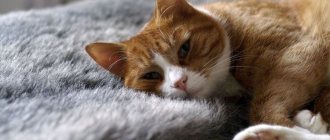
Preparing a cat for rhinoscopy
Despite the safety of rhinoscopy, a prerequisite for this endoscopic examination technique is general anesthesia.
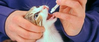
To reduce the risk of complications that arise when putting an animal into deep sleep (anesthesia), the diagnostic procedure is performed on an empty stomach. The cat is kept on a starvation diet for 12-24 hours. Access to drinking water is limited 3-4 hours before. Such preparation is necessary so that during the administration of veterinary drugs the animal does not choke on vomit.
Before setting a date for rhinoscopy, the veterinarian conducts a complete examination of the cat. To minimize risks, we recommend showing your pet to a veterinarian-cardiologist or anesthesiologist.
After the procedure, we do not recommend taking the cat home immediately, since one should not exclude possible complications caused by anesthesia (lethargy, anaphylactic shock, decreased temperature). If necessary, the veterinarian will provide emergency assistance to the animal.
Rhinoscopy as the main method for diagnosing pathologies of the nasal cavity in dogs and cats
The author of the article is a veterinarian, Ph.D. Kuleshova O.A.
The nasal cavity in animals is the first link in the formation of the respiratory system, playing an important role in cleansing, moisturizing, warming and conducting air into the upper and lower respiratory tract. The structural features of the nasal cavity entail difficulties in diagnosing diseases of this localization and, as a result, making incorrect diagnoses and choosing the wrong treatment tactics.
Rhinoscopy is the most informative method of instrumental diagnostics, allowing real-time assessment of the condition of the nasal mucosa and the anatomical structures of the nasal cavity (nasal passages, turbinates, nasopharynx). If changes are detected in the mucous membrane itself or when neoplasms of the nasal cavity of various origins are detected, a targeted biopsy from the pathological focus can be performed simultaneously for cytological and histological examination. Thus, rhinoscopy not only allows you to diagnose the condition of the nasal cavity, but also make a final diagnosis taking into account the study of mucosal tissue obtained during rhinoscopy, which distinguishes this method from MRI and CT.
IMPORTANT!!! Rhinoscopy is performed on an animal in the absence of an infectious process. If, upon examination, the animal has a clinical picture of an obvious respiratory infection (this is especially true for young and unvaccinated animals), then at the first stage, the mucous membrane is washed off to exclude an infectious cause of the disease.
The main indications for rhinoscopy are unilateral or bilateral discharge of various types from the nasal cavity, difficulty in nasal breathing, sneezing, as well as the lack of persistent positive dynamics in the drug treatment of rhinitis.
Very often, animals are diagnosed with RINITIS. Rhinitis is an inflammation of the nasal mucosa. The effectiveness of drug treatment directly depends on the cause of this inflammation in the nasal cavity. But this inflammation may not be the main cause of nasal discharge. For example, a dog on a walk actively sniffs everything around and when inhaling, an organic (a piece of a spikelet) or an inorganic (a fragment of stone or metal shavings) particle may enter the nasal cavity, which will cause inflammation in the nasal cavity, which will be manifested by discharge. In this case, only removal of this foreign object from the nasal cavity will resolve this disease. Another striking example is tumors of the nasal cavity, which also occur with the appearance of rhinitis. In some animals, rhinitis can be of a special nature. This type of rhinitis can only be determined by histological examination of the nasal mucosa. We are talking about autoimmune rhinitis. In all the described cases, antibiotic therapy and immunotherapy can give positive dynamics, but after discontinuation of drug treatment, discharge and sneezing will resume again or after some period.
Thus, if respiratory infections have been ruled out for your animal and rhinitis is being treated, but this treatment does not provide lasting positive dynamics, then rhinoscopy is indicated for your animal. Since rhinitis is not a diagnosis, it is a manifestation of the underlying disease. It is necessary to establish the cause of rhinitis, and only influencing the cause will cure the animal.
The main causes of nasal discharge:
- Respiratory infection;
- Foreign body in the nasal cavity;
- Fungal rhinitis;
- Bacterial rhinitis;
- Autoimmune rhinitis;
- Aneurysmal cyst;
- Nasal polyp;
- Tumor of the nasal cavity.
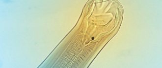
Rice. A. Normal nasal mucosa. Rice. B. Tumor of the nasal cavity (Esthesioneuroblastoma in a dog). Rice. B. Performing a targeted biopsy from the pathological focus.
Rhinoscopy is performed under general anesthesia (gas anesthesia) with the simultaneous use of local anesthesia. During rhinoscopy, the nasal cavity is examined, the mucous membrane and anatomical structures are assessed. When pathological tissue is identified, a targeted biopsy is performed using special punches to obtain a piece of tissue for its cytological and histological study. According to indications, the mucous membrane is washed from the depths of the nasal cavity to determine specific microflora, with the selection of antibiotics sensitive to it.
Rhinoscopy is an atraumatic method of examination, but when performing a biopsy from pathological tissue, minor nosebleeds may occur. To relieve it, at the end of rhinoscopy, a temporary turunda is installed in the animal’s nasal passage, which is removed before discharge. The animal wakes up in the center's hospital under the supervision of a veterinarian. The animal is discharged only in a fully awake state.
Video 1. Foreign body in the nasal cavity of a dog.
Video 2. Polyp in the nasal cavity of a cat.
If you have any questions, or your animal needs rhinoscopy, you can consult by calling the center’s reception at 8 (962) 952-66-77 , the doctor on duty will answer all your questions.
BE HEALTHY!!!
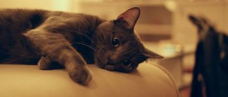
Contraindications to rhinoscopy in cats
Rhinoscopy has a number of contraindications, including:
- severe nosebleeds;
- chronic pathologies of the cardiovascular system;
- intolerance, impossibility of putting an animal under anesthesia (age);
- injured ENT organs;
- pregnancy, lactation;
- severe pain in the sinuses.
How is it carried out?
Magnetic resonance imaging is used to detect polyps that may appear due to inflammatory processes.
If local treatment is not effective, then the next method for examination is MRI. This diagnosis shows the presence of polyps or other formations that appear after chronic inflammation, inflammatory processes in the nasal passages. After the MRI results are obtained, the animal is prepared for rhinoscopy. Having discovered an infection with pathological flora in the sinuses and formations in the nasopharyngeal space, this is eliminated surgically. When a polyp is found in the nasal cavity or there is simply inflammation, rhinoscopy is performed along with histology, cytology and bacterial culture.
When performing posterior rhinoscopy, the animal’s mouth is fixed with a special mouth dilator. If a foreign body is detected, it is removed immediately if the size and shape allows this to be done without injury. If this is not possible, a planned surgical operation is prescribed. A mandatory examination before surgery includes a visit to a cardiologist and an anesthesiologist to determine the existing risks.
Not every veterinary clinic has equipment for performing rhinoscopy. If the behavior and condition of the animal has changed, it is necessary to urgently consult a doctor for early diagnosis of pathological processes. Therefore, rhinoscopy in cats is not only about diagnosing the condition of the nasal and oral cavities, but also about obtaining a complete diagnosis with the study of the tissues of the mucous membrane. This is where the method differs from MRI.
Rhinoscopy is a modern method for diagnosing nasal breathing disorders in animals.
The ability to breathe is one of the most important components in the life of any living organism. But, unfortunately, it often happens that owners encounter nasal breathing problems in their pets.
Sneezing, sniffling, discharge of purulent or bloody discharge from the nose - if you notice such symptoms in your pet, you should pay attention to this and be sure to see your doctor, as this may indicate serious problems with the animal’s nasal passages.
There are quite a few reasons for impaired nasal breathing in small pets and it is worth understanding them. Respiratory disturbance can be acute or chronic, there can also be discharge of a different nature - purulent, hemorrhagic (blood), serous, and can be manifested by frequent sneezing, sometimes “grunting”, snoring during sleep. The animal may or may not experience any discomfort if nasal breathing is disrupted. In any case, it is necessary to understand the cause of the nasal breathing disorder in your pet, which will allow you to prescribe more targeted treatment, and therefore help a quick recovery.
There are frequent cases of impaired nasal breathing in animals due to the presence of foreign bodies in them. Foreign bodies enter the nasal passages of animals when they are inhaled. Removal of these objects is quick and simple under endoscopic control and helps to immediately restore nasal breathing.
The cause of impaired nasal breathing can be congenital anomalies in kittens and puppies. For example, cysts and polyps of the nasal passages. Very often in our practice we encounter young patients, under the age of one year, with breathing problems and various kinds of discharge from the nasal passages. It should be remembered that first of all, especially in unvaccinated animals, viral infections in kittens and puppies that can cause rhinitis should be excluded. Cysts and polyps in young animals are easily diagnosed and easily removed using endoscopic technologies. Timely diagnosis and removal of cysts and polyps is important, since over time they can lead to deformation of the bones of the facial skull and severe chronic purulent rhinitis.
Rhinoscopy is an endoscopic examination of the nasal passages and nasopharynx using an endoscope. To examine the nasal passages, rigid endoscopes are most often used, which are inserted through the nostrils into the nasal passages. To examine the nasopharynx, it is more convenient to use flexible endoscopes; the examination is carried out through the oral cavity. The rhinoscopy procedure itself is carried out with active sanitation with saline solution, which also has a therapeutic effect, especially for chronic and purulent rhinitis. During the study, all nasal passages (ventral, middle, dorsal), as well as the nasopharyngeal cavity, are examined, the color of the mucous membrane, the nature of its damage are assessed, patency and the presence of foreign objects, cysts and neoplasms are assessed. If suspicious areas of the mucous membrane are detected, in case of chronic rhinitis, neoplasms, material is taken for histological examination. Histological examination makes it possible to make a more accurate diagnosis and exclude the oncological nature of the disease.
During rhinoscopy, in addition to examination, the removal of polyps, cysts and neoplasms is also carried out. Removal of formations is performed using special endoscopic instruments; it is very important that the removal is carried out under endoscopic control.
Currently, diagnosing problems of the nasal passages is impossible without the use of endoscopic technologies, since they allow us to examine all structures in detail, take a biopsy, establish an accurate diagnosis and help the patient in a minimally invasive and less traumatic manner.
It is worth noting that rhinoscopy in animals is performed under general anesthesia, so you should prepare for this procedure in the same way as for any operation. In breeds at risk for cardiac diseases, as well as in older animals, we recommend seeing a cardiologist and, if necessary, performing an echocardiogram. The necessary list of preoperative tests (blood tests, x-rays, ECHO, etc.) for your pet can be discussed at a preliminary appointment and examination by an anesthesiologist.
If your pet has any breathing problems, be sure to see your doctor, who will prescribe the necessary tests.
We wish you and your pet good health!
Sincerely, Anastasia Nikolaevna Gerasimova, veterinarian, endoscopist surgeon, SVK Svoy Doctor, Lyublino branch
Cysts of the nasal passages
large nasopharyngeal polyp
removed polyp