Radiography is an indispensable diagnostic method in both medicine and veterinary medicine. Most often, radiography is used when there is a suspicion of diseases of the musculoskeletal system (bone fractures, joint dislocations, arthrosis, etc.).
This is due to the physical characteristics of X-ray radiation, which, passing through the tissues of the body, allows a good assessment, first of all, of dense bone structures.
Why do cats and dogs need X-rays?
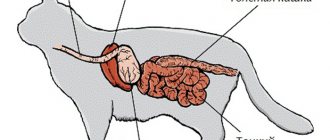
X-raying a dog or cat is necessary to confirm a diagnosis or to examine internal injuries that are not noticeable during visual examination. X-rays pass unhindered through the animal's body, displaying the entire anatomy on a special film.
The image is black and white, but the X-ray film, like an overexposed photograph, shows the internal structure of the pet's body. The image appears due to the fact that bones and soft tissues absorb the X-ray beam differently, creating a unique imprint.
As a result of such diagnostics, the specialist sees not only joints and internal organs, but can also detect hernia, tumors, and fractures.
Images are usually taken in several projections, which allows you to more accurately identify the problem and select the optimal treatment regimen.
Associated symptoms with vomiting hair
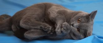
If a cat repeatedly vomits hair, you should take a closer look at the condition of the animal and assess the presence of alarming symptoms (lethargic condition, complete or partial refusal of favorite food, drinking too much, lack of interest in communication, prolonged sleep, rare urination, diarrhea, changes in body temperature , discharge from the eyes or nose, signs of a cold).
Combinations of these signs along with frequent vomiting with hair are a reason for an urgent visit to the veterinarian.
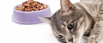
If the cause of vomiting is large trichobezoars that cannot leave the stomach (too dense and large hair ball), and the cat is young and healthy, then the symptoms may be as follows:
- the cat eats often, but in small portions;
- is constantly near the bowl (there is a feeling of hunger against the backdrop of a full stomach);
- deterioration of coat quality;
- frequent urge to vomit;
- cough;
- weight loss.
If you experience such alarming symptoms, you should not self-medicate with Vaseline or vegetable oil, as the hairball may slip into the intestines and cause a blockage. The right decision is an urgent consultation with a veterinarian.
Digital veterinary x-ray at the Panvet clinic
The Panvet veterinary clinic is equipped with the latest and most modern equipment and offers digital veterinary x-ray services to animal owners in Moscow. Currently, this is the most innovative diagnostic method.
We provide services around the clock, the price of the procedure starts from 1000 rubles.
The use of digital x-rays for animals provides the following advantages compared to conventional photographs:
- Higher resolution – allows you to accurately diagnose internal pathologies.
- Speed – digital photographs are ready for viewing instantly, and do not require film development.
- Safety – the level of X-ray radiation is reduced several times and does not pose even the slightest threat to pets.
- Practicality – you can store pictures on any storage medium.
- Visibility – pet owners can immediately see the result on the monitor.
Once digitally processed, the image will be ready for interpretation within minutes, providing significant time savings in emergency situations. In addition, the digital format allows you to instantly send the results to other specialists via email or the clinic’s internal local network.
General information
A gastric bezoar (stomach stone) is a calculus of varying density that is formed when substances are swallowed that are not digested in the stomach. The name of the stomach stone is associated with the breed of mountain bezoar goats, in whose stomachs grayish-blue stones of wool, mucus and leaves were often found.
In Russian gastroenterology, the first mentions of pathology are found at the beginning of the 19th century in the works of the Russian surgeon V.M. Mouse. Gastric stones occur rarely, as of the early 1990s. About 400 cases of the disease have been described. Concretions can be single, reaching a mass of up to 1 kg, or multiple. The latter are small in size and prone to migration into the duodenum and jejunum.
Stomach bezoar
X-ray of internal organs of dogs, cats and other animals
Our clinic conducts the following diagnostic tests for cats and dogs:
- X-ray of the spine.
- Chest X-ray.
- X-ray of the abdominal cavity.
- X-ray of the skull.
- X-ray during pregnancy.
- X-ray of joints.
- X-ray of teeth.
- X-ray of the heart.
- X-rays of light.
- X-ray of the stomach.
- X-ray of paws.
Causes
A gastric bezoar occurs when objects of organic or inorganic origin are repeatedly swallowed, most often with food. There are states and conditions that increase the likelihood of the formation of bezoars:
- Mental disorders. Neuroses, trichotillomania, schizophrenia, mental retardation can cause uncontrolled absorption of inedible substances (plasticine, glue, hair) in large quantities.
- Stomach diseases. Insufficient secretion of hydrochloric acid, slow evacuation of contents into the duodenum due to gastroenterological pathology (gastroparesis, secretory insufficiency) causes the gradual formation of stones. Excessive proliferation of Candida fungi in the stomach initiates the formation of fungal bezoars.
- Stomach operations. Previous surgical interventions (gastric resection, vagotomy) contribute to a decrease in secretory function, digestive disorders, and the accumulation of indigestible products.
- Violation of food culture. Poor chewing of coarse, fibrous heavy foods and swallowing fruit seeds can provoke the formation of a gastric bezoar.
Is it possible to x-ray reptiles, rodents, and birds?
Yes it is possible. Veterinary x-rays are considered general diagnostics and are therefore suitable for all pets without exception.
The clinic staff includes specialists of a narrow profile: herpetologists and ornithologists, who also need to conduct internal research on their patients. Therefore, x-rays are used quite often for exotic animals.
Classification
Gastric bezoars can have different composition and consistency (loose, dense, hard, elastic). Depending on the origin, the following main types of gastric calculi are distinguished:
- Phytobezoars. They make up up to 70% of all bezoars. Formed by eating the skins, seeds, peels of berries and fruits (persimmons, cherries, grapes, figs, etc.). Plant substances gradually become overgrown with mucus, fat and mineralize. The stones have different consistencies, a foul odor, and are dark green or brown in color.
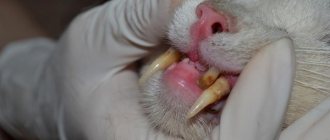
- Trichobezoars. They are formed when hair regularly enters the stomach. More often they occur in neurosis-like conditions and mental disorders with an irresistible urge to bite off hair.
- Stibobesoars. Formed by frequent consumption of fatty foods of animal origin (lard, lamb). Once in the stomach, food is transformed and compacted to form fatty stones.
- Shellacobesoars. They occur when using inedible products of chemical origin (varnish, paint) as food. When entering the stomach, substances interact with water and precipitate. Shellac stones have a viscous consistency, a slightly rough surface, and a dark brown color.
- Stones of embryonic origin. They are formed from exo- and endoderm at the stage of intrauterine development. They are dermoid cysts or teratomas of the stomach.
- Lactobezoars. They are formed in newborns who are fed an artificial mixture with a high content of lactose and casein. Premature babies are prone to stone formation.
There are rare types of gastric stones that occur in isolated cases in medical practice. Pyxobesoars are formed by consuming resin or pitch. Hemobezoars are formed during prolonged swallowing of blood in patients with nasal and esophageal bleeding. Stones can occur when taking poorly soluble and insoluble drugs (sucralfate, aluminum hydroxide, activated carbon). Mixobezoars consist of threads, wool, and pieces of wood.
Preparing dogs, cats and other animals for x-rays
In most cases, x-rays for dogs and cats do not require any special preparation. For individual cases, the doctor may give additional recommendations:
- X-ray of bones and joints - done without preparation; if the animal experiences acute pain, anesthesia is administered before the procedure begins.
- X-ray of the abdominal cavity - to obtain clear images, defecation and emptying of the bladder is desirable.
- Chest X-ray – without any features, according to general requirements.
- A study using barium - a contrast agent helps to make a more accurate diagnosis; it is administered without prior preparation.
General recommendations: fasting diet before the procedure for dogs and other animals. You can give your pets water without restrictions.
X-ray contrast of the gastrointestinal tract
The purpose of this study is to isolate an organ or organ system from surrounding tissues. Such examination is useful in determining the shape, position, location, and functioning of an organ. The resulting images using a contrast agent complement or confirm the suspected diagnosis.
The purpose of survey images before using contrast agents is to obtain the initial image, set the necessary modes, and prepare the patient. Sometimes plain films can answer questions, eliminating the need for contrast agents. Survey photographs of the area of interest should be taken in two projections. Contrast studies should not be used instead of plain films. The contrast agents used in gastroenterology are, as a rule, X-ray transparent
appearance (white), but radiolucent substances (air, oxygen, carbon dioxide) are also used, they have a black appearance.
A contrast study of the gastrointestinal tract requires a series of images.
,
to control the passage of the contrast agent
through the gastrointestinal tract. Typically, the first images are taken immediately after contrast injection to rule out esophageal pathology (a slightly oblique ventrodorsal view should be used to avoid overlap of the spine with the esophagus.)
Next, pictures are taken in a certain sequence: immediately, after 15 minutes, 30 minutes, 1 hour, 3 hours, 6 hours, 12 hours, 24 hours.
The obtained images are numbered, indicating the volume of contrast agent injected, what substance was injected, and the time intervals when the images were obtained.
Types of radiocontrast agents used for contrasting the gastrointestinal tract.
There are positive contrast agents
- water-soluble (ionic and non-ionic iodides), Ionic (Sodium diatriosate, Meglumine diatrizoate), Non-ionic (Iopamidol, Yohexol, Iodixanol)
Used in the diagnosis of the gastrointestinal tract when perforation is suspected, they are hypertonic solutions that attract fluid into the lumen of the gastrointestinal tract, as a result of which the contrast agent is diluted, reducing the quality of the final image. Fluid loss may complicate hypovolemia in dehydrated animals. It is not recommended to use organic iodides instead of barium sulfate, only if gastrointestinal perforation is suspected.
- water-insoluble (Barium sulfate)
They have a high atomic weight (high density), so they absorb more x-rays and fewer x-rays pass through the patient, causing certain areas to appear white on the x-ray. Most often, when examining the gastrointestinal tract, the positive, water-insoluble substance barium sulfate is used from contrast agents; it is available in the form of powders, pastes, and suspensions. Since it is water insoluble, the body eliminates it naturally. When administering a contrast agent orally, avoid contact with the respiratory tract as aspiration pneumonia may occur.
Gastric torsion.
An X-ray image shows a dilated stomach with a contrast agent (barium sulfate).
The axis of the stomach is changed, there is no evacuation of the contrast agent.
Indications for contrast examination of the gastrointestinal tract:
- megaesophagus,
- tracheoesophageal fistula,
- esophageal dysfunction,
- indomitable vomiting (in this case, the use of antiemetics is justified),
- occasional vomiting,
- vomiting blood,
- palpable abdominal formations in the abdominal cavity,
- acute abdominal pain, the cause of which is not established on a plain x-ray,
- unexplained cachexia,
- melena (tarry, spotty stools),
- bloody stools
- gastrointestinal obstruction,
1)
spastic,
2)
mechanical,
- change in the wall (expansion or narrowing of the walls of sections of the housing complex),
- displacement of abdominal organs,
- intestinal perforation.
Studies with radiopaque contrast agents are not helpful in patients with diarrhea without vomiting.
Patient preparation.
The animal is kept on a 24-hour diet, water is allowed, and a cleansing enema is given 3 and 1 hour before the test.
The patient must be accompanied by two people over 18 years of age to securely secure the animal.
Methodology.
The contrast agent is administered into the cheek pouch or through a gastric tube
Table 1
Upper gastrointestinal studies are DYNAMIC
.
The studies are recorded using numerous radiographs. Photographs of the abdominal cavity in the lateral and ventrodorsal projections are repeated 15, 30 and 60 minutes after injection
. Repeat images of the abdomen continue to be taken every hour until the contrast agent has reached the colon.
Recommended time intervals between x-rays are given in Table 2 (for dogs)
table 2
Recommended time intervals between x-rays are given in Table 3 (for cats)
Table 3
To evaluate the results
In this study,
standardization of the procedure is very important
.
For example, reducing the amount of medicine may lead to a decrease in the time of gastric emptying, which may be misinterpreted. Standardization must be observed at least within one clinic.
An adequate amount of mixture ensures physiological stretching of the gastrointestinal tract and visualization of the lumens. After barium administration, the stomach is completely visualized no later than 15 minutes.
Accelerated gastric emptying is not a sign of pathology, but delayed gastric emptying is a pathognomonic sign
.
Standards for conducting X-ray examinations with contrast agents adopted in our clinic
1.
If possible, you should avoid using any medications.
2.
To avoid misinterpretation of the study results, before drinking the mixture, the patient must be protected from contact with the contrast agent on his skin and fur.
3.
It is necessary to avoid introducing the mixture into the trachea.
4.
The finished mixture is administered in the quantities given in
Table 4
.
Table 4
Attention!!!
1.
If the administration
of tranquilizers
, the drug of choice
for dogs
is
acetylpromazine
and
ketamine
for
cats
.
2.
Enough contrast material is injected to fill the stomach.
If gastric or intestinal perforation is suspected, water-soluble organic iodides are used
.
3.
Water-soluble organic iodides are contraindicated in dehydrated animals, as they have hypertonic properties.
4.
All photographs should indicate the time since insertion.
Pneumogastrograms. Negative contrast gastrography. Gastrography with double contrast without serial examination of the upper gastrointestinal tract.
help in determining the position of the stomach. On gastrograms with double contrast, ulcerations of the gastric mucosa and damage to its neoplasms are better visible.
Indications:
- suspicion of the presence of tumors in the stomach,
- radiolucent foreign bodies,
- obstruction of the outflow tract.
Contraindication:
- if there are internal contents,
- diarrhea,
- patient with diabetes mellitus,
- patient with pheochromocytoma.
Methodology:
Pneumogastrography
. A gastric tube is inserted to introduce air. When using effervescent granules, they are injected into the cheek pockets using a syringe. This prevents foam from being lost before it reaches the stomach. After introducing a negative contrast agent into the stomach, the probe is removed. Take pictures of the anterior abdominal region
- in the right side,
- left side,
- ventrodorsal,
- dorsoventral projection.
Negative contrast gastrography
. There is also a double contrast method. Where positive and negative contrast agents are used simultaneously. For better visualization of the structural forms of the organ under study. When most of the barium sulfate suspension has left the stomach, a gastric tube is inserted, followed by a negative contrast agent. Remove the probe and roll the patient from side to side so that the contrast agent is properly deposited on the mucous membrane.
Take pictures of the anterior abdominal region
- in the right side
- left side
- ventrodorsal
- dorsoventral projection.
Gastrography with double contrast without serial examination of the upper gastrointestinal tract
. The stomach is paralyzed with glucagon. A gastric tube is inserted and then a positive contrast agent is injected. Leaving the probe in place, a negative contrast agent is injected. The probe is removed and the patient is rolled from side to side so that the contrast agent settles on the mucous membrane.
Take pictures of the anterior abdominal region
- in the right side
- left side
- ventrodorsal
- dorsoventral projection.
Table 5
Barium enema
Barium enema
is a procedure used to study the position of the colon contour. Serial examination of the anterior gastrointestinal tract is not suitable for examination of the large intestine.
Indications:
- diarrhea of the large intestine,
- tenesmus,
- fresh blood in stool
- neoplasia of the colon or rectum,
- colitis,
- diseases of the mucous membrane,
- ileocolic intussusception.
Contraindications:
- suspicion of gastrointestinal perforation,
- proctoscopy within 12 hours,
- cleansing enema within 4 hours before the study. Table 6
Table 6
Survey radiograph.
Photographs of the abdominal cavity in ventrodorsal and lateral projections before enemas.
Patient preparation:
The large intestine should be free of feces. Animals are kept on a fasting diet for 24 hours. The evening before, a cleansing enema is performed. General anesthesia is required.
Methodology:
Before administration of the contrast agent, photographs of the abdominal cavity are taken in the lateral and ventrodorsal projection. The catheter is inserted into the rectum and the cuff is inflated to prevent leakage of the solution. Three-quarters of the calculated dose is administered and the animal is x-rayed in a ventrodorsal view to check the amount of distension of the large intestine. If the colon is not completely distended, add 5 ml of contrast agent and repeat the image. Continue until the correct stretch is achieved. With proper stretching, a lateral view of the abdominal cavity is obtained
.
Leaving the catheter in place, the contrast material is removed and replaced with negative contrast material
.
Images of the abdominal cavity are obtained in lateral and ventrodorsal projections
. Before removing the boat, remove as much contrast material as possible.
For the negative staining method, air can be used as a contrast agent. Avoid overstretching the large intestine. Use higher voltage settings for positive counterstaining. To dilute the contrast agent to 20% concentration, it is necessary to use a saline solution.
Complications
Prolonged stay of a bezoar in the stomach leads to the formation of bedsores of the organ wall. Local blood circulation is disrupted, ischemia and necrosis develop with the formation of ulcerations. Erosion and gastric ulcers can lead to perforation of the muscle layer and the occurrence of peritonitis and sepsis. A rare complication is intestinal obstruction. When a bezoar stone enters the small intestine and blocks the intestinal lumen, obstructive intestinal obstruction develops.
Treatment of stomach bezoar
Treatment of the disease depends on the type, consistency, composition of the stone and concomitant pathology. If the size is small, the gastric stone may pass on its own. Conservative treatment methods are effective for bezoars of soft and medium consistency, often of plant origin. Warm alkaline solutions based on soda, mineral water, and proteolytic enzymes are prescribed orally. A light massage of the epigastric region has a positive effect. Patients are advised to follow a gentle diet with limited fruits, meat and fatty foods. Small-density stones are removed endoscopically. Using special instruments, the stone is crushed and removed under the control of a gastroscope. In case of obsessive states, consumption of inedible foods, consultation with a psychiatrist is indicated.
If therapy is ineffective, large and dense bezoars require surgical removal of the stone. The operation consists of cutting the stomach (gastrotomy) and removing the bezoar stone. Indications for surgical intervention are conditions caused by complications of the disease (intestinal obstruction, peritonitis).