The eye is a complex but rather fragile organ. Impairments in work are manifested by loss of clarity of vision. If a film appears on the eye, this may indicate the presence of a serious disease. Therefore, it is necessary to consult a doctor to clarify its nature. This pathology is called pterygium. At the initial stage, the tumor covers only part of the cornea, without preventing the person from seeing well. The danger of this disease is that the pterygium gradually grows, closing the pupil. This leads to blindness. The film can form on one eye or on both eyes at once.
Symptoms of diseases
The film on the eye causes a feeling of discomfort. It may vary in intensity and duration. Most often, this condition occurs in the morning or after hard work at the computer. In some cases, the unpleasant symptom disappears if you blink your eyes. Moisturizing the organs of vision and wiping are often required. Such conditions are often accompanied by other symptoms, including:
- itching and unpleasant burning sensation;
- increased sensitivity to light;
- the appearance of spots and “flies”;
- general weakness.
A film on a person’s eyes, which is accompanied by similar signs, may indicate the development of diseases not only of the visual organs, but also of the nervous system. That is why you should seek help from ophthalmologists. Specialists will make an accurate diagnosis and prescribe appropriate therapy. Often the development of a film on the eye is observed in older people. The neoplasm consists of an overgrown conjunctival membrane.
Symptoms of pterygium
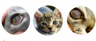
- Slight clouding of the periphery of the cornea, which is the first sign of developing pterygium. At this stage the patient has no complaints. There is only a mildly expressed cosmetic defect.
- The appearance of a growth on the cornea that has an opaque consistency. This growth is quite noticeable and usually grows from the side of the nose.
- The sensation of a foreign body in a particular eye, which occurs because the pterygium begins to rise above the surface of the cornea, irritating the nerve endings receptors located on the inside of the eyelid.
- Persistent eye irritation. The reason for this is the absence of a tear film on the surface of the neoplasm, as well as a violation of the formation of such a film on a healthy area of the cornea. Persistent feeling of dryness in the eye.
- Gradual decrease in visual acuity. This symptom occurs when a pterygium grows on the center of the cornea, as a result of which the passage of light into the eyeball is disrupted.
- If the pterygium is inflamed, hyperemia of the eyeball, itching, swelling of the conjunctiva, and increased lacrimation are observed.
Cause of the disease
There are many known factors that provoke the appearance of a film on the eye. Among them it is worth highlighting:
- Prolonged exposure to ultraviolet radiation. This disease is common among residents of southern countries. This is where high intensity radiation is present, as well as many sunny days.
- Mechanical impact of dust, sand and small particles. These negative factors are amplified by the wind, which irritates the mucous membrane of the organs of vision.
- Hereditary predisposition. Pathology is often transmitted between relatives.
- Conjunctivitis. With frequent inflammatory processes, the mucous membrane of the organ is affected by bacteria, adenoviruses, and so on. Frequent conjunctivitis leaves scars, which leads to changes in eye tissue.
Other internal reasons
Other reasons include the following:
- Inflammation (neuritis) of the optic nerve;
- Migraine. The appearance of a film in the eyes is one of the signs of an approaching migraine;
- Transient ischemic attack or stroke. Problems associated with blood circulation in the vessels, in particular, the brain, can be characterized by such a sign as the appearance of a film on the eyes;
- Neoplasms in the brain. As a rule, in this case the film “attacks” only one eye;
- Medicines. Their frequent use can lead to such circumstances as the appearance of a film in the eyes. Such drugs include antidepressants, corticosteroids, oral contraceptives, drugs for the treatment of heart disease, anticholinergic medications;
- Frequent conjunctivitis, as well as the presence of a predisposition to such.
Other factors
Working at a computer for several hours causes dry eyes and irritation of the mucous membranes. The conjunctiva reacts very sharply to almost any impact, because it contains many blood vessels, as well as nerves. The mucosa consists of several layers: epithelium and submucosa.
The conjunctiva of the eyes performs several functions at once. Inside it is a gland that produces tear fluid. The mucous membrane moisturizes the eyes and protects them from the negative effects of the environment. With prolonged exposure to an irritating factor, transformation of the epithelium occurs. As a result, connective tissue begins to grow and a film closes the eyes.
Treatment of pterygium
Pterygoid pleurisy is treated using conservative and surgical methods. If the film does not affect the pupil, it is usually not removed. The patient is prescribed eye drops and other medications. When the tumor has grown greatly, surgery is performed to remove it. Film removal is carried out using various methods, including using a laser. The operation is quick and painless.
Diseases that cause pterygium
Why does it feel like there is a film in my eyes? The following diseases may be the cause of this phenomenon:
- Refractive diseases, such as astigmatism, farsightedness. Laser therapy and wearing corrective glasses or lenses can help get rid of such disorders.
- Dry eye syndrome. If there is not enough hydration, the cornea begins to dry out, and the clarity of vision gradually decreases. The reason is the deterioration of the mucous membrane.
- Cataract. The disease causes clouding of the crystal. Most often, the disease occurs in older people.
- Glaucoma. In this case, the film on the eye occurs due to a violation of eye pressure.
- Inflammation of the optic nerve.
It is worth noting that a white film on the eyes can occur as a result of taking certain medications, such as corticosteroids, oral contraceptives.
Treatment
Treatment for veils in the eyes directly depends on the cause of this symptom:
- Retinal detachment is treated with eye medications that improve retinal metabolism and vascular patency. In case of severe pathology, laser coagulation (a kind of “gluing”) of the retina to the choroid can be performed.
- If keratitis is detected, drug treatment is prescribed in the form of antimicrobial and anti-inflammatory drops, as well as in the form of tablets and injections that improve the supply of nutrients to the cornea.
Floxal is an antibacterial agent that is used as local therapy during ophthalmic diseases
- If the cornea is damaged, a corneal transplant may be necessary.
- For immature cataracts, the doctor prescribes drops with eye vitamins and nutrients to slow down the clouding of the lens. When the cataract matures, surgery to replace the lens will be required.
- Glaucoma is treated with drops to reduce intraocular pressure. If medication methods do not have an effect, surgical treatment is also performed to relieve acute attacks of glaucoma.
Read about laser iridotomy in the article.
- Dry eye syndrome is treated with moisturizing drops, which are artificial tears (Systane Ultra drops, Hilamax, Hilo-comod).
- A brain tumor is subject to surgery, as well as radiation and chemotherapy.
- In case of a stroke, bed rest is necessary; if it is caused by a blood clot, then in hospitals it is dissolved.
- For anemia, therapy consists of identifying the cause of low hemoglobin and treating it.
- A hypertensive crisis is treated by emergency doctors, and treatment takes place in the cardiology or therapy department.
- Diabetic retinopathy is treated in tandem by an ophthalmologist and an endocrinologist, who selects insulin doses for the patient. The ophthalmologist, in turn, prescribes medications that allow the vessels to more effectively nourish the structures of the eye.
Read more about Systane Ultra drops in the material.
Treatment of the identified disease, giving up bad habits, playing sports (within reason) and proper nutrition can prolong eye health for many years. The effectiveness of treatment directly depends on the correct diagnosis.
Blindness before the eyes is a symptom that often gives an unfavorable prognosis, so it is very important to stop the disease at an early stage, while vision can still be preserved.
Preparations from the “Artificial tear” series
What to do?
What to do if your eyes get covered with a film? Experts distinguish several stages of pathology development:
- Initial. There are no manifestations or symptoms of the disease.
- Second stage. Symptoms such as blurred vision, irritation, swelling, unpleasant burning, and film growth appear.
It is worth noting that it is impossible to get rid of such a disease with drugs or folk remedies. Medicines that can remove the film have not yet been created. The only way to get rid of this pathology is through surgery. At the initial stage, the film is not removed. At this stage, the growth of the tumor is monitored.
If the size of the film or its condition changes, surgical intervention is immediately prescribed. Tightening is not recommended. After all, the operation is very complicated. In addition, there is a risk of relapse.
Stages and forms of pterygium
There are two stages of this disease. At the first stage there are no symptoms, the tumor does not cause any discomfort. In the second stage, itching, burning, and swelling appear. The eyes become red and watery. At the same time, vision deteriorates. A person constantly feels the presence of a foreign object in the eye. Pterygium can be stationary, when it does not grow for a long time, and progressive, in which rapid growth of the film is observed.
How is the operation performed?
If the eye is covered with a film, then surgery is performed immediately. The day before the operation, the patient must prepare. During this period, it is prohibited to take aspirin, as well as other drugs that affect the condition of the blood.
Surgery is performed using local anesthesia. The film is excised to the sclera. Removal of the tumor takes no more than half an hour. After surgery, a bandage is applied to the patient's eye. The specialist should prescribe drops to prevent the occurrence of an inflammatory process. After surgery, the patient remains under observation for several hours. If during this time severe pain occurs, the doctor should prescribe painkillers. You cannot rinse your eyes with water for several days. It is worth considering that pterygium may begin again. If a film appears, you should immediately contact an ophthalmologist.
Treatment of the disease
When such a disease appears, drug treatment is powerless and is not prescribed by a doctor. If the film on the eyes is of insignificant size and it is not worth operating on it, then it is monitored, it is determined whether the tumor is growing or not, and how quickly it is progressing. If the film begins to grow, it must be removed. This can only be done surgically.
This operation is performed by a qualified doctor under local anesthesia. The patient is first instilled with a special solution into the affected eye.
Surgical intervention involves the surgeon removing the growth itself on the visual apparatus. To prevent the patient from having a relapse, a special graft is inserted into the treatment area instead of the excised area. It is cut out from another section of the conjunctiva.
In order to avoid relapse, the doctor may prescribe antitumor antibiotics. The rehabilitation period after such surgery does not last long. For 48 hours, the affected area should be covered with a special bandage. In order for swelling to go away faster, you can use drops that your doctor will prescribe.
Pterygium is usually eliminated using a laser. This lengthy procedure takes no more than half an hour. The principle of surgery is very simple. It is necessary to remove the growing film down to the sclera with a laser. The advantage of the operation is that the laser not only removes the growth, but also cauterizes the ruptured blood vessels, blood stops flowing to the film, and the likelihood of relapse is minimized.
In this case, the patient also needs to walk with a bandage for some time and be sure to use anti-inflammatory drops. It is necessary to prepare for the operation. On this day, the patient is prohibited from taking aspirin and any coagulants. After the operation is performed, the person needs to stay in the hospital for several hours.
The surgical procedure is performed as follows:
- The operation is performed using local anesthesia. First of all, they introduce it. As a rule, drops with an anesthetic effect are used. A special solution with an anesthetic effect is injected into the thickness of the film itself.
- The defect is excised using a blade, after which the conjunctiva is sutured.
- After the operation, the surgeon applies a bandage with antiseptic medications to the eyes.
As a rule, surgical treatment is carried out quickly, and the patient is usually not hospitalized. After surgery, drops with anti-inflammatory effects are prescribed.
Typically, the patient does not need to follow any special measures during rehabilitation. It lasts about 2 weeks, after which the person can return to normal life.
Features of the recovery period that the patient should know about
- Painful sensations that are quite pronounced. When surgical treatment is performed, the cornea is necessarily affected, and it is the most sensitive eye shell. It is because of this that a person experiences pain, which will subside as soon as the wound heals. The pain may be accompanied by tearing, a feeling that there is a foreign body in the eye that is preventing it from opening or closing;
- During the first hours after surgery, bleeding from the conjunctival blood vessels is possible. This is a normal reaction to surgery. You just need to change the bandages in a timely manner;
- Redness in the eyeball, caused by possible blood flowing behind the conjunctiva. Such redness subsides within a couple of weeks at most;
- Due to the sutures, there may be a feeling of speck in the eye. Within a week (maximum 2 weeks), the sutures dissolve, resulting in a feeling of comfort returning.
Unfortunately, the film can reappear, and if it recurs, surgery will be required again.
Film excision methods
Surgery can be performed with a laser or scalpel. Each method has its own advantages and features. However, the use of a laser is the most popular way to remove film from the eyes. Its advantages include:
- Cauterization of blood vessels. This avoids bleeding.
- There is no need for stitches after surgery.
- The patient's rehabilitation proceeds faster.
- Painful sensations are less pronounced.
Treatment options
Before using any type of treatment, you should consult with a qualified ophthalmologist to determine the cause of the clouding and discuss the best ways to eliminate it.
Traditional medicine
If vision problems are not accompanied by a visible manifestation of a veil, the patient is prescribed vasoconstrictor or vasodilator drops or ointments that reduce fatigue, swelling, and wash the membrane of the eye from mechanical irritants.
For eye problems as a concomitant symptom of other diseases (migraine, allergies), medications are prescribed that directly eliminate the causative agent of the disease.
Vitamin preparations in the form of drops
If the clouding becomes visible to those around the patient, a treatment chart is developed. The clouded area is examined, its behavior is observed, and therapy is prescribed depending on the disease and symptoms.
In acute cases, vision surgery is required. About a day before, the patient begins to prepare for surgery, excluding blood thinning factors. Surgical intervention is performed using a scalpel or laser, which has certain advantages:
- exclusion of internal bleeding due to timely cauterization of vessels with a laser;
- there is no need for sutures;
- the rehabilitation process takes less time;
- pain sensations are smoothed out.
Stages of laser vision correction
Removal of the visible area of opacity is possible only with surgery; there are no mechanisms for its drug treatment.
Video: What to do if a veil appears before your eyes?
Available means
Decoctions of plants with medicinal properties help relieve inflammation, rinse and soothe the eye:
- chamomile;
- yarrow;
- Linden;
- herbal collection;
- yarrow root.
Yarrow
They are taken in the following forms:
- washing;
- baths;
- compresses.
Typically, packages of medicinal herbs contain a printed recipe for their use for the visual organs.
Linden decoction
If you don’t have the above substances at home and you need to wash your eyes urgently, you can use pure black tea. It is necessary to brew strong tea without sugar, moisten a cotton pad with it, rinse the eye or apply it to the eyelid for a while. If the tea is bagged, apply a freshly brewed bag to the eyelid.
Film on a newborn's eyes
If the eye is covered with a film in a small child, then the cause may be hidden in obstruction of the lacrimal canal. Dacryocystitis is a disease that occurs in 5% of babies. The disease occurs when:
- absence of the lacrimal canal;
- anomalies in the development of ducts;
- injury to the face with obstetric forceps.
During intrauterine development, the nasolacrimal ducts of the fetus are clogged with a gelatinous film. Thanks to this, amniotic fluid does not enter the respiratory tract. After birth, the film breaks through with the baby's cry. If this does not happen, then the duct remains closed. As a result, this pathology leads to stagnation, causing an inflammatory process in the lacrimal sac.
Causes associated with diseases
If a floating film appears in the eye, this may be a sign of the development of pathological processes in the organs of vision. Therefore, if such symptoms appear, you should definitely consult a doctor. Especially if they do not go away for a long time.
Losing Focus
Impaired focusing occurs against the background of astigmatism, myopia, and farsightedness. A person sees blurry images at different distances. There may be a splitting of objects and their vagueness. Each of these pathologies requires correction and specific treatment.
Presbyopia
In most cases, this disorder occurs in patients over 40 years of age. In this case, a person has difficulty reading near. Presbyopia develops gradually. Initially, vision in one eye deteriorates. If treatment is not taken promptly, damage to both eyes will occur. The patient is prescribed wearing optical products and supportive therapy.
Cataract
Cataracts are a dangerous ophthalmological disease.
If you do not consult a doctor in a timely manner, this can lead to complete loss of vision. Cloudiness of the lens gradually occurs. Lack of therapy leads to complete clouding. The severe stage of this disease can be cured only by a single method - surgery.
Glaucoma
Glaucoma is also a dangerous eye disease. Initially, a short blurred vision appears. The duration of such unpleasant symptoms gradually increases. Visual impairment is only an accompanying symptom. Glaucoma is dangerous due to increased intraocular pressure. It is not recommended to ignore such manifestations. Timely diagnosis will help preserve vision.
Macular degeneration
This pathology occurs in elderly patients over 60 years of age. The reason lies in long-term work with small objects nearby. For example: needlework, long reading, working with documents and at the computer. Initially, slight clouding appears, objects do not have clear outlines.
Hemophthalmos
The disease is very dangerous because it is accompanied by internal bleeding in the eye. In this case, the cloudiness is red. Patients who suffer from diabetes and hypertension are at risk of developing hemophthalmos.
Neuritis
Neuritis has different etiologies. Accompanied by inflammatory processes in the nerves of the visual organs.
The reason lies in excessive loads. There is visual impairment in the form of blurred objects and the inability to focus on them. If a relapse of the disease occurs, the film tends to be very dense and may appear unexpectedly.
Migraine
Migraine attacks are accompanied by painful sensations when focusing vision. Pain may occur when exposed to bright light. Migraine is an unpleasant disease.
Signs of pathology
Not only a specialist, but also the child’s mother can notice a pathology such as dacryocystitis. This disease is characterized by:
- lacrimation without crying;
- purulent discharge accumulating in the corners of the eyes;
- swelling and redness of the lower part of the eye.
To prevent film from growing on the child’s eyes, appropriate therapy is carried out. For this disease, rinsing the vision organs with an antiseptic, massage and eye drops are prescribed. By the age of one year, the film should break through on its own. If this does not happen, then surgery is performed.
Diagnostics
The doctor conducts a comprehensive examination, which will consist of the following stages:
- slit lamp examination;
- measurement of intraocular pressure;
- ophthalmoscopy (examination of the fundus of the eye).
In some cases, an ultrasound of the eyeball may be needed.
If the cause of the white veil in the eyes is not associated with eye diseases, then the ophthalmologist will refer the patient for Doppler sonography or magnetic resonance imaging of the head and neck.
Dry eye syndrome: what is the cause and what to do about it
Sometimes it happens that it’s as if sand has been poured into the eyes... There is a feeling of dryness and redness, photophobia, increased sensitivity to wind, tobacco smoke, and the like. Where does this feeling come from, why do the eyes “dry”?
The simple and unobvious cause of most cases of dry eye syndrome will surprise you. It is quite natural that you will not need drops and such things.
First, a little theory:
The tear film is a thin layer of fluid, approximately 7-10 microns thick, covering the outer surface of the cornea.
The tear film in the eyes performs a number of important functions. Once you get acquainted with them, it will immediately become obvious to you that it is simply impossible to replace all of them with any drops.
- The tear film is the first barrier to aggressive external factors. It washes away dust particles, harmful agents and inflammatory products, and prevents the cornea from being damaged by small foreign bodies.
- The surface of the cornea consists of stratified epithelium. For the cornea to function normally, like an optical lens, its surface must be perfectly flat and smooth. Covering the surface of the cornea, the tear film performs an optical function, providing a perfectly smooth optical surface.
- Among other things, tear fluid performs a trophic (nutritive) function, delivering biological substances and oxygen to the corneal tissue. It is needed to maintain adequate metabolism of the corneal and conjunctival epithelium. Oxygen directly dissolved in the tear film ensures adequate respiration of the surface epithelium of the eye.
- The tear film acts as a lubricant during eyelid movements.
- Due to the presence of antibacterial substances in the tear fluid (secretory immunoglobulin proteins IgA, lysozyme, lactoferrin, ceruloplasmin, beta-lysine, etc.), it creates protection that is very resistant to various infectious agents, preventing the development of infections.
- Serves as an accessible pathway for the movement of leukocytes involved in the “repair” of scratches of the cornea and conjunctiva.
Our tear film is made up of three components:
Tear film (macro enlargement)
The outer layer in contact with air is a thin lipid film secreted by the meibomian glands, the mouths of which are located along the edges of the upper and lower eyelids.
The middle layer , the main part, consists of aqueous tears coming from the main and accessory lacrimal glands.
The inner layer is a mucous film adjacent to the outer epithelial surface (hairy surface). This mucilaginous gel prevents the aqueous film from contacting the surface of the cornea. Mucin is primarily produced by conjunctival goblet cells, but can also be produced by corneal or conjunctival epithelial cells.
Glands and glandular structures involved in the formation of tear fluid (Polunin G.S., Polunina E.G., 2007)
The outer layer of the tear film is composed mainly of lipids, which are products of the meibomian glands of the eyelids. It consists predominantly of phospholipids, neutral fat and cholesteryl esters, which dissolve at human body temperature and vary to some extent in thickness depending on the width of the palpebral fissure.
Lacrimal glands
The middle water layer is 5-7 microns thick. - occupies almost 90% of the thickness of the entire tear film and is formed mainly due to the secretion of the accessory lacrimal glands of Henle, Manz, Krause, Waldeer, and Wolfring.
The impulses that cause the activity of the main lacrimal gland are more associated with emotional (when crying) or reflex factors, as a response to irritation of the receptors on the surface of the eye and some other areas (for example, in the nasal cavity) innervated by the first branch of the trigeminal nerve.
The aqueous layer has a low viscosity and includes all the water-soluble components of tears - inorganic salts, glucose, urea, proteins, glycoproteins, etc.
The inner layer of the tear film (mucin layer) is represented by acidic mucopolysaccharides, found in gel and salt forms. The source of mucin is the Becher goblet cells of the conjunctiva.
Stinging, pain, feeling of a foreign body, sand in the eyes, as well as unstable vision are all typical symptoms of a defective tear film. When the form is advanced, the sensations can be quite debilitating, and the prescribed treatment is ineffective, forcing a person to consult a doctor again and again.
First, you need to make sure that nothing foreign (dust, sand, makeup or hand cream) gets into your eyes. I think that there is no particular need to explain that in such cases, you can wash your eyes yourself with water or strong tea (after washing your hands well beforehand).
Contact lenses, by their nature, are a foreign object and often themselves cause discomfort in the eyes, including irritation of the cornea.
It is necessary to understand that the so-called “dry eye syndrome” (as it is commonly called in medicine) can occur for two reasons:
Evaporation of tear fluid and the logical rupture of the tear film that arises from this.
This is the most common and practically harmless cause, which must be excluded first. To do this, you just need to observe yourself by filming your eyes on your mobile phone for 10-15 minutes during the main lesson.
If your day is dominated by, say, computer work, you can turn on webcam recording for this.
After this, you need to evaluate the frequency of your eye blinks. You'll likely find that you blink very rarely as you work. It may even turn out that periodically (during special concentration on a task) you do not blink at all. This is especially common during long car trips and among people working at a computer.
Make it a rule (and set yourself a visible reminder) to blink once every 1-2 seconds. This must be done relaxed, in a normal manner.
Within 1-2 days of repetition, the discomfort in the eyes should go away.
Normally, with the eyes open, the tear film is able to maintain its integrity for 10-15 seconds, after which it ruptures.
The integrity of the tear film is restored when blinking. The number of blinks, with a normal level of general well-being, should not be less than 10-15 per minute. If its quality changes due to damage in individual layers, the film preservation time can be reduced to 1-2 seconds, and at least 60 blinks per minute are required to restore its integrity.
With a very high visual load, the number of blinks per minute is sharply reduced, up to their complete absence. At the same time, the cornea is “bare”, and its superficial nerves are very sensitive and then react even to contact with air, which causes the unpleasant sensation of “sand” in the eye.
Important: the normal blinking mode corresponds to a frequency of at least 1 time every 2 seconds!!!
This simple daily rule will help you forget about the feeling of dry eyes forever!
If you have completely ruled out this cause, but the discomfort in the eyes persists, it means that you have a high probability of disorders associated with the second cause (discussed later) and you should consult a specialist.
Insufficient amount of tears, decreased production of tear fluid.
The main reasons for this are infections, immune disorders and the use of eye drops with preservatives, including benzalkonium chloride. Prolonged exposure to an aggressive environment, for example, excess heat radiation, dust, gases or solvent vapors, swimming in chlorinated water, can also cause tear film deficiency.
Often such a violation occurs while wearing contact lenses.
There are three main forms of tear production disorders:
1️⃣ Lipoid deficiency form, in which the structure of the surface lipid layer is disrupted. This form is observed with dysfunction of the meibomian glands, with diseases of the eyelids - meibomitis, blepharitis of various etiologies, including allergic, infectious and demodectic;
2️⃣ Water deficiency form, in which there is a deficiency of the internal water layer of the tear film, which is caused by damage to the main lacrimal glands, most often these are immune-mediated diseases, including Sjögren's syndrome;
3️⃣ Mucin deficiency form, in which the structure of the mucin layer of the tear film is disrupted and it cannot ensure its adhesion to the surface of the cornea of the eye. This form develops from lesions of the goblet cells of the conjunctiva and epithelial cells of the cornea and conjunctiva, which often develop when wearing contact lenses, when using eye drops with toxic preservatives, and when taking certain medications.
Due to a violation of the optical properties of the cornea, when the tear membrane ruptures, sometimes even visual impairment is noted, which becomes “floating”. It either becomes clear, or all objects are seen as if through “uneven glass.”
Quite often, ophthalmologists cannot immediately understand what the reason is when referring their patients to a neurologist. Although the cause of such disorders is purely ophthalmological.
If you think that you may have any of the above, the best option would be to immediately consult an ophthalmologist rather than go to the pharmacy for new drops.
Did you like the article? Share with your friends and subscribe to the weekly digest of the Academy of Vision to keep track of useful materials.
Read: 233
#Dry eyes#Dry eye syndrome
Posted By
Lila Ungvari
Lila Ungvari, a professional ophthalmologist with more than 25 years of experience in European ophthalmological centers, 10 of which were involved in the certification of airline flight personnel. European vision restoration instructor.
You might also like
189
Vision correction Visual impairment
Eye drops: are they good for vision
09/02/2019
More from Ophthalmologist's column
420
Contact lenses: how to minimize harm
Published on December 23, 2019 by Lila Ungvari 0
Lenses have several harmful factors, the minimization of which determines which lenses will be best for you. To be honest, I consider contact...
308
I wanted the best: complications after laser correction
Published 12/06/2019 Lila Ungvari 0
Let’s take a look at what complications may await you immediately after laser vision correction surgery. As a rule, you are not told about this...
46
Contact lenses: why you definitely shouldn't swim in them
Published 10/30/2019 Lila Ungvari 0
The most unpleasant thing about swimming in lenses is not that they can float away. Swimming with lenses is dangerous due to complications that can...