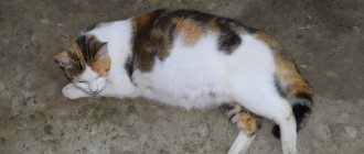
Mastocytoma in cats is treated based on where the tumor is located. It can affect both the skin and internal organs. They resort to radiation, chemotherapy or surgery. The tumor is predominantly malignant, but it can also be benign or with a high risk of metastasis.
Etiology of the disease
Mast cells are present in all body systems and take part in allergic and inflammatory processes. Mastocytoma is the formation of these elements, which has acquired a malignant nature. To this day, scientists have not identified the exact reasons that cause the transformation of benign mast cells into malignant ones.
The tumor can affect any organs, but is mainly localized on the skin. Diseases are often diagnosed in pets that are over 4 years old. Veterinarians note that Siamese cats are most susceptible to the disease.
Post-vaccination sarcoma in cats
Post-injection sarcoma in a cat
Feline post-vaccination sarcoma is a malignant tumor that appears in areas usually following subcutaneous or intramuscular injections. The tumors have a low metastatic effect but tend to recur locally unless excised with a very wide and deep excision. One of the distinguishing features is a latency of onset of months or even years between injection and tumor development, followed by extremely rapid growth from a growth point to a diameter of several centimeters within a few weeks. Injection site sarcoma develops at the injection site of vaccines, especially feline leukemia virus and rabies. Most sarcomas resulting from vaccination appear in the subcutaneous fat layer of the shoulder blades, along the dorsal and lateral rib cage, and in the thigh muscles. Vaccines containing adjuvants cause an acute inflammatory reaction at the injection site, which is the main trigger in the development of sarcomas.
Types of sarcomas:
- Rhabdomyosarcoma
- Myxosarcoma
- Chondrosarcoma
- Malignant fibrous histiocytoma
- Undifferentiated sarcoma
- Fibrosarcoma (most common and aggressive)
Etiology and pathogenesis
Vaccine injection typically causes an acute inflammatory response that varies in severity and duration depending on the vaccine and adjuvant. Presumably, post-vaccination fibrosarcoma results from inappropriate or extreme inflammatory or immunological reactions associated with the presence of vaccine components in the vaccination site, which led to uncontrolled growth of fibroblasts and myofibroblasts.
Localization:
- Shoulder blade area
- Thigh muscles
Metastasis is predominantly hematogenous, mainly to the lungs, especially when the tumor recurs.
Diagnostics
X-ray examination of a cat with injection site sarcoma
The diagnosis of injection site sarcoma in cats is relatively simple and is based primarily on clinical signs. Visual examination reveals dense, lumpy, clearly defined, partially encapsulated, mobile and slightly painful formations. As a rule, they are detected by owners at sizes from 2 cm. As a laboratory diagnosis, the method of fine-needle biopsy or biopsy through an incision is effective. To confirm the diagnosis and clarify the specific location of the tumor, they resort to X-ray diagnostics or computed tomography of the chest and the site of damage. Information about the general condition of the animal can be provided by complete clinical and biochemical blood tests, tests for FIV and FeLV. The average age of onset of post-vaccination sarcoma in cats is lower than that of non-vaccination sarcoma and begins at approximately 6-7 years, with a secondary peak at approximately 10-11 years. Typically, cat owners report sudden, rapid growth of the tumor. Anamnesis usually reveals that vaccination was carried out one to three months ago. Sometimes this time can be up to one year.
Differential diagnosis
The diagnosis is usually simple and fairly obvious because granulomas and other epithelial cancers such as basal cell carcinomas (often cystic in cats) have a slower growth rate.
Treatment
It is now recognized that the best chance of cure is offered by a multimodal approach that combines extensive surgery and radiation therapy.
Surgery
Cat after surgical removal of sarcoma
Surgery for post-vaccination sarcoma in cats is currently based on radiological and CT findings. The mass is removed, including healthy tissue 3-5 cm of macroscopically healthy tissue from the tumor and at least one fascia under the tumor mass. These criteria are not always easy to meet, given that the tumor is located in the interscapular region. Sometimes it is necessary to remove part of the spinous vertebrae, perform a partial scapulotomy, or complete removal of the scapula, remove part of the chest wall, or amputate a limb. In all cases, good, adequate pain relief is required.
Radiotherapy
Along with surgery, radiation therapy is the main treatment for feline injection site sarcoma.
Chemotherapy
Chemotherapy is used primarily to control metastases, but it can also be used before and after surgery to reduce the size of the tumor mass. Chemotherapy may be used if cat owners refuse radiation therapy.
Forecast
An integrated approach combining surgical and therapeutic methods can reduce the rate of recurrence at the surgical site by 41-44% within two years, while recurrence of metastases (mainly in the lungs) is about 12-24%. Median survival is 23 months, with median disease-free survival of 13 to 19 months.
Prevention of relapse
- Early diagnosis. The prognosis depends on the size of the tumor. Cats with tumor sizes of 2-3 cm have the best chance.
- Early terms of adjuvant chemotherapy using high-dose regimens and further administration of antimetabolites.
- Wide excision of the tumor using ablastics and antiblastics methods.
Prevention of occurrence
Given the iatrogenic etiology of the tumor, prevention plays an important role. Currently, it is recommended for any type of subcutaneous injection (a tumor may develop after an intramuscular injection, and then the diagnosis will be made later) such places as the lateral parts of the abdominal cavity, from the spine and the limb area. It is better to vaccinate in the distal part of the limb, taking into account that tumors can lead to amputation of the entire limb. This is more effective than removing the deep part of the abdominal wall. In any case, given that sarcoma can begin not only after an injection of a vaccine, but also after any injection of a drug, a veterinarian should give injections to animals in extremely necessary cases.
What symptoms indicate pathology?
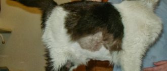
As the disease progresses, growths appear on the pet’s skin that have a dense consistency and a dark or pink tint. At this point, the animal’s fur begins to fall out and an ulcer appears. The skin on the scalp is often affected. If several tumors are observed at once, this condition mainly indicates that a well-differentiated histiocytoma has also affected the internal organs. Such symptoms pose the greatest danger to the cat’s life. Siamese cats that are less than 4 years old sometimes develop tumors on the skin; they resolve on their own within 2-3 months.
Bloating in a pet develops when internal organs are affected by formations.
If we talk about those situations where mastocytosis is localized on internal organs, then most often it affects the spleen. Moreover, this type of pathology is dangerous because metastases can spread to the liver, respiratory organs, gastrointestinal tract, lymph nodes and bone marrow. If we talk about the defeat of the latter, then systemic damage to the body, which in veterinary medicine is called mastoleukemia, cannot be ruled out. When internal organs are damaged, the following symptoms develop:
- severe weight loss;
- vomit;
- diarrhea;
- bloating;
- lethargy;
- breathing problems.
Treatment of histiocytoma in dogs
Treatment of this disease is possible only after an accurate diagnosis has been made. The most acceptable methods of treatment: excision (excision) or cryosurgery. If the tumor is inoperable due to its size or shape, the use of hormonal drugs is necessary. They are used systemically in an immunosuppressive dose or locally in the form of blockades. The essence of this technique is to introduce a large concentration of hormonal agents into the area of the tumor, which block the growth of the tumor and promote its regression. The prognosis is favorable. If systemic histiocytosis develops, a course of chemotherapy is prescribed and the prognosis worsens. The main drugs for chemotherapy are antitumor antibiotics of the anthracycline series and alkylating agents in recommended regimens.
Diagnostic measures
If pet owners suspect mastocytoma, it is important to contact a medical facility as soon as possible. At the appointment, the veterinarian will conduct a survey of the owners, during which he will find out how long ago the unwanted symptoms began. Then the doctor begins a visual examination of the animal and assesses the condition of the skin. To confirm a preliminary diagnosis, a general and biochemical blood test is performed, and a urine test is also required. Using the results, it is possible to determine whether the tumor has affected internal organs. Then an x-ray of the peritoneum and sternum is performed, as well as an ultrasound examination, which allows one to detect the localization of the formation and identify metastases.
The final stage of diagnostic measures is cytology.
Prevention of mastopathy
In order not to endanger the pet, the owner needs to take care of its health in advance:
- provide a balanced diet and dosed drinking to increase immunity weakened by childbirth and form proper lactation;
- carefully inspect and feel the milk bags of a nursing cat; if it is very swollen or painful, express;
- Trim kittens' claws to reduce the risk of nipple injury;
- timely deworming, getting rid of fleas, antiseptic treatment of scratches and injuries;
- stop using medications containing hormones and hormone-like substances;
- exclude contact between your pet and infected animals;
- Use products made from natural materials as a lounger.
If mastopathy is suspected, veterinarians recommend unscheduled mating. If the owner is not committed to caring for the offspring, the best solution is to sterilize the cat before the first heat. We must not forget about regular preventive examinations with a veterinarian. A simple procedure will save the owner’s nerves and the health of the four-legged household.
How is the treatment carried out?
Most often, the disease is treated by performing surgery on the animal.
The choice of treatment method for mastocytoma in cats is made by a physician and is directly related to the location of the tumor. Most often, surgical intervention is performed, during which a physician removes the tumor. To do this, as doctors from the Zoostatus veterinary clinic note, is required along with 3 centimeters of healthy tissue, which is not always possible. In addition, the possibility of relapse of the disease cannot be excluded.
Before the operation, the cat is prescribed injections of glucocorticosteroids, which can help reduce the size of the tumor. When test results indicate the presence of metastases near the excision border, the animal is prescribed additional treatment methods, for example, radiotherapy. It also helps in situations where it is impossible to completely eliminate the tumor during surgery. Chemotherapy is sometimes prescribed, but the effectiveness of the technique has not been confirmed.
general information
This is the name of a tumor formed by mast cells. They are formed in the bone marrow, but complete their maturation in peripheral tissues. Mast cells contain a lot of heparin, histamine and proteolytic enzymes.
Mast cells release these substances, attacking antigens, which can be bacteria, viruses, fungi, and other foreign bodies. They not only destroy them themselves, but also act as “guides” for lymphocytes and other cells of the immune system. They are very important and play a leading role in allergic reactions, wound healing and other physiological processes in the body.
Tumors arising from accumulations of mast cells can be malignant or benign. Very often they appear in the form of “broods”; local, single neoplasms are almost never found. Fortunately, malignant forms of mastocytomas are very rare in cats. Even such tumors, oddly enough, practically do not metastasize in these animals, and therefore, with timely treatment, the chances of recovery are very high.
Recovery prognosis
When the tumor is located exclusively on the skin, does not metastasize, and it is possible to remove it during surgery, veterinarians make a positive prognosis. However, if the tumor is observed in the internal organs or metastasis has occurred, the prognosis is not always favorable. Often, the doctor sends cat owners for a consultation with an oncologist, who offers additional therapy methods.
Veterinarians remind that detection of histiocytoma and mastocytoma in the initial stages of development makes it possible to carry out timely treatment and avoid serious consequences for the health of the animal. Therefore, it is important for owners to systematically take their pet for medical examinations and not self-medicate if unwanted symptoms occur.
Symptoms
Conventionally, mastopathy is divided into 3 types: fibrous (tissue grows in the gland and compaction occurs), cystic (cysts with liquid inside appear), diffuse (formations fill the cavity in the form of nodes).
External signs depend on the type, form, and neglect of the pathology:
- at first, a slightly enlarged gland (one or several) is painless, and the state of health is normal;
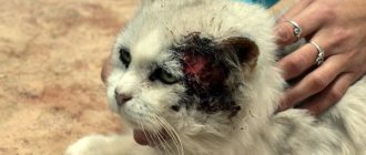
- As the inflammation develops, subcutaneous bumps are already felt in the area around the nipple, it will become painful, the female will meow and not allow the kittens to come to her, lick the nipples, sometimes to the point of severe damage, as in the photo:
- and then the body temperature rises (above 39 degrees), the lymph nodes enlarge, the nose becomes dry and hot, the mucous membranes turn red, ichor with pus is released from the nipples, the hind legs swell, the cat becomes lethargic and sometimes aggressive.
Diagnostics
- Fine needle biopsy - a needle is inserted into the tumor and a sample is taken for analysis.
- Complete clinical blood test, blood biochemistry and urine analysis. These tests help determine the animal's overall health and, in some cases, indicate whether tumors have spread to internal organs.
- X-ray. X-rays of the abdomen and chest help identify tumors and their spread within the body.
- Ultrasonography. May provide more detailed visualization of the tumor and help detect metastases.
- Biopsy - removal of the tumor and its subsequent examination.