Pulmonary edema in a cat or acute pulmonary failure is not an independent disease, but a complication of certain ailments of a cardiogenic or non-cardiogenic nature. It is accompanied by acute suffocation, progressing as the swelling increases. If veterinary care is late, the animal dies from asphyxia. In the article we will look at the causes of pulmonary failure in animals, answer the questions: why the disease is fatal, whether the pathology can be cured, what preventive measures exist.
Causes of pulmonary edema in cats
An attack of cardiac asthma can be triggered by the following factors:
- radiation sickness;
- bronchial asthma;
- drug overdose;
- hernia;
- neoplasms, lung tumors can cause compression;
- airway obstruction;
- exposure to toxins;
- traumatic injuries;
- swelling due to kidney disease;
- feline leukemia;
- congestive heart failure;
- pneumonia;
- feline infectious peritonitis;
- blood clot formation;
- parasitic infection;
- chylothorax;
- viral infection;
- anemia;
- overdose of anesthesia during castration/sterilization.
Abnormal accumulation of fluid in the alveoli
For your information! Most often, heart disease provokes the formation of edema.
A characteristic sign is that only the hind limbs can swell.
How to determine the disease
Epilepsy in cats: symptoms, treatment, how to stop seizures
Not only a medical professional, but also a person without special education can recognize the symptoms in time to notice that the lungs are swelling. Responsibility for the health of the pet lies primarily with the owners. They must carefully monitor the slightest changes in their pet's condition and behavior.
Even a brief description of the cat's condition, which can be done based on observations of it, will help the healthcare professional make a diagnosis and prescribe appropriate treatment.
Pulmonary edema can be diagnosed in any cat.
Causes of swelling:
- disorders accompanied by left ventricular failure;
- damage and prolonged pressure in the chest, pleurisy;
- liver and kidney diseases;
- allergy.
Cats of these breeds are hereditarily predisposed to heart disease:
- Maine Coon;
- Scottish fold;
- Turkish van;
- sphinx;
- rex;
- British Shorthair;
- American Shorthair.
Prevention
There are no special preventive measures to protect against pulmonary edema. Prevention consists of careful monitoring of animals that are at risk for cardiovascular and colds:
- Regularly undergo preventive examinations with mandatory vaccinations.
- Prevent the risk of allergic reactions.
- When purchasing food, be guided not by price, but by quality.
- Do not restrict the cat's movements. The more she runs and plays, the better for her health.
- Avoid contact with electricity and toxic substances.
- Avoid hypothermia and overheating.
- Do not let the animal outside unattended.
The main rule is simple - constant monitoring of the cat’s condition. This is especially true for owners of purebred cats. You need to pay more attention to your pets, and they will be healthy.
Pulmonary edema in cats is treatable provided that the animal is constantly monitored and timely contact a specialist if the slightest signs of the disease are detected. Relapses occur mainly in cats with heart disease or due to age. You should carefully monitor your pets to prevent a recurrence.
Types of disease
Anemia in cats: symptoms and treatment, causes
There are three options for the occurrence of fluid in the lungs of a cat; in any form, the alveolocapillary wall is damaged.
Pulmonary edema is divided into cardiogenic, non-cardiogenic and toxic.
Cardiogenic pulmonary edema is a complication of various diseases characterized by a rapidly developing decrease in the pumping function of the left ventricle or its filling with blood, leading to acute venous stagnation of blood. Causes:
- impaired contractility of the left ventricle (coronary heart disease, cardiomyopathy);
- severe dysfunction of the mitral or aortic valve.
Non-cardiogenic pulmonary edema
Non-cardiogenic edema is not associated with disorders of the cardiovascular system and is not caused by the presence of toxins in the body. Veterinarians identify the following diseases and causes of edema:
- bacterial or viral pneumonia;
- allergic reaction;
- kidney diseases;
- severe chest injury;
- foreign body of the upper respiratory tract;
- drowning;
- acute pancreatitis, fat embolism.
Toxic pulmonary edema is the body’s reaction to the penetration of toxic substances. There are several stages during the course of the disease:
- reflex (initial);
- latent phenomena, lasts 6–24 hours;
- clinical manifestations (cyanosis of lips, gums), lasts up to 2 days;
- reverse development lasts 4–6 days.
Toxic pulmonary edema takes 4-8 hours, but can last up to 24 hours.
Main causes of the disease
When analyzing the issue of any disease, first of all they talk about the causes of the disease in order to protect and prevent its occurrence in the animal. Pulmonary edema in a cat does not occur out of the blue and has certain factors for its occurrence. Let's take a closer look at what these factors are.
There are three main causes of the disease:
- Due to increased pressure in the capillaries, which occurs in the presence of heart disease (heart failure), or intravenous infusions. In this case, blood circulation inside the body is disrupted, and blood, penetrating through the walls of the capillaries, can enter the lung.
- A decrease in oncotic pressure occurs with the development of certain diseases.
- Inflammation of the lungs (pneumonia). In this case, the protein protection of the capillaries may be disrupted, which will provoke the spillage of liquid.
These are, so to speak, reasons associated with disturbances in the functioning of the body.
There are also other factors that can provoke pulmonary edema :
- electric shock. No one is immune from an accident, but you always need to monitor where and what the animal comes into contact with;
- Heat stroke can cause swelling. During the warm season, animals are especially susceptible to heatstroke. The cause of the blow may be being in a stuffy room, overheating, etc.;
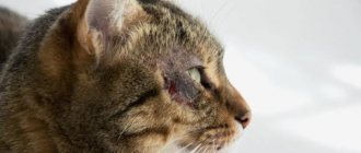
- any mechanical damage that may cause harm to organs;
- Snake bites can very often serve as a factor for the development of edema.
- Pulmonary edema in cats can also be divided into two types:
- Cardiogenic - associated exclusively with the presence of heart disease.
- Non-cardiogenic - this includes edema due to all other causes.
Symptoms
So that the owner can quickly respond to the presence of pulmonary edema in cats and immediately consult a doctor
, he needs to know and be able to see the presence of certain symptoms of a particular disease. It is important to know that there are procedures that, if poorly tolerated, can endanger internal organs. For example, if your pet has a predisposition to heart disease, anesthesia may have a negative impact on his body. When performing operations using anesthesia, monitor the cat’s behavior after the procedure.
And now more about the symptoms, what you need to pay attention to
:
1decrease not only in physical activity expressed by games, but also in general. The cat begins to move less and lie down more . React poorly to your interactions with her. This is caused by the fact that when there is a lack of oxygen, the cat experiences shortness of breath, thereby saving the animal energy to avoid discomfort; 2you may notice how the cat, for no apparent reason, begins to breathe with its mouth open , sticking out its tongue, like a dog. While dogs cool their bodies in this way, this is not typical for cats. Of course, sometimes cats can breathe with their mouths open after strenuous and intense exercise. But this lasts no more than two or three minutes. If this symptom appears in the absence of activity, you should worry; 3 change in the breathing process itself. A healthy pet should breathe fully from both the chest and stomach. If there is any pathology of the lungs, the animal will breathe only with its stomach ; 4 wheezing when breathing . When the airways are damaged due to fluid accumulation or swelling, the airways narrow. At the same time, when the animal inhales, you can hear whistles and wheezing; 5 cough . Cough can occur with a variety of diseases and does not always carry very dire consequences. Sometimes your pet may cough to clear the foreign body from the airways. With pulmonary edema, the cough may be accompanied by the presence of sputum and blood clots during expectoration; 6cyanosis is the symptom that is the most pronounced among all the others. With cyanosis, the mucous membranes take on a blue tint . This is due to a lack of oxygen.
Diagnostics
Knowing what symptoms an animal may experience with pulmonary edema, the owner has the opportunity to respond in time to the onset of the disease and seek help from a veterinary center. Under no circumstances should you attempt to diagnose yourself, because an incorrect diagnosis can worsen your pet’s condition. And pulmonary edema is directly related to breathing, which can lead to death if it is disturbed. Diagnosis should only be carried out by a qualified doctor
.
The most common types of diagnosis of pulmonary edema are radiography and listening to the lungs. Blood tests (general and biochemical) are also taken. When listening, as a rule, they try to establish the respiratory rate and the presence of extraneous noises and wheezing. X-rays show the presence of pathologies and changes in the lung tissues. If necessary, additional examination is prescribed to see the full picture.
The doctors at our veterinary center are highly qualified and have extensive experience. This can guarantee you the quality of the services provided.
Signs and symptoms of pulmonary edema in cats
Viral peritonitis in cats: symptoms and treatment
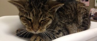
Fluid in the lungs of a cat causes severe shortness of breath due to a decrease in the lumen of the alveoli, infundibulae and bronchioles, as well as a decrease in their mobility and ability to expand. Animals stand, lie only on their chests, or take a sitting position.
Note! Physical signs of cardiogenic edema include tachypnea and respiratory failure. The heart rate is usually fast and the pulse is weak. In cases of biventricular heart failure, distension of the jugular vein may be observed.
Symptoms
What should alert the owner and become a signal that the cat is developing pulmonary edema:
- Loss of interest in food and games, lethargy, anxiety, restlessness.
- The cat tries to remove the fluid by coughing and swallowing.
- The animal has difficulty breathing: shortness of breath, breathing with effort, “belly”, rapid breathing.
- A cat breathes like a dog: with its mouth open and its tongue hanging out.
- While breathing, wheezing and gurgling sounds are heard.
- When coughing, blood may be observed in the liquid secretion.
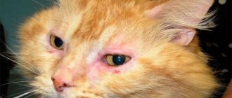
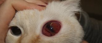
- The tongue and gums appear blue.
- Mucus is discharged from the nose.
- The cat tries to expand its chest and takes a pose with its limbs widely spaced.
- If the condition worsens, the position changes to a lateral recumbent position.
- Interruptions in heart rhythm - alternation of high and low pulses.
- A sharp drop in pressure and body temperature, cold extremities - collapse.
If one or more symptoms appear, you must immediately show your cat to a doctor. Pulmonary edema is a rapidly developing pathology that threatens the life of the animal. You can lose a pet in a few days or hours.
Stages of cardiac pulmonary edema in a cat
Pulmonary edema due to myocardial insufficiency develops rapidly. There are several stages of the process:
| Stage | Radiography |
| Pre-edema: increased blood flow in the vessels. | Strengthening of the pattern in the upper lobes of the lungs. |
| Interstitial edema: fluid in the perialveolar spaces. | Fluid in the interstitium, enlarged roots. |
| Alveolar edema: fluid in the alveoli. | Strengthening shadows in the roots of the lungs. |
Clinical signs and symptoms of non-cardiogenic pulmonary edema:
- tachycardia;
- cyanosis;
- cough;
- heavy shortness of breath.
First aid before going to the veterinary clinic
If there are signs of pulmonary edema, first aid must be provided to the animal:
- ensure patency of the upper respiratory tract;
- establish inhalation of oxygen from an oxygen cushion;
- If possible, give a diuretic such as furosemide;
- It is better to transport a cat to the hospital in a position lying on its side, but do not put it down forcibly (if it wants to sit, let it sit);
- The animal should be provided with complete rest, limiting physical activity.
What it is?
In general, such a disease does not exist in nature. This term refers to a set of symptoms that indicate severe damage to the entire respiratory, and sometimes circulatory, systems. Simply put, the effects of pneumonia that cause difficulty breathing, coughing and shortness of breath could very well be pulmonary failure. Speaking more generally, this term should refer to a condition in which the body receives an insufficient amount of oxygen due to dysfunction of the respiratory system.
Diagnostics in a veterinary clinic
Many veterinarians use chest x-rays to confirm the diagnosis of pulmonary edema. Once diagnosed, your doctor may recommend additional testing to determine the nature of the fluid and explore possible causes of the condition.
The doctor may insist on additional examination
Note! If the animal’s condition is serious, even before a detailed examination, treatment is prescribed aimed at stabilizing the condition.
Methods of laboratory and instrumental examination
- general blood analysis;
- electrocardiography, ultrasound examination of the heart;
- culture of sputum, urine;
- arterial blood gases;
- X-ray of the chest organs.
Treatment in hospital
For all forms of pulmonary edema, the diagnostic test of choice is a chest x-ray. It will demonstrate interstitial and alveolar infiltrates. The spread of infiltrates will help in identifying the etiology and treatment.
- To ease the work of the heart, reduce excessive blood pressure and blood supply to the pulmonary vessels, as well as to reduce cyanosis and shortness of breath, copious bloodletting is indicated.
- To improve heart function, cardiac medications are prescribed into a vein or under the skin.
- To reduce the permeability of cell membranes and blood vessel walls and to reduce the phenomena of sweating, a 10% calcium chloride solution is needed intravenously, and a 40% glucose solution is needed intravenously.
- In case of severe cyanosis, oxygen inhalation is prescribed.
- Rubbing the chest is indicated as a distraction therapy.
- If the heart function is satisfactory, atropine sulfate is used subcutaneously.
- Rest, fresh and clean air (not draft), nutritious and easily digestible food, and general skin massage are recommended.
- Bedridden patients are turned over to avoid hypostasis every 3–4 hours from one side to the other.
- You will have to treat not only the swelling, but also its root cause.
Atelectasis. Emphysema
Atelectasis. In newborn calves that die in the first days of life, congenital atelectasis of the anterior and middle lobes of the lungs is often observed. This happens due to insufficient respiratory movements in weak animals, when not all parts of the lungs receive air and the anterior and middle lobes are less ventilated; they do not straighten out and remain collapsed, like a fetus. Atelectasis can also be acquired as a result of blockage of the bronchial tube by exudate, a foreign body, a tumor, parasites, when the part of the lung located below the blockage does not receive air, and the previously existing air is absorbed, and the alveoli collapse.
The portion or areas of the lungs that are in a state of atelectasis are much thinner and smaller in volume compared to normal lung tissue. They are red because there are significantly more blood vessels per 1 mm3 than normal. The consistency is meaty, the pieces sink in water. Under a microscope, the lumens of the alveoli are almost invisible. The interalveolar septa are closely adjacent to each other. There is a small amount of mucus in the bronchi.
Emphysema is the filling of the lungs with air. Occurs after the movement of animals, as well as during pneumonia; in the latter case it is called compensatory. Emphysema can be alveolar, interstitial, or bullous. With alveolar emphysema, the lungs are larger in volume and lighter in color than normal, anemic, with a fluffy consistency. When cutting, a faint cracking sound is heard. Under a microscope, the cavities of the alveoli are wide. The interalveolar septa are thinned, torn in places, and several alveoli form a common cavity. The respiratory epithelium is flattened. There is little blood in the capillaries (Fig. 25).
In chronic bronchitis, alveolar emphysema can be chronic, with interalveolar septa atrophying, the number of elastic fibers decreasing, and interstitial connective tissue increasing. As a consequence of emphysema, enlargement or hypertrophy of the right ventricle of the heart appears. With significant ruptures of the alveoli, blisters are formed that are visible to the naked eye - blistering emphysema. The size of the bubbles varies, often reaching more than 2 cm in diameter. Under a microscope, the walls of the bubbles are fringed, with fragments of alveoli. Interstitial emphysema is observed with abnormal body position of the animal, traumatic injuries, and severe coughing. In this case, the alveoli rupture and communicate with the interlobular connective tissue. The air, spreading through the cracks of the interstitial tissue, pushes its bundles apart and forms
Rice. 25. Chronic alveolar emphysema.
cavities. The interstitial tissue with air bubbles in it often becomes like beads.
Venous congestion in the lungs is noted with weak heart function, difficult outflow of blood from the lungs, valve defects of the left half of the heart, and flatulence. When opening the lungs, they are dark red on the surface and on the cut; a significant amount of blood flows from the cut. Large vessels are filled with loosely coagulated blood. When pressed, a reddish, slightly foamy liquid is released. Under a microscope, the interalveolar septa are thickened, the blood vessels are dilated, and filled with red blood cells.
Prognosis for recovery
When the animal is young, with a strong immune system, recovery is possible, but there is a risk of relapse. Weakened, elderly pets, as well as kittens, usually experience the disease more severely, recover more slowly, and the risk of complications is higher.
For your information! If repeated swelling is observed over several months, this leads to death in almost 100% of cases.
When pulmonary edema occurs as a result of trauma (such as a head injury or strangulation), it can be permanently treated.
Treatment method and prognosis
There is no single treatment regimen for pulmonary edema; it will depend on the cause that caused the pathological process and, accordingly, is aimed at eliminating it.
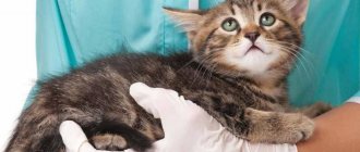
If the condition is critical, the animal must be given first aid. To do this, the cat is given an intramuscular injection of a drug that relieves swelling and eliminates hypoxia - Prednisolone, Dexamethasone, Diprospan, Hydrocortisone.
Oxygen replenishment is carried out by placing the pet in a pressure chamber, using an oxygen mask, and ventilation procedures.
To remove excess fluid, diuretics are used (injected or orally).
To eliminate the infectious diseases that caused the swelling, a course of antibiotic therapy is prescribed. For viral etiology of edema, antiviral agents are indicated.
During illness, cats usually experience severe stress, so they are prescribed sedatives.
If, after carrying out the necessary medical procedures, the cat’s condition has stabilized and his health is no longer in danger, the fluffy cat is sent home to recover.
If all first aid measures and further treatment were carried out correctly, the prognosis is positive. However, if a sick pet is brought to the clinic too late, the risk of death is very high.
Preventive actions
Prevention is based on preventing edema in patients at risk. It includes:
- restriction of activity of patients with severe pathology of the kidneys and liver;
- timely treatment and diagnosis of arrhythmias, heart failure, hypertension;
- compliance with the doctor’s prescriptions and recommendations to avoid relapse of the disease.
Following the veterinarian's instructions will help avoid relapses.
The respiratory system is the most vulnerable place in cats. Therefore, any breathing disorder requires diagnosis and treatment. No disease that causes shortness of breath can be cured at home. Only emergency care can be provided to your pet at home.