A swollen and hard belly is a rather alarming symptom that accompanies many pathologies. One of them is ascites, or dropsy, which is often found in cats. It is not an independent disease and develops as a complication of pathological processes affecting the abdominal organs. To eliminate it, it is very important to determine the root cause, since relieving existing symptoms will only bring relief for a limited time.
What is abdominal ascites in cats and cats?
Normally, there is a small amount of fluid in the abdominal cavity of cats and kittens. It washes the internal organs, facilitating the movement of intestinal loops and helping the immune system. With its help, toxins and harmful microorganisms smoothly leave the body without having time to penetrate healthy cells and cause significant harm.
As a result of impaired absorption and blood circulation, the amount of fluid increases due to transudate or exudate. Unusual volume leads to increased pressure. The internal organs, shrinking, gradually lose their functionality, so without timely help the animal may die.
How to diagnose ascites
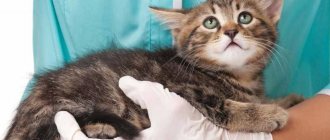
Diagnosis of the disorder is carried out by a veterinarian.
Diagnosis of ascites
The following methods are used to determine pathology:
- palpation of the cat's abdomen;
- general analysis of urine and blood;
- X-ray;
- Ultrasound of the abdominal and thoracic cavities;
- biopsy and analysis of fluid from the abdominal cavity.
Additional diagnostic testing is performed to determine the underlying disease. Methods depend on the suspected disease.
Causes of fluid accumulation
The risk group includes animals with congenital and acquired pathologies, which are the main causes. These include:
- Diseases of the cardiovascular system. Heart failure disrupts natural blood circulation, which causes an increase in internal pressure and increased vascular permeability.
- Abdominal injuries that provoke the development of swelling, inflammation and internal bleeding.
- Liver and kidney diseases. Disturbance in the functioning of the urinary system organs leads to acute intoxication with decay products and inflammation.
- Parasitoses and infections of various etiologies. Toxins released by pathogenic microorganisms irritate the receptors of the mucous membrane. To destroy them, the body launches an inflammatory reaction, accompanied by abundant production of exudate.
- Disturbances in the functioning of the endocrine system. Provoking factors are diabetes, excess weight and high cholesterol.
- Malignant neoplasms. Cancer cells clog lymphatic vessels, disrupting natural circulation and increasing the permeability of the walls.
Sometimes the owner is to blame for the excess amount of fluid in a cat’s stomach because he does not pay enough attention to the balance of the diet and the frequency of feedings. Complications can occur due to insufficient amounts of proteins or vitamins, as well as sodium abuse.
Abdominocentesis
Aspiration of fluid in the encysted form of ascites, when the effusion is localized in small areas, should be carried out under ultrasound control, which is not necessary with significant fluctuating volumes of fluid. In the latter case, the ascitic fluid sample is aspirated blindly using a needle or, more preferably, using a needle catheter. For diagnostic purposes, a small volume of peritoneal fluid (1-5 ml) is required. The fur in the puncture area must be shaved and the skin treated with an antiseptic; infiltration anesthesia can be performed.
To perform an abdominal puncture, the animal is usually fixed in a lateral lateral position, since this position causes the intestinal loops to be displaced from the abdominal wall. According to theoretical data, when puncturing the abdominal cavity, the needle is best passed through the virtually vascular-free linea alba, caudal to the umbilicus and below the crescentic layer of adipose tissue.
However, with significant ascites after abdominocentesis, the animal may experience constant leakage of ascitic fluid from the puncture site. In such cases, it is recommended to puncture the abdominal cavity away from the midline of the body, since the muscle tissue after puncture will help close the resulting hole. Diagnostic peritoneal lavage is performed if the volume of ascitic fluid, even under ultrasound guidance, is too small to allow sampling to determine the composition of the effusion (see BSAVA Manual of Canine and Feline Emergency and Critical Care).
What does ascites look like in a cat: main symptoms
Dropsy in cats develops gradually and responds well to treatment in the early stages. A rapid increase in alarming symptoms and a sharp deterioration in condition are typical for pets with weakened immune systems.
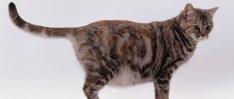
One of the main signs of pathology is an enlarged abdomen. It swells and loses its elasticity. When pressing inside, you can feel the movement of water.
These symptoms are similar to ordinary overeating, so to confirm your guesses, do a small test. Lift the animal up, holding it by the armpits. With stagnation, all the contents will rush down, giving the stomach a pear-shaped shape. After placing the cat on four paws, the shape will again become round.
In addition to changes affecting the peritoneum, it is necessary to highlight several more signs:
- yellowing of mucous membranes;
- dulling of fur;
- swelling of the limbs, groin, chest and ears at the base.
All other symptoms will depend on the cause of the illness. In addition to lethargy, apathy and poor appetite, the pet may experience vomiting, stool disturbances, urinary retention, fever, shortness of breath and cough. The advanced form is accompanied by severe pain, so the mustachioed pet should be taken to an appointment at the veterinary clinic before they appear.
Good to know
- Sialocele
- Short bowel syndrome
- Irritable bowel syndrome
- Biliary vomiting syndrome
- Sclerotic encapsulating peritonitis
- Cececocolic intussusception
- Special feeding method
- Gut protectants
- Stomatitis
- Strongyloidiasis
- Tenesmus
- Digestibility and Absorption Tests
- Trichomoniasis
- Cytological analysis of stool
- Intestinal strangulation
- Congenital weakness of the esophagus
- Ulcers and erosions of the gastrointestinal tract
- Physical examination
- Hiatal hernia
- Chronic gastritis
- Septic peritonitis
- Schistosomiasis in dogs
- Sialadenitis, salivary gland necrosis
Diagnostics in a veterinary clinic
To make a diagnosis, it is necessary to verify the presence of the suspected pathology and find out the cause of its occurrence. For this purpose, the following studies are used:
- urine analysis to determine pathologies of the urinary system;
- blood biochemistry, revealing the presence of toxins that poison the liver;
- Ultrasound confirming the presence of effusion and its exact volume, the functionality of internal organs and the presence of tumors;
- X-ray, which helps to detect the consequences of mechanical injuries and monitor the situation not only in the peritoneum, but also in the chest;
- puncture (puncture with collection of a biological sample) used to determine the composition of the fluid (exudate or transudate).
Also, the veterinarian must palpate (feel) the abdomen and collect anamnesis. After receiving the results of all studies, individual treatment is selected for the mustachioed patient.
Types of ascites and when they occur
In the practice of a veterinarian, the following types of ascites are encountered, developing when:
- malignant tumor. Happens in 80% of cases due to liver tumors or multiple metastases in other organs;
- pancreatitis, pancreatic tumors;
- injuries;
- fibrosis, cirrhosis of the liver;
- portal vein embolism;
- presence of portocaval shunts.
Often the pathological condition develops with increased pressure in the vena cava. Also, with intestinal perforation, infected ascites occurs, and its bacterial form turns into peritonitis due to the penetration of gram-negative bacteria into the fluid. Dropsy is diagnosed with feline infectious peritonitis.
Is hydrops of the abdominal cavity curable in cats?
For your pet to recover, the underlying disease must be eliminated. If it is diagnosed in a timely manner and can be treated, the prognosis is positive.
If the pathology is incurable or detected too late, then treatment is reduced to maintenance therapy. It helps prolong life, but does not eliminate the problem. This situation is typical in oncology, cirrhosis and advanced peritonitis.
If the cause of the disorder lies in an immune failure, then the likelihood of relapse is high. In this case, the animal will have to regularly take anti-inflammatory drugs and diuretics, and also visit a veterinarian to pump out excess transudate.
Type of ascitic fluid
- Study Results
- Transudate of the blood OCA Moderate (microcytic) anemia in pSS
- HD serum analysis Hypoalbuminemia (
- Increased liver enzyme levels in liver disease
- Bile acid levels are elevated in pSS
- Urinalysis Proteinuria in NPP
- Protein ratio; creatinine in urine The value is increased with NBP
- Determination of Iα1-P in feces The value is increased in EPD
- Visual methods for examining the abdominal cavity
- Liver sizes
- PSS
- Portal venography Portal vein obstruction, PSS
- Chest X-ray Hydrothorax
- Modified transudate
- TCA of blood Moderate (microcytic) anemia in liver diseases
- Inflammatory picture in the leukogram in septic peritonitis
- Bile acids Levels are elevated in liver disease
- Abdominal ultrasound Primary liver diseases
- Congestive secondary liver diseases
- AV fistula of the liver
- Liver arteriogram AV fistula
- Diagnostic laparotomy Liver biopsy
- Detection of neoplasm
- Chest X-ray Heart disease
- Type of ascitic fluid
- Study Results
- Modified transudate
- Ultrasound of the heart Cardiac tamponade
- DKM
- Right-sided heart failure
- ECG Cardiac tamponade
- Right-sided heart failure
- Angiography Pathological changes in the CPV
- Exudate of OKA blood IPC: neutrophilia, lymphopenia
- Inflammatory picture in the leukogram with pancreatitis
- Serum globulins The level is increased with IPC > 70 g/l
- Cultivation of ascitic sample Septic peritonitis
- Serological test for coronaviruses Indicates that the animal has had a coronavirus infection (but does not allow a diagnosis of IPC)
- In the blood serum and ascitic fluid, the level of amylase and pancreatic lipase may be increased and the IRI and TPI may be increased
- Pancreatitis (the most specific test is to assess
- IRI in blood serum. The sensitivity and specificity of ascitic fluid testing is unknown)
- Ultrasound Pancreatitis
- Diagnostic laparotomy Identification and elimination of the cause of septic peritonitis
- Detection of carcinomatosis
- Blood Tests to assess blood clotting (OSPT, APPV, PRF, PIVKA, d-dimer level)
- Disorders of the blood coagulation system
- Assessment of the animal’s condition from the point of view of the development of shock
- Bleeding due to injury
- Rupture of hemangioma/hemangiosarcoma
- X-ray of the abdomen New growths of the spleen or liver
- Chest X-ray Metastasis
- Ultrasound neoplasm of the spleen or liver
- Diagnostic laparotomy For bleeding due to trauma - lack of response to infusion therapy
- Detection and surgical removal of the tumor
- Bile Diagnostic laparotomy Identification of the area of bile leakage and surgical treatment
- Lymph Vascitic fluid cholesterol level is lower than in serum
- Lymph detection
- Triglyceride levels in ascitic fluid are higher than in serum
- Lymph detection
- Lymphangiography Detection of area of lymphatic leakage or lymphatic vessel obstruction
- Diagnostic laparotomy Identification of the area of lymph leakage followed by ligation of the vessel or removal of part of the vessel with obstruction
- Urine Creatinine concentration in ascitic fluid is higher than in serum
- Detection of the presence of urine in the abdominal cavity (urea can penetrate the peritoneal membrane, so assessing its concentration in ascites is less informative)
- Intravenous urography
- Cystography
- Identifying the area of urine leakage
- Diagnostic laparotomy Identification of the area of urine leakage and surgical treatment
^Top
Therapy methods
In addition to eliminating the root cause, treatment of abdominal ascites in cats involves relieving the main symptoms and removing accumulated fluid. In most cases, therapy is limited to taking medications, but sometimes it is necessary to resort to surgery.
Traditional drug treatment
The basis of drug treatment are diuretics (diuretics), which eliminate congestion by stimulating urination. All other drugs will depend on the diagnosis. These may include:
- analgesics necessary to suppress acute pain;
- cardiotonics that increase heart rate;
- antiemetics, weakening the gag reflex;
- potassium chloride and vitamin K, which reduce the permeability of blood vessels and strengthen their walls;
- antipyretics and sedatives;
- cephalosporin antibiotics that fight infection;
- colloidal solutions that increase the percentage of protein;
- immunomodulators that protect against secondary infection.
It is recommended to take all medications strictly as prescribed by your veterinarian. Incorrect dosage calculation or ending treatment too early can result in worsening of the condition and even death.
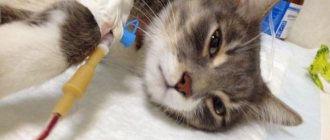
Drainage and surgery
Pumping out using puncture of the abdominal wall is resorted to in case of ineffectiveness of drug therapy or when there is dangerously high pressure on the internal organs. The procedure is carried out under anesthesia and repeated at least 2 times a week. This frequency and the need to use an anesthetic increases the risk of complications.
Surgery may be required if there is internal bleeding due to injury or oncology, as well as with purulent or necrotic processes. Depending on the problem, the surgeon excises pathological tissues and restores the integrity of damaged organs.
Folk remedies and help at home
The use of traditional medicine is permissible only as additional therapy and under the strict supervision of a veterinarian. Infusions and decoctions cannot eliminate the disease itself, but will alleviate its symptoms.
Self-medication using recipes found on the Internet or received from friends is unacceptable. Herbs often cause allergic reactions and are not always compatible with doctor-approved medications. Such help can result in the death of your pet, so do not try to solve the problem on your own.
If you find yourself far from the city and cannot get to a veterinary clinic in the near future, prepare an infusion of birch leaves. Pour boiling water over them in a ratio of 1:10, let it brew for 6 hours and strain. Give the resulting medicine twice a day, 1 teaspoon.
Despite the harmlessness of such an anesthetic, it is better to contact your doctor at least by phone and clarify the safety of the actions you are performing.
Nutrition correction and diet
Once the condition has stabilized, hydrops of the abdomen in cats is treated at home. In addition to taking medications, it is necessary to review your current diet:
- The amount of water and salt is kept to a minimum.
- The daily menu is adjusted in favor of meat and dairy products enriched with proteins. Twice a week, chicken, turkey or beef can be replaced with sea fish.
- The lack of vitamins and minerals is compensated by special additives to dishes.
If your pet suffers from diarrhea, then to strengthen the stool, he needs to be given rice porridge or a decoction. It is recommended to boil or steam meat and fish. This processing method will make it easier to digest.
When dry feeding, the usual food will have to be changed to veterinary food. Your doctor will help you decide on the exact brand, since a sudden switch to a new manufacturer can cause digestive upset.
Features of kitten treatment
Ascites in a kitten is treated according to general recommendations with minor differences. They consist of lower dosages and a ban on the use of immunomodulators. The first feature is explained by lower weight, and the second by the need to develop independent immunity. Taking such drugs for up to 1 year can lead to a malfunction in the immune system, resulting in the inability to distinguish healthy cells from infected ones.
Diagnosis of the disease
Making a diagnosis based only on external signs is wrong. This is why a sick cat needs to be taken to the vet. The specialist will make his verdict only after the animal has been examined and taking into account the test results. This may require:
- palpation and tapping;
- medical history (it is important that the cat owner can provide the doctor with as much information as possible about the diseases the pet has suffered);
- biological blood and urine tests;
- ultrasound examination of the abdomen;
- abdominal x-ray;
- laparoscopic examination (rarely used);
- computed tomography (practised only in elite veterinary clinics);
- fluid collection using puncture.
Examination of accumulated fluid is one of the most important measures in diagnosing ascites. Only from this material can one understand what caused the dropsy. It is a mistake to believe that water can be pumped out independently, although the intake is made through a puncture in the stomach. Firstly, the animal must be immediately given an antibiotic so that when the fluid is pumped out, an infection does not occur. And secondly, this procedure is only possible with the use of anesthesia. In order for the cat to endure the pumping itself, as well as anesthesia, such an event should be trusted only to the best veterinarian.
For diagnosis, it is necessary to at least conduct a study of ascites fluid (cytological study, PCR test for viral peritonitis).
Maria Lvovna Soloshek, veterinarian
https://www.zoovet.ru/forum/?tid=7&tem=821877
An X-ray of the abdominal cavity is needed in order to understand what abnormalities there are (increased size or incorrect position of organs, whether they are compressed, etc.)
I heard that not all veterinary clinics are equipped with special equipment (tomograph, x-ray, etc.). Many cat owners turn to several clinics at once for specific tests, and then with all the collected papers they go to their veterinary clinic. I once saw a message on some forum that a cat with ascites was abandoned. The veterinarian simply told the owner that the animal had ascites, there was no cure for it, and he would certainly die. But the owner of the poor cat did not give up on her friend, she took tests at different veterinarians, and then turned to a good specialist. The cat was cured (however, it later turned out that the first clinic simply did not have the necessary equipment).
How to distinguish it from other diseases with similar symptoms
If it becomes clear that the animal has fluid in its stomach and not something else, then this also may not only indicate ascites. For example, fluid accumulates in the abdominal cavity during exudative peritonitis. Exudative (wet) peritonitis is inflammation of the abdominal wall caused by infection. With peritonitis, ascites fluid may accumulate in the cat's abdomen, but the symptoms will vary:
- High body temperature (with ascites it is normal).
- Painful reaction to touching the belly (cats with ascites calmly respond to stroking).
- Rapid development of the disease - from 1 to 6 hours (ascites develops slowly - sometimes a week or more).
- Enlarged lymph nodes.
- Different chemical composition of the liquid.
With exudative peritonitis, the fluid contains more protein and leukocytes.
Video: how to do an ultrasound when diagnosing ascites
How to suspect pleural effusion?
Signs of the presence of pleural effusion are inspiratory dyspnea - rapid breathing with difficulty exhaling and rapid heartbeat - tachycardia. There may also be signs of cardiac and respiratory failure - cyanosis of the mucous membranes of the oral cavity and tongue. If these symptoms are present, it is imperative to examine the animal to confirm the presence or absence of pleural effusion and to determine the reasons that provoked it.
Since the accumulation of fluid in the chest cavity causes compression of vital organs such as the heart and lungs, it is necessary to remove this fluid as quickly as possible. The amount and nature of the fluid may vary, which determines the severity of symptoms.
In our clinic, there were cases when it was necessary to remove 150-180 ml of fluid from the right and left halves of the chest cavity, respectively, and in total it reached 350 ml. In such conditions, due to strong compression, the work of the animal’s heart and lungs is significantly hampered, creating a life-threatening situation.