Lentigo simplex: dangerous or not skin defect
The formation of lentigo (lentigo simplex) is associated with the presence in open areas of a large number of melanocytes, cells responsible for the production of melanin, which colors the skin pigment dark. The more there are, the darker the pet’s color.
This is called “acromelanism” and can be observed in:
- Burmese cats;
- Siamese;
- Himalayan
Kittens are born pure white, but under the influence of air temperature the tail, head and paws change to a dark color. It is noteworthy that the lower the air temperature, the darker the color will be. This is due to tyrosinase, an enzyme that is involved in the synthesis of melanin and has a temperature dependence on the cat's external environment.
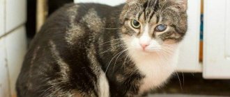
Vitiligo in cats
“My name is Scrappy. I was born in 1997 completely black, and at the age of 11 I began to turn white. I think I got this color because of vitiligo."
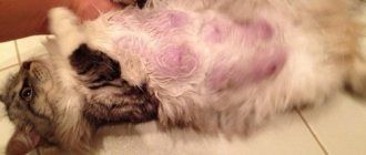
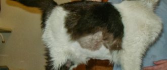
The owners began to notice white spots on the cat’s fur, which over the course of several years completely changed the pet’s appearance.
Vitiligo is called a disease, although many experts consider it a “feature of the body”: changes in skin color or hair matrix do not harm overall health. The cause of vitiligo is considered to be the disappearance of melanin. Most often, the process is irreversible: treatment can stop the appearance of spots, but a way to restore the affected areas has not yet been found. The disease occurs in both humans and animals.
This is what the cat looked like at the age of five and now.
And a few more photos of the marbled cat:
Sources used: pikabu.ru
YOU MAY BE INTERESTED in: Black cumin oil treats vitiligo Vitiligo after 40 years
Pathological conditions and their danger
Lentigines look like flattened, hyperpigmented macules; their appearance on the skin is associated with the accumulation of melanocytes (melanosomes) in one place. An examination by a dermatologist at the RosVet VC is required to differentiate from melanomas, dark plaques caused by papillomavirus.
Hyperpigmentation of the skin can be associated with a severe inflammatory process on the skin; this most often occurs in dogs, but in cats it occurs with atopic dermatitis or pemphigus foliaceus. The color of the skin changes to dark in places of constant mechanical irritation associated with the constant licking of a limited area of skin, friction of the collar on the neck (flea or regular, if too tight).
Sometimes pigmented areas appear where the skin has been damaged by a fungal or bacterial infection, and they can cause light areas of the coat to turn a reddish-rusty-brown color.
VPV (viral pigmented plaques) in cats are caused by the action of papillomavirus, skin lesions can look like dark macules, so it is important to accurately determine the nature of the neoplasms.
Important! A consultation with a veterinary dermatologist is necessary to rule out the malignant nature of hyperpigmentation. The course of ISP plaques is unpredictable and they can either disappear completely or degenerate into squamous cell carcinoma.
According to the observations of doctors at the RosVet Center, owners have a lot of myths about changes in coat or skin color due to certain components of a cat’s diet. It is believed that beets and carrots can give a reddish or yellowish tint to the coat in light-colored individuals. This is fundamentally wrong; root vegetables do not change color in any way.
In dachshunds, acanthosis nigricans is not caused by genetic abnormalities, but by a consequence of the dog having atopic dermatitis or systematic rubbing of the skin area, or secondary infections.
What needs to be distinguished from?
If spots similar to pigmentation are detected, you should show your pet to a veterinarian to rule out papillomavirus skin lesions.
A condition where a cat's paws, ears and tail tip are darker than the overall color is called acromelanosis. It is explained by the dependence of melanin synthesis on temperature. It is necessary to exclude diseases such as allergies, ectoparasites, and hormonal disorders. Cats, like people, have papillomavirus skin lesions. In the initial stages, they can masquerade as hyperpigmentation. The course of the disease is unpredictable. The formation may disappear on its own without a trace, or it may develop into a malignant tumor. If you have doubts about your pet's health, it is better to visit a doctor. In controversial cases, the veterinarian will take a biopsy (tissue sample) from the pathological focus to clarify the diagnosis.
The skin defect has nothing to do with melanoma and is not predisposed to malignancy.
There are cases when pigment spots form against the background of an inflammatory process of various etiologies. They are often located in places of mechanical friction when a bacterial, viral or fungal component is attached. At the same time, the color of the coat changes and becomes dull. In other cases, hyperpigmentation is associated with areas of baldness (alopecia). In these places, the dermis becomes dark because it tans in the sun.
Clinical signs of lentigo in cats
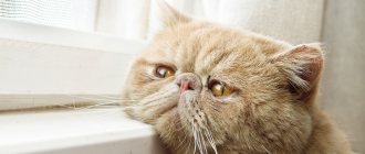
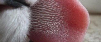
Lentigo in cats in the photo looks like “freckles” abundantly sprinkled on the nose, edges of the lips and oral mucosa. At first they are small, resembling black splashes of paint, but with age they will gradually increase in size and become numerous. But they remain clearly defined and do not blur together.
The surrounding skin and mucous membranes remain normal, there is no peeling, redness or inflammation. The color also does not change. The cat is not bothered by lentigo, there are no lifestyle changes, ailments, lack of appetite or difficulties with breathing and eating food.
The size of the spots varies from 1 mm to 1 cm; the older the animal, the larger the spots, but in extreme old age they can become lighter.
The owner who discovers lentigo in a cat should visit a veterinarian with the pet and make sure that the “freckles” are safe, since neoplasms with similar clinical signs can be the result of other pathologies and pose a danger.
If necessary, a dermatologist at the RosVet VC will take a biopsy (a piece of the tumor) and send it to the laboratory to rule out melanoma.
Causes
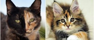
Lentigo is a condition of the skin and mucous membranes of an adult animal, which is characterized by the formation of dark spots of a certain localization. The causes of the phenomenon have not been established, but the hereditary factor of its development has already been confirmed. Lentigo is more common in dogs than cats. The latter are carriers of orange, cream or tortoiseshell coats.
The cosmetic oversight is harmless, so no treatment is necessary.
How does the defect appear?
During intrauterine development, cells of a neurogenic nature (melanocytes) enter the skin, iris, mucous membranes, and other tissues. These cells form the pigment melanin, which is deposited in the deepest layers of the epidermis and in the hair follicle. The presence of a coloring pigment determines the color of the coat and skin. There are only 2 pigments:
Only two pigments are responsible for the color of the coat - eumelanin (black) and pheomelanin (red).
- Eumelanin - carries a brown or black color.
- Pheomelanin is responsible for the red color.
As a result, 4 basic colors are distinguished: white (no pigment), black, orange and brown. The rest of the color palette is represented by shades of the basic ones. The accumulation of a large number of melanocytes in certain areas of the integument explains limited hyperpigmentation, which is called lentigo. A large affected area with multiple foci is lentigo profuse, and if there is one or several spots, then lentigo simplex.
Features of pigmentation
- Localization - on the lips, gums, eyelids, nose, sometimes on the paws and conjunctiva of the eyes.
- Appears before the age of one year and can progress.
- Appearance - small black dots, similar to freckles with clear boundaries or confluent dark spots (macula).
- Flat or slightly raised above the surface of the integument.
- They do not cause pain, itching, burning and do not affect the cat’s vital functions.
- The skin and mucous membranes around the defect do not change.
- Over time, pigmentation weakens.
Treatment of lentigo and prognosis
Such freckle spots are not a disease. They are a purely cosmetic defect in ginger cats, which some owners consider to be a highlight, since pets with “freckles” have a rather funny appearance. There are no known cases of disqualification of such animals at exhibitions.
Conclusion! Lentigo is not dangerous, harmless and does not require treatment. It is only important to rule out a different nature of the formations by visiting a veterinary dermatologist.
If you have any doubts about your cat’s pigmentation on any part of the body, do not hesitate - call +7 (495) 256-11-11, 24 hours a day. Make an appointment with a dermatologist, bring your pet for examination and rule out dangerous diseases.
Vitiligo in cats and dogs
Description and reasons
Vitiligo is an acquired skin disease characterized by the destruction of melanocytes and accompanied by melasma (skin discoloration) and/or leukotrichia (hair discoloration).
The exact causes of vitiligo in cats and dogs have not yet been determined; there are several theories with multiple pathophysiological mechanisms. In humans, vitiligo is a multifactorial disease in which various chromosomal loci (at least 16) may be responsible, most of them responsible for regulating the immune response, and some of them are also associated with other autoimmune diseases. The study of the pathogenesis of vitiligo in cats and dogs is less advanced than in humane medicine, however, when examining 17 dogs of the Belgian Tervuren breed, antibodies to melanocytes were found (they were not found in healthy individuals). Also, antibodies to melanocytes were discovered in a study of vitiligo in a small number of Siamese cats. Overall, scientists have concluded that the development of vitiligo is caused by certain genetic abnormalities that make melanocytes more susceptible to the effects of antibodies and oxidative damage from free radicals.
Clinical signs
Vitiligo is quite rare in dogs, and even rarer in cats. Vitiligo has been described in dogs, with possible hereditary transmission in breeds such as the Belgian Tervuren, Rottweiler, and Old English Cattle Dog. Cases of vitiligo have also been described in dog breeds such as collie, Doberman pinscher, giant schnauzer, bullmastiff, Newfoundland, German cattle dog and dachshund with the development of diabetes in adulthood. There is speculation that some cases described as idiopathic leukotrichia or nasal depigmentation are actually forms of vitiligo, so the list of predisposed dog breeds may be expanded. In cats, vitiligo has only been described in Siamese breeds, but it is believed that the previously described disease periocular leukotrichia is also a form of vitiligo. In Siamese cats with vitiligo, the disease has a slight sex predisposition to develop in females.
Techniques to improve pigmentation
The struggle for the ideal coat color is mainly faced by those who exhibit their young pets for the purpose of participating in breeding. Special shampoos and conditioners have been developed for each color to enhance the natural color of the coat. This type of washing is especially important for white cats, which should not have even a hint of yellowness.
We suggest you read: Is it possible to sterilize a pregnant cat?
For white and black individuals, special coloring powders are used before the exhibition. They can be dry or liquid. For especially problem areas in white cats (chin, tear ducts), gels with a whitening effect have been developed:
- All Systems;
- Super whitening gel.
Tinting is a harmless trick by breeders, giving an effect for no more than a day. To correct color for a long time, special nutritional supplements are used.
Suitable for red fur:
- complex Beaphar Algolit with seaweed (Beaphar Algolit Vitamin{amp}amp;mineral food supplement);
- GAC-carotene – granules from pure carrots containing carotene – provitamin A.
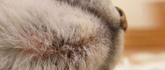
Feline acromelanosis
In cats of some breeds, the coat color is regulated by special heat-labile enzymes. In other words, the synthesis of melanin in such animals depends on the ambient temperature. Heat promotes the appearance of light hair, and cold promotes dark hair.
Breeders should know this feature when preparing their pets for an exhibition. Changes in color under the influence of temperature are typical for cats with “colored” paws:
- Siamese and Thai;
- Burmese;
- Himalayan;
- Balinese and some other breeds.
It is known that in these breeds the darkening of the coat increases with age, which is especially noticeable in some colors (color point).
Hypopigmentation (lightening)
There are several genetic diseases in cats that result in a lack of pigment in the hair and skin. They are quite rare and cannot be treated:
- In cats with a bluish-smoky color and yellow eyes, this is Chediak-Higashi syndrome. This disease causes not only discoloration of the coat, but also partial albinism of the eyes, photophobia and cataracts.
- Siamese cats (more often females) have vitiligo. In young animals, progressive patchy depigmentation of the nose, lips, eyelids, paw pads, and perianal area is observed. Although these cats cannot be used for breeding, vitiligo does not prevent pets from leading full lives.
Acquired lack of pigment can occur under the influence of any factors that destroy melanocytes:
- injuries;
- burns;
- frostbite;
- or fungal skin infection;
- use of certain medications (glucocorticoids).
Dangerous cancers - lymphoma, squamous cell carcinoma - can look similar to vitiligo.
Which doctor should I contact?
At the first stage of diagnosing pigmentation disorders, you need to consult a dermatologist. He will make a diagnosis and refer the patient to other specialists: oncologist, surgeon, endocrinologist. Often an examination by an allergist, neurologist, gastroenterologist, immunologist, or infectious disease specialist is required. If pigmentation disorders are a sign of a hereditary disease, the patient may need to consult a geneticist to determine the risk of passing the disease on to offspring.
With such pigmentation disorders, which are observed with neurophyromatosis, coffee spots and freckle-like elements of a brown tint appear in the axillary and groin areas. Their diameter can reach several millimeters or centimeters. The spots are present from birth or appear in the first year of life.
Skin pigmentation disorders: causes of age spots on the body
Each person has their own skin tone, which is determined genetically. Normally, human skin pigmentation is determined by the following four main components:
- epidermal melanin;
- carotenoids;
- oxygenated hemoglobin;
- deoxygenated hemoglobin.
It is melanin, located between the keratinocytes surrounding melanocytes, that is the main factor that determines skin color. In fair-skinned people, the skin most typically contains the light brown type of melanin (pheomelanin) in small quantities. And dark-skinned people have dark brown melanin (eumelanin) in large quantities. It is the ratio between pheomelanin and eumelanin that determines skin tone.
Most people experience pigmentation disorders during their lifetime. In most cases they are benign, limited and reversible. A striking example of such temporary disorders can be hyper- or hypopigmentation of the skin in inflammatory dermatoses. They exist for several months, but then completely disappear on their own. But some pigmentation disorders may be irreversible, can only be corrected with surgery, or cannot be cured.
In our article we will introduce you to the main types of skin pigmentation disorders and those diseases that are characteristic of this or that pathology.
Formations that are mistaken for moles
The formations that are mistaken for “hanging moles” and considered completely harmless have a very different nature. Some neoplasms really do not pose a threat to the life of the animal, while others are very dangerous and insidious.
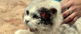
Melanoma
One of the most insidious malignant tumors that “masquerades as moles” is melanoma. It also consists of melanocytes, like moles, but is not as harmless as they are. Melanomas are very aggressive tumors; they easily grow into deep tissues and even bone structures. They are difficult to treat, including surgical removal. When they appear, the prognosis is very doubtful. Fortunately for cats, such formations are rare.
Papillomas
These are benign tumors of viral etiology. Animals owe the growth of these formations to the papillomavirus. Papillomas are papillary-like skin formations or growths on the mucous membrane 1-3 cm in diameter. In cats, papillomas are most often colored black and have an uneven, spongy surface.
Most often, papillomas appear on the head in cats. Papillomas themselves are not dangerous. Their potential threat lies in their tendency to malignancy. Veterinary practice indicates that papillomas quite often become malignant. The risk of malignancy increases if papillomas form in the animal's mouth.
Hemangiosarcoma
This is a malignant neoplasm containing the walls of blood vessels, usually it is dark red, actually black. Hemangioma protrudes above the surface of the skin. Tumors are most often found on the animal’s head. This is explained by the effect of direct sunlight on this area. Fortunately for cats, this is a rare disease in cats, as the tumor is very aggressive and prone to metastasis.
Squamous cell skin cancer (carcinoma)
A malignant formation that is most often observed in the ear, nose and eyelids of an animal. It is believed that the appearance of carcinoma is due to prolonged exposure to ultraviolet radiation on the skin of the animal.
A red spot forms on the affected area of the animal, sores may appear, and hair may fall out. Ulcers heal with rough tissue. Usually the tumor is limited to healthy tissue. But metastases to the nearest lymph nodes and lungs are not excluded.
Lipoma
This is a benign neoplasm of the color of the skin of an animal, mobile, soft and painless. Consists of adipose tissue. As long as the tumor does not grow and does not compress surrounding tissues, it is not dangerous for the animal. In this case, a trip to the veterinarian is justified by the fact that a more aggressive formation can be mistaken for a lipoma or wen.
Mite
The head of the tick that has burrowed into the animal’s skin is not visible, and its body, swollen with the cat’s blood, resembles a papilloma or lipoma. Usually, experienced cat owners still distinguish ticks from neoplasms.
Tumors in a cat's mouth.
Tumors in the mouth and throat are less common in cats than in dogs. Unfortunately, tumors that do arise are often malignant.
Benign tumors.
Gum fibroma
(gingival fibroma) a benign (non-growing) growth that usually occurs near the gum line. The growth is relatively insensitive and hard, having either the color of normal gums or a slightly paler color. The amount can be large enough to completely cover the surface of several teeth. The usual treatment is surgical removal of the fibroid. After the operation, daily rinsing is prescribed until the cat recovers completely.
Epulis
(supragingival, Epulides) is another type of benign tumor-like formation that occurs on the gums. In practice this is rare. This type of tumor usually affects only the area around one tooth. A biopsy of tissue samples may be performed for proper diagnosis and treatment.
Malignant tumors.
Squamous cell carcinoma is the most common malignant tumor in the oral cavity of cats. Usually occurs on the gums and tongue, then quickly spreads throughout the mouth.
Symptoms depend on the location and size of the tumor. Typically, bad breath, refusal to eat, and excessive drooling are observed. If the swelling affects the back of the mouth or throat, swallowing may be difficult. The tumor often has ulcers and bleeds. Your cat's face may become swollen as the tumor enlarges and invades surrounding tissue. Nearby lymphatics often enlarge before the tumor itself becomes noticeable. For diagnosis, a biopsy of tissue samples is usually performed.
Treatment and prognosis depend on the type of tumor and its stage. Malignant melanomas are highly invasive and grow rapidly, so the prognosis is poor. Surgical removal improves the chances of survival and may even eliminate the tumor, but recurrences are common. Squamous cell carcinoma has a poor prognosis and survival is only possible with early diagnosis and treatment. Removing a tumor often requires removal of the lower jaw.
Videos and Illustrations
Moles in animals
Moles
Lentigo
Papiloma
Hemangiosarcoma
Mites mistaken for moles
Melanoma
Squamous cell carcinoma of the ear
Description of moles
Unfortunately, it is difficult even for a specialist to visually distinguish a benign formation, that is, a mole, from more serious formations. In addition, analyzing the questions of cat lovers addressed to veterinarians, it becomes clear that non-specialists mistake formations on the skin of a pet of varying degrees of malignancy for moles: papillomas, melanomas, hematosarcomas, squamous cell carcinoma. And even small lipomas and a tick attached to the animal.
Moles can be:
- congenital and acquired;
- vascular and pigments;
- flat and slightly rising above the surface;
- By color, brown, black, and sometimes moles of other colors are also found;
- By size they are divided into: small (0.5-1.5 cm), medium size (1.5-10 cm), giant (more than 10 cm).
Moles are benign neoplasms; they rarely become malignant (malignant), but it is still possible. In this sense, giant moles are considered the most dangerous.
The fight for a healthy color
So, if you notice a change in the pigmentation of your nose outside of the summer, it makes sense to analyze the possible causes and try to eliminate them. Remove all strong-smelling substances, in particular household chemicals, from your reach. If a change in the color of the nose is accompanied by lethargy or the animal shows anxiety, it makes sense to contact a veterinarian. Take a general blood test, which will help to accurately determine the cause of the phenomenon.
If no deviations are identified, vitamins and microelements will help maintain health and return the usual color of the nose. Zinc and vitamin E care for the skin as a whole, which means they have a positive effect on the nose. It is worth giving vitamins in accordance with the instructions for the medicinal product, where the intake rates per kilogram of weight are prescribed. The course of treatment should be stopped after a clear restoration of the color of the nose. It also makes sense to introduce essential fatty acids Omega 3 and Omega 6 into the diet in the form of dietary supplements. Their dosage is also calculated taking into account the weight of the animal.
Once nasal pigmentation is restored, medications can be discontinued. It is worth making it a rule to monitor the health of your furry pet, because any disease is easier to prevent than to treat. A healthy cat is the key to a good mood for its owners.
Sudden changes or uneven pigmentation of the nose in cats can signal health problems for your pet.