General information
Cholangitis syndrome begins its development in the bile ducts and can spread to the tissues of the liver and intestines. At the same time, various diseases of these organs can provoke cholangitis syndrome. In the case of an advanced condition, it is quite difficult to determine which disease was primary and which was secondary.
In many cases, cholangitis is accompanied by pancreatitis and inflammatory bowel disease. Therefore, their combination is usually called a “triad”.
Cholangitis can occur as a result of bacterial infection entering the bile ducts, or as a result of the activity of a parasite, or have an autoimmune origin.
There are three main types:
- neutrophilic (purulent);
- lymphocytic (non-purulent);
- lymphoplasmacytic.
Most often, the development of the neutrophilic type is promoted by a bacterial infection of the intestines or liver, which penetrates the bile ducts. Often accompanied by inflammation of the pancreas.
Lymphocytic and lymphoplasmacytic types are still under study. Presumably, they arise as a result of a malfunction of the body's immune system.
The neutrophilic type is more common in young cats, while the lymphocytic and lymphoplasmacytic types are more common in mature and elderly cats. A hereditary predisposition to cholangitis is observed in Persian cats.
Definition of disease, spread
Among the many vital roles performed by the liver is its contribution to the digestion of food that a cat typically consumes. This digestive process relies heavily on the liver's efficient production and secretion of bile, a powerful, greenish-brown fluid that moves from the liver through the biliary system (a complex network of small channels called bile ducts) into the gallbladder. The bile is then stored in the gallbladder—a small spherical sac—until it is called to work in the intestinal tract. In response to hormonal signals, the gallbladder contracts and expels bile through the common bile duct into the small intestine, where it performs essential digestive processes such as emulsifying dietary fats so they can be absorbed from the cat's intestines and processing harmful toxins so they are expelled from the body through defecation.
Clinical picture
Symptoms of cholangitis in cats will depend on the type of pathology. The purulent variety is characterized by an acute onset with a rapid increase in symptoms. The cat has vomiting, loss of appetite, abnormal bowel movements, jaundice, and general lethargy.
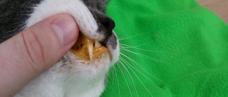
Jaundice in cats is manifested by changes in the color of the skin and mucous membranes. You can notice this sign in areas of the body that are minimally covered with hair (ears, belly, groin). Icterus is also clearly visible on the sclera, mucous membrane of the eyes and mouth: a pronounced yellow tint to the tissues will be noted.
Important! Neutrophilic (purulent) cholangitis progresses quickly and is especially dangerous for the animal’s body. In the absence of timely help, the disease can be fatal.
The non-purulent variety of the disease is characterized by a sluggish course, slow progression and chronicity. Most often, this condition is observed in older cats and it is not always possible to immediately suspect the development of pathology. The animal's appetite worsens, frequent vomiting appears, rapid weight loss is noted, and jaundice gradually develops.
Important! The chronic course of the pathology is dangerous due to the occurrence of complications in the form of abdominal dropsy.
Diagnostics
When the first signs of disease development appear (vomiting, loss of appetite, lethargy, jaundice), you should immediately consult a veterinarian.
Diagnostic methods include examination, laboratory and instrumental examination methods. Based on the clinical picture data, the veterinarian carries out differential diagnosis with the following types of pathological conditions:
- poisoning with toxic and medicinal substances with liver damage;
- infectious peritonitis;
- liver lipidosis;
- hepatic trematodosis;
- liver neoplasms.
To make a diagnosis, your cat may undergo the following tests:
- general and biochemical blood test;
- Analysis of urine;
- X-ray examination of the abdominal cavity;
- Ultrasound;
- percutaneous liver biopsy;
- laparoscopy.
A blood test will show increased levels of bilirubin, anemia, leukocytosis, and increased levels of bile acids in the blood serum. Ultrasound and x-rays show a characteristic enlargement of the liver, obstruction of the bile ducts and stagnation of bile.
With the help of laparoscopy, the veterinarian will be able to thoroughly examine the liver, bile ducts, gallbladder, and also obtain biological materials for biopsy. However, despite the high information content of this method, it is rarely performed.
Liver biopsy, which is performed percutaneously, is of great importance for making the correct diagnosis. The procedure is carried out after the animal’s condition has stabilized.
Therapy for cholangitis in cats involves the use of medications. If the bile ducts are obstructed, surgery is performed. Emergency surgery is performed if signs of peritonitis appear.
Among medications, antibiotics are of primary importance. They are prescribed for the treatment of any type of cholangitis. In therapy, amoxicillin (for the activity of anaerobic bacteria) and aminoglycosides (for the activity of anaerobic infection) are most often used. Contraindication is the use of tetracycline, which has hepatotoxic properties.
In the treatment of lymphocytic and lymphoplasmacytic type, immunomodulators (prednisolone) are used.
Vitamin K is prescribed in case of increased blood clotting time.
To maintain liver function, it is possible to use hepatoprotectors. They prevent the destruction of cell structures and stimulate their regeneration.
During the treatment process, it is necessary to improve the cat's nutrition. It is recommended to use easily digestible food (or natural food) with a low protein content.
Treatment of cholangitis takes a long time. Its duration can range from several weeks to several months. At this time, additional tests will be required to monitor the condition.
Every 2 weeks a repeat biochemical blood test with liver enzyme testing is required. If the disease does not resolve within 4-6 weeks, an additional liver biopsy is performed.
This pathology requires stable drug therapy. In case of inadequate or delayed treatment, there is a risk of complications (ascites, hepatic encephalopathy).
The purulent type of pathology, despite its acute course, has a more favorable prognosis. Other types of disease quite often lead to cirrhosis of the liver.
Problem with gums in a cat, neutrophilic bacterial inflammation
Hello. The cat is outbred, approximately 2-3 years old. Taken from the street in February 2020. 1. In April, the lower lip was significantly swollen, and a diagnosis of Eosinophilic granuloma was made. Glucortin was administered 3 times. The lip is back to normal. 2. A big problem with the gums is inflamed, red. There are only 3 incisors on the upper jaw, one of them is loose. On the lower jaw there are also 3 incisors, outer and in the middle. Treatment (for 2 months) with miramistin, dentavedin gel and Canina anti-inflammatory gel did not produce results. The teeth themselves are white, there is no plaque or stone. Tests taken: Feline viral leukemia and viral immunodeficiency (Elisa, snap test and PCR) - negative. Calcivirosis, PCR - negative. 30.11. They did a fine-needle biopsy, the cytology results came back, I am attaching a description and blood tests. X-rays (spot shots) are not taken. Can you please tell me the diagnosis based on cytology and what is the reason for this condition of the gums? Or can it not be considered final? The doctor prescribed Sinulox for a course of 10 days.
If an infection gets into the bile ducts, cholangitis develops in cats. The pathology has 3 types and is accompanied by vomiting, yellowness of the mucous membranes, and weakness. At the first signs of deterioration in the pet’s health, the owner must take it to a veterinarian, who will conduct a diagnosis, prescribe medications and a therapeutic diet, and give preventive recommendations.
According to veterinarians, animals that are female, obese, and older than 8 years are most prone to cholangitis and liver inflammation.
Feline cholangitis syndrome
Feline cholangitis syndrome
Cholangitis is an inflammation of the bile ducts, manifested by indigestion and jaundice.
The main cause of inflammation of the biliary tract is microflora that penetrates from the duodenum through the bile duct, portal vein and hepatic artery. Often the disease occurs as a complication of infectious (colibacillosis, salmonellosis) and parasitic (fascioliasis, dicroceliosis, eimeriosis) diseases. Inadequate diets, especially in vitamin A, as well as feed toxicoses contribute to the development of the disease.
Cholangitis in cats is relatively common and is very different from liver disease in dogs. It can be quite difficult to treat, especially if the cause of the disease is not precisely known.
There are three main types of cholangitis in cats:
Neutrophilic cholangitis is caused by a bacterial infection in the liver, leading to inflammation. Usually develops as a result of migration of bacteria to the bile ducts from the small intestine. The disease is sometimes observed simultaneously with inflammation of the pancreas and intestines.
Lymphocytic cholangitis is non-infectious in nature, although it also leads to inflammation. The exact cause is unknown, but the disease may be related to disturbances in the cat's immune system (immune-mediated disease).
Lymphocytic cholangitis presents with typical chronic tissue changes progressing over months or years and may initially be asymptomatic. The disease is more common in younger and Persian cats. The pathophysiological mechanisms of its development are unknown, but they are assumed to be immune-mediated.
In the third case, the disease develops against the background of opisthorchiasis, a parasitic disease.
Endocrine glands
Clinically, the disease can manifest itself as drowsiness, loss of appetite, vomiting, weight loss, and bloating due to effusion into the abdominal cavity. On palpation and percussion there is severe pain in the liver area. In most cases, signs of obstructive jaundice appear due to obstruction in the outflow of bile. However, sometimes the cat’s condition does not correspond to the diagnosis at all, and the appetite even increases.
Definition, terminology
Cholangitis in cats
- inflammation concentrated in the biliary tree (bile ducts), a common form of liver disease, and apparently the second most common after lipidosis. A new simplified classification scheme proposed by the WSAVA Liver Diseases and Pathologies Standards Study Group suggests 3 different forms of cholangitis in cats:
- Neutrophilic (Bacterial) Cholangitis
- Lymphocytic Cholangitis
- Chronic Cholangitis
The new proposed classification scheme also prefers the term "cholangitis" to "cholangiohepatitis" since inflammatory parenchymal lesions are not always present and, if present, are a complication of primary cholangitis.
Cholangiohepatitis in cats
Cholangiohepatitis
is an inflammation of the liver and bile ducts.
The wide prevalence of cholangiohepatitis in cats is associated with the peculiarities of their anatomy: the pancreatic duct and the gallbladder ducts connect before emptying into the duodenum. Therefore, inflammation of the small intestine or pancreatitis
(inflammation of the pancreas) also leads to inflammation of the bile ducts (
cholangitis
).
Cholangiohepatitis can manifest itself in acute and chronic forms.
Acute form
occurs more often in young cats.
It begins with a sudden refusal to feed and lethargy. Vomiting appears, body temperature often rises, and the abdominal area is painful. With acute hepatitis, dehydration quickly occurs. After this, the so-called “jaundice” or icterus
(yellowish tint of the skin and mucous membranes), which is noticeable on the sclera of the eyes and gums. During this period, the activity of liver enzymes, bilirubin and the number of leukocytes in the animal’s blood increases.
Chronic form
Cholangiohepatitis is more common than acute, and older cats are more prone to it. Symptoms with this course appear and disappear in periods, with periods of exacerbation often associated with stress.
Depending on the type of cells that are found during microscopy of liver samples, chronic cholangiohepatitis may have different names. If lymphocytes predominate, it is called lymphocytic cholangiohepatitis
;
if neutrophils - then neutrophilic
;
if other defense cells (macrophages, plasmacytes) – then granulomatous
.
All forms of cholangiohepatitis can ultimately lead to liver atrophy ( cirrhosis)
).
The causes of acute cholangiohepatitis are often bacterial infections that pass to the liver from the small intestine (duodenum) and pancreas. In addition, acute cholangiohepatitis can be caused by coronavirus infection, intoxication, or feeding with low-quality or unbalanced feed.
Among the causes of chronic cholangiohepatitis, genetic predisposition comes first; it can also be due to an autoimmune disease, helminthiasis, cystoisosporosis, or malnutrition.
- Hepatic lipidosis
. Cats do not tolerate periods of not eating well (anorexia). At this time, their liver often begins to store fat, which leads to lipidosis; the functional liver tissue is irreversibly replaced by fatty tissue. Cats with a lack of appetite due to cholangiohepatitis are at risk. - Hepatic encephalopathy
. Due to increased levels of ammonia and other unwanted components in the blood, brain damage occurs. - Portal hypertension
and the formation of free fluid in the abdominal cavity (ascites). - Sometimes chronic cholangiohepatitis progresses to cancer. In humans, there has been a proven connection between chronic stimulation of lymphocytes and the occurrence of malignant lymphoma. Therefore, it is likely that chronic lymphocytic cholangiohepatitis in cats can provoke lymphoma and malignant abnormalities of lymphocytes.
- General clinical examination of the animal.
- General clinical and biochemical blood test. The presence of hepatitis or chronic inflammation of the intestines and pancreas is indicated by a high level of GGT, an increase in ALT and alkaline phosphatase with normal levels of thyroid hormones. The level of bilirubin and globulins also increases, cobalamin and folic acid decrease.
- Serological studies. Used for suspected viral infections (feline leukemia, feline immunodeficiency, feline viral peritonitis), as well as toxoplasmosis.
- X-ray examination.
- Ultrasound of the abdominal cavity (is important in diagnosing cholangiohepatitis or obstruction (blockage) of the bile duct).
- Liver biopsy. A needle is inserted through the abdominal wall into the animal's liver and material is collected for further research. The best way to diagnose hepatitis is by examining small pieces of the animal's liver. They are obtained by exploratory laparotomy (surgically) or by biopsy. Both procedures pose a certain risk and must be carried out with extreme caution, because in severe cases of the disease, the affected organs may bleed during puncture, and anesthesia poses a risk to the sick animal.
- Bacterial culture of liver and bile. If liver and bile samples can be obtained for pathological examination, it may be possible to test them for the presence of bacteria.
- stabilization of the body's condition in critical cases (intravenous therapy with electrolyte solutions). If necessary, artificial or parenteral nutrition and antiemetics are prescribed;
- antibiotics;
- choleretics and hydrocholeretics (substances that help the passage of bile from the liver to the intestines). They are prescribed to prevent bile stagnation, because this is one of the main phenomena of cholangiohepatitis;
- anti-inflammatory drugs;
- immunosuppressants (prescribed for lymphocytic portal hepatitis);
- vitamins K, E, B12. With cholangiohepatitis, the ability to absorb these vitamins through the intestines is reduced. In some cases, the use of taurine, folic acid and L-carnitine is also indicated.
The cat should be switched to a diet containing a reduced amount of sodium, carbohydrates and an increased amount of protein. It is not advisable to feed your animal food containing sucrose or fructose.
What it is?
This is the name for inflammation of the bile ducts . If in this case the entire liver is inflamed, the disease can be characterized as “cholangiohepatitis”. The disease is quite serious, as it often leads to obstruction of the bile ducts . Bile is extremely important for the digestion of fats, so a sick cat will quickly develop digestive problems. If nothing is done, cholemia will occur, which can be fatal.
There are three main types of cholangitis in cats:
- Neutrophilic variety . When examining a liver sample, it is discovered that the organ tissue is literally saturated with neutrophils. It can occur in both acute and chronic forms.
- Lymphocytic type . Accordingly, when examining tissue samples, a large number of lymphocytes are found.
- In the third case, the disease develops against the background of opisthorchiasis , a parasitic disease.
Prognosis and prevention
Timely detection and treatment of cholangitis gives a chance for a favorable prognosis. However, the chronic form can cause cirrhosis. In advanced cases, the purulent type of the disease leads to the death of the animal. Undiagnosed opisthorchiasis is also dangerous, when helminths can settle not only in the bile ducts, but also in vital organs, which provokes death.
To avoid cholangitis in a cat, it is recommended not to give your pet meat and fish that have not undergone heat treatment, and to regularly deworm it. The weight of the animal should be monitored and the correct diet should be ensured. This will help prevent the formation of stones. All pathologies that can cause the disease must be treated in a timely manner.
Causes and clinical manifestations
The acute neutrophilic form is caused by a bacterial infection. Microorganisms enter the liver from the intestines, moving up the bile ducts. It is believed that the chronic neutrophilic form is a consequence of the acute variety. The cause of the lymphocytic form has not yet been precisely identified, but experts suggest that autoimmune diseases are to blame, when the body begins to attack itself.
There is a group of factors called the “trinity” or “triad”. In this case, cholangitis is accompanied by pancreatitis and inflammatory bowel diseases. It is difficult to say which disease occurs first and “pushes” the development of other pathologies.
What are the symptoms of cholangitis in cats? Signs vary depending on the type of disease. The acute neutrophilic type causes a rapid deterioration in the animal’s condition and can be fatal. The cat vomits, has no appetite, and develops profuse diarrhea. Intermittent fever is possible. The acute form of the disease is more common in relatively young cats; cats get sick much less often.
On the contrary, chronic neutrophilic cholangitis is more often detected in cats, and older ones at that. The animal suffers from persistent vomiting, the skin and all visible mucous membranes turn yellow. The chronic variety is dangerous due to the development of ascites (edema of the abdominal cavity). The parasitic type manifests itself in approximately the same way, but it is more characterized by severe pain in the right hypochondrium. The diagnosis is made based on the following studies:
- Complete medical examination of the animal, taking blood and urine tests. Blood biochemistry often shows an increase in liver enzymes and bilirubin.
- An ultrasound examination will help determine what exactly is affected - the liver, its ducts, or the entire tissue of the organ. A biopsy is very desirable, since with its help it is possible to accurately identify the type of disease and prescribe treatment.
- If parasitic cholangitis is suspected, a stool sample is taken.
- Therapeutic techniques
Treatment for cholangitis in cats depends on its type. Because acute neutrophilic cholangitis is associated with a bacterial infection, antibiotics are a key component of therapy. Unfortunately, the course of treatment often lasts several weeks. In chronic neutrophilic and lymphocytic cholangitis, the goal is to reduce inflammation. are used , and sometimes more powerful immunosuppressants are used. For cholangitis associated with liver flukes, praziquantel and drugs based on it are used.
Regardless of the type of disease, replacement therapy is necessary to allow the organ to fully recover.
Various hepatoprojectors are prescribed for this purpose. In addition, treatment involves:
- Vitamin K and E injections.
- Painkillers.
- Liquid broths without fat as food.
- Ursodeoxycholic acid. Used to stimulate bile secretion and reduce the intensity of inflammation.
- If the flow of bile is blocked by something (stones, adhesions), surgical intervention is necessary.
How is the treatment carried out?
Use of pharmaceuticals
Medicines are prescribed only by a veterinarian; self-medication of the pet is prohibited. Therapy includes a set of drugs shown in the table:
| Medication group | Veterinary medicine |
| Antibiotics | "Amoxicillin" |
| "Cephalexin" | |
| "Clavimox" | |
| Choleretic | "Ursofalk" |
| "Actigall" | |
| Corticosteroids | "Prednisolone" |
| "Dexa" | |
| Vitamins | Injections of vitamins E, K |
| Antiemetics | "Cerucal" |
| "Serenia" | |
| Hepatoprotectors | "Divopride" |
| "Hepatovet" | |
| "Vet Expert" | |
| Anthelmintic | "Praziquantel" |
Medical nutrition
An animal with this diagnosis should not eat fried foods.
With cholangitis, the owner must introduce a strict diet for the pet. It is forbidden to give food “from the table”, smoked, fried and salty dishes. Meals should be fractional. If the cat cannot feed on its own, a feeding tube is used. The following foods are useful for a sick cat:
- lean fish or meat broth;
- oatmeal or rice porridge;
- rice broth;
- minced chicken or beef, steamed or boiled;
- low-fat dairy products;
- boiled carrots or potatoes.
Veterinarians also recommend special medicinal foods:
- Hills Diet Felin;
- Royal Canin Hepatic;
- Royal Canin Gastro Interstinal.
Symptoms: how to recognize the disease?
The cholangitis/cholangiohepatitis complex is a series of associated inflammatory or infectious diseases of the liver as well as the biliary tract. Cholangitis refers to infection or inflammation of the bile duct, and cholangiohepatitis is inflammation of the biliary system and, in particular, the liver parenchyma around the bile ducts.
The clinical signs of these diseases in this complex are similar and include:
- Jaundice (yellowing of the skin, sclera of the eye and mucous membranes)
- Lethargy
- Anorexia
- Vomit
- Fever is common in acute form but rare in chronic form.
- Dehydration
- Diarrhea
- Ascites (fluid in the abdominal cavity) sometimes occurs due to cirrhosis
There are 3 types of the disease:
- Neutrophilic, provoked by bacteria and characterized by an acute onset and course.
- Lymphocytic, which has a non-infectious etiology, is characterized by a slow course and chronic form.
- Parasitic, developing against the background of infection with helminths, mainly opisthorchid.
Treatment of chronic cholangiohepatitis
Histopathological features of acute cholangiohepatitis include the presence of infiltration of large numbers of neutrophils into the portal areas of the liver and cholangitis (that is, inflammation of the bile ducts). Destruction of the periportal limiting plate leads to disruption of the boundary at the portal sites, periportal necrosis and infiltration of neutrophils into the hepatic lobes. Bile duct hypertrophy and fibrosis are usually mild or absent.
Acute cholangiohepatitis begins as a bacterial infection ascending through the biliary tract. In some sick cats, microorganisms such as Escherichiacoli, Clostridia, Bacteroides, Actinomyces and alpha-hemolytic Streptococcus can be isolated. Diseases of the biliary system, including gallstone formation, and anatomical abnormalities of the gallbladder may predispose cats to cholangiohepatitis.
In one retrospective study, 83% of cats with cholangiohepatitis had associated inflammatory infiltrates in the duodenum, jejunum, or both, and 50% had pancreatic lesions. Thus, inflammatory bowel disease, pancreatitis, impaired gallbladder stricture or function, and cholelithiasis may predispose the animal to temporary cholestasis and allow reflux of pancreatic secretions or retrograde bacterial invasion, or both.
Clinical signs of acute cholangiohepatitis are not specific. These include anorexia, weight loss, weakness, somnolence, vomiting and fever (Table 1).
In approximately half of the cases, hepatomegaly can be detected. 90% of cats with acute cholangiohepatitis had neutrophilia or a left shift in the blood count due to an inflammatory response. Serous biochemical abnormalities included normal or slightly elevated alkaline phosphatase (ALP) activity, moderate to markedly elevated alanine aminotransferase (ALT) activity, and moderate to markedly elevated total bilirubin concentrations. Bile acid levels in fasting or post-feeding serum, or both, were also abnormal in affected cats.
Treatment of lymphocytic portal hepatitis
Lymphocytic portal hepatitis differs from acute and chronic cholangiohepatitis by the infiltration of lymphocytes and plasmacytes, but not neutrophils, into the portal areas, the absence of cholangitis, and the presence of an intact periportal limiting plate. The number of lymphocytes and plasmacytes in the portal areas ranges from a small number (10-20) to a large number (more than 100).
In some cases there are large lymphoid accumulations. Bile duct hypertrophy and portal fibrosis are often observed, but do not progress to pseudolobular formations or biliary cirrhosis. Because the inflammatory infiltrate consists primarily of lymphocytes and plasmacytes, some authors have suggested that lymphocytic portal hepatitis is an immune-mediated disease (Jones, 1989; Center and Rowland, 1994; Gagnee et al., 1996).
Common clinical signs of lymphocytic portal hepatitis are anorexia and weight loss (see Table 1). Vomiting, weakness, drowsiness, fever and diarrhea are less common. In sick cats, the size of the liver increases by about half. In most cats with lymphocytic portal hepatitis, the results of a clinical blood test do not differ from normal ones, with the exception of poikilocytosis, which is observed in 2/3 of affected cats.
Features of the neutrophil phase of inflammatory processes
The neutrophilic phase of inflammatory processes has its own characteristics. When bacteria or fungi enter the body, the process of programmed cell death is triggered, with neutrophils releasing a special extracellular trap. This phenomenon is called "netosis".
After pathogenic microorganisms have entered the body, neutrophils rush to the site of the lesion.
After this, the following happens:
- Calcium is released from the intracellular organelle (endoplasmic reticulum).
- The internal membranes of the neutrophil are destroyed.
- Massive decondensation (unwinding of the molecule) of chromatin (the basis of chromosomes, consisting of protein, RNA and DNA) occurs.
- The level of ROS (reactive oxygen species) increases sharply.
- Antibacterial proteins, nucleoplasm and cytoplasm contained in the neutrophil are mixed, resulting in the formation of a future trap inside the cell.
- The plasma membrane ruptures and the resulting trap is thrown outside the neutrophil.
The trap that is formed as a result of netosis has the appearance of a three-dimensional network. Its basis is DNA strands on which antimicrobial proteins are located. Such threads can be woven into thicker “cables”, resulting in a large ball. Pathogenic microorganisms (bacteria and fungi) get stuck in this network and die under the influence of proteins.
Such traps quickly and effectively trap bacteria that have entered the cat's body and then destroy them. However, this process has a peculiarity - in autoimmune diseases, asthma and some inflammatory diseases, the networks released by neutrophils injure neighboring cells, and inflammation progresses, but pathogenic microorganisms die.
Scientists compare the mechanism of action of neutrophils with spiders. Immune cells, like spiders, anchor the web and swarm away from it. DNA continues to stretch and unravel behind the neutrophil. Other immune cells stumble upon these networks and also begin to “weave a network.”
As a result, even a small number of neutrophils are able to jointly weave a fairly large trap into which foreign particles fall.
Inflammatory liver disease is the second most common cause of liver disease in cats (26%). Other common causes of liver disease are hepatic lipidosis (49%) and lymphosarcoma (7%). The terminology used to describe the histopathological signs of liver disease in cats can be confusing. These terms include: suppurative cholangiohepatitis, chronic cholangiohepatitis, interstitial cholangiohepatitis, pericholangiohepatitis, chronic lymphocytic cholangitis, progressive lymphocytic cholangitis, sclerosing cholangitis, lymphocytic-plasmacytic cholangitis-cholangiohepatitis, lymphocytic cholangitis-cholangiohepatitis, lymphocytic port al hepatitis and biliary cirrhosis.
A number of retrospective studies have classified inflammatory liver diseases (Jones, 1989; Center and Rowland, 1994; Gagnee et al., 1996). The authors examined 175 feline liver biopsies collected at the University of Minnesota Medical Clinic (Gagnee et al., 1996). The results of this study show that inflammatory liver diseases can be divided into two groups: cholangiohepatitis and lymphocytic portal hepatitis. Cholangiohepatitis is divided into acute (purulent) and chronic forms.
