If the receptors that perceive light die in a cat's eyes, retinal atrophy develops. The disease has several forms and affects young animals, and is also accompanied by progressive loss of vision and clouding of the lens. At the first symptoms of visual impairment, you should take your pet to a veterinarian, who will diagnose and prescribe treatment.
According to statistics, retinal atrophy in a cat leads to the death of 96% of the receptors.
Causes of pathology
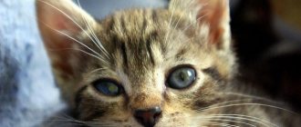
Progressive retinal atrophy is characterized by the gradual death of rods and cones—the tissues of the inner lining of the eyeball that are capable of receiving and refracting light rays, as well as seeing in the dark. In cats, the disease occurs mainly in kittens and young animals due to a gene mutation. The pathology is observed mainly in purebred cats. The disease is also preceded by retinal detachment, which develops due to injuries, blood clots, and diabetes.
When the rods die, oxygen accumulates in the tissues, which also destroys the cones. The probable causes and features of the forms of retinal atrophy are shown in the table:
| Form | Peculiarities |
| Autosomal dominant | There is rod and cone dysplasia |
| Appears due to a malfunction of the gene responsible for the development of photoreceptors | |
| Occurs in kittens of the Somali and Abyssinian breeds | |
| Leads to complete blindness when the cat reaches 1 year of age | |
| Autosomal recessive | Photoreceptor cells gradually lose function and die |
| Complete loss of vision occurs by age 3 | |
| Occurs due to a mutation in the gene that encodes a special protein responsible for the function of rods | |
| Found in Munchkin, Ocicat, Singaporean, Bengal, Persian breeds | |
| Acquired | Long-term use of antibiotics or overdose |
| Lack of taurine, which causes retinal dystrophy and blindness |
Bibliography
- Crispin, Sheila M.: Notes on veterinary ophthalmology. Blackwell Publishing.Ames. 2005. 371 p.
- Gelatt KN. Veterinary Ophthalmology 5ed. Wiley-Blackwell. Ames. 2013.2170 p.
- Maggs DJ, Miller PE, Ofri R. Slatter's fundamentals of veterinary ophthalmology 5ed. Elsevier. St. Louis. 2013, 506 p.
- Petersen-Jones S, Crispin S (eds): BSAVA Manual of Small Animal Ophthalmology, 2nd ed. Gloucester, BSAVA, 2002; 316 p.
- Bjorn Ekesten, Andras M. Komaromy, Ron Ofri, Simon M. Petersen-Jones, Kristina Narfstrom. Guidelines for clinical electroretinography in the dog: 2012update. Doc Ophthalmol (2013) 127:79–87.
- Grozdanic SD, Helga Kecova, Tatjana Lazic. Rapid diagnosis of retina and optic nerve abnormalities in canine patients with and without cataracts using chromatic pupil light reflex testing. Veterinary Ophthalmology 2012: 1–12.
- Grozdanic SD, Matic M, Sakaguchi DS et al. Evaluation of retinal status using chromatic pupil light reflex activity in healthy and diseased canine eyes. Investigative Ophthalmology & Visual Science 2007; 48:5178–5183.
Symptoms: how does pathology manifest itself?
With this pathology, going down stairs becomes a problem for the animal.
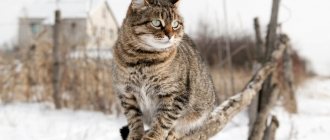
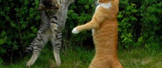
With retinal atrophy, a cat has no pain syndrome, so the owner may not know for a long time that the pet is sick. In addition, the cat has an excellent memory, which the animal uses when its vision decreases. Numerous vibrissae also help to navigate. However, in an unfamiliar environment the animal becomes helpless. Veterinarians recommend identifying that a cat has retinal atrophy based on the following signs:
- reluctance to go down stairs;
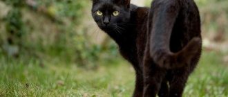
- bumping into an obstacle, especially if there was none before;
- lack of movement in the dark;
- dilated pupils;
- reflection of light rays from the back of the organ;
- cataract.
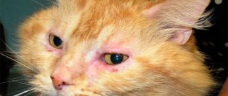
Wounds of the eyelids
The cat may injure its eyes. All wounds are usually divided into several types. Among them are such damage as superficial, deep and through , which results in damage to all layers of the eyelids.
Symptoms
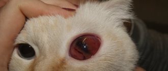
Depending on the type of injury, your cat's eyes may appear red, swollen, or lightly bleeding. Over time, the wound can develop into inflammatory edema and exudate appears. If there is a rupture of the eyelid, then, as a rule, this is not accompanied by bleeding, but the uneven edges of the wound are striking.
Such injuries may be caused by an unfortunate collision between the cat and an object or as a result of a fight with another cat. The wound can be caused by claws or teeth.
Giving help
Small, shallow wounds do not require special treatment; the cat can easily tolerate them. They heal over time. But if the wound is through and very deep, then the cat should be bandaged over the eye and immediately taken to the veterinarian.
Diagnostic measures
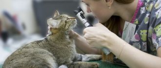
A veterinary ophthalmologist can determine retinal atrophy in a cat. If pathology is suspected, especially in breeds that are at risk, genetic tests are performed. Diagnostic procedures are also carried out, such as:
Using an ophthalmoscope, the doctor must examine the animal's eyes.
- ophthalmoscopy;
- electroretinography;
- computed tomography or magnetic resonance imaging if total blindness is suspected and to establish the etiology;
- Ultrasound of the heart, peritoneal organs and sternum, if the onset of the disease is associated with a lack of taurine;
- radiography.
Video
More photos Author(s):
S.A.
Boyarinov is a veterinary ophthalmologist at the IVC MBA, head of the treatment and preventive department of the SBBZh in Pushkino, postgraduate student of the department of the Federal State Budgetary Educational Institution of Higher Education MGAVMiB - MBA named after. K.I. Scriabin, member of RVO, ESVO, Russian Geographical Society. Organization(s):
Federal State Budgetary Educational Institution of Higher Education “Moscow State Academy of Veterinary Medicine and Biotechnology - MBA named after K.I.
Scriabin" (FSBEI HE MGAVMiB - MBA named after K.I. Scriabin) Journal:
No. 1 -2017
Introduction
The retina is a unique organ with a complex structure and functionality, providing visual perception of the surrounding world in dogs and cats. Since eye pathologies are often associated with various somatic diseases in animals, it is necessary to take into account the possibilities of primary diagnosis of pathology and, accordingly, prognosis. One of these visual diseases is retinal detachment (RD).
Normally, the retina of the eye is tightly adjacent to the underlying layers, the pigment epithelium (RPE) and choroid. This condition is due to the gentle pressure exerted on it by the vitreous body (VT), which holds the retina in a physiological position. The retina is tightly attached to the underlying layer only in a few places: along the dentate line and near the optic nerve. In other areas, the connection is caused only by gentle pressing of the CT, which, accordingly, allows us to conclude that the probability of developing detachment in these places is the highest [16, 42].
OS is an eye pathology in which there is a complete or partial separation of its 9 layers (neuroretina) from the RPE and choroid (choroid). Normally, these structures are tightly adjacent to each other, providing trophic functions [44].
With OS of the eye in animals, vision decreases to the point of complete blindness, and in advanced cases OS leads to the death of the eye. Therefore, this pathology is an emergency condition and requires immediate contact with a veterinary ophthalmologist.
Etiology
This eye disease occurs in both dogs and cats, but often has different causes. For example, hypertensive retinopathy with both total and local OS of an exudative nature is most typical for cats [37].
Most often, the following factors and pathologies can lead to OS in dogs and cats.
- — Congenital developmental anomalies such as retinal dysplasia (RD) [11, 29, 41], collie eye anomaly (CEA), and primary hyperplastic persistent TS syndrome (PHTVL/PHPV) [3, 20].
- — Eye injury leading to retinal rupture and hemorrhage [4].
- — Inflammatory processes (chorioretinitis), leading to the accumulation of exudate or blood in the subretinal space [4, 15, 26].
- — Degeneration and dysplasia of CT [21, 25].
- — Neoplasms of the posterior segment of the eye, including the choroid [6].
- — Buphthalmos in glaucoma, leading to stretching of the membranes of the eyeball [1, 28].
- — Pathologies leading to damage to the vascular bed: systemic hypertension, blood hyperviscosity syndrome, diabetes mellitus [5, 8, 22].
Based on the reasons leading to OS, several types of this pathology are distinguished.
Serous OS occurs as a result of the accumulation of fluid under the retina and, accordingly, its separation from the underlying layer. There are two types of serous detachment: the first is the exudative type, characterized by the accumulation of inflammatory fluid (exudate) as a result of infectious diseases [15, 26], the second is the hemorrhagic type, characterized by the presence of blood under the neuroretina due to systemic arterial hypertension, coagulopathies, thrombocytopenia [ 5, 8, 10].
Tractional detachment occurs as a result of tension on the retina from the side of the CT, to which it fits tightly. This condition is possible due to posterior uveitis, the formation of moorings and cords during degeneration of the CT [43], as well as when it is displaced forward as a result of luxation of the lens and displacement of the iridolenticular diaphragm [24].
Rhegmatogenous OS is associated with thinning and formation of retinal breaks as a result of degenerative changes, especially in older animals. Through these breaks, CT can penetrate under the retina, leading to detachment [13].
Traumatic OS is the result of injury to the eyeball (contusion, penetrating injury). In these cases, trauma can lead to both acute detachment as a result of retinal rupture, displacement of the retina, subretinal hemorrhages, and to detachment in the long term (chronic inflammatory process, destruction of the retina, hypotension) [4, 15].
It is also worth mentioning the possible iatrogenic OS after intraocular manipulations, in particular phacoemulsification of cataracts and vitrectomy. Thus, with phacoemulsification of 290 eyes in dogs and their three-year follow-up, postoperative complications in the form of OS amounted to 1–2% [33], although in the works of other researchers they range from 4 to 9% [7]. Despite the small percentage of detachments after phacoemulsification of cataracts in dogs, it is necessary to regularly assess the condition of the retina by ultrasound in the immediate and late postoperative periods [39].
According to the degree of prevalence, it is customary to distinguish the following types of OS: local, total, subtotal.
As a result of detachment of the neuroretina from the RPE and choroid, the following disorders occur [17]:
• decreased metabolism in neuroretina; • disruption of retinol transport from the RPE to the neuroretina; • disruption of the blood supply to the neuroretina from the choriocapillaris; • development of atrophy of the photoreceptor layer of the neuroretina; • release of vascular endothelial growth factor (VEGF) by hypoxic neuroretina [30].
It should be noted that OS is a condition that requires emergency treatment by the animal owner to a veterinary specialist and provision of immediate assistance to the patient. In many cases, when treatment is timely, and depending on the type and cause of OS, the prognosis for vision can be favorable. However, the lack of treatment, the inability to diagnose this pathology, as well as late treatment can lead to complications that can arise after OS - retinal atrophy, glaucoma, hemophthalmos, etc. In such cases, irreversible blindness develops and there is a high risk of losing the eye as an organ [28].
Risk factors
Risk factors for the development of OS in dogs and cats include the following [16]:
• high blood pressure (hypertension); • old age; • presence of overripe cataracts; • luxation of the lens; • phacoemulsification of cataracts; • genetics.
Clinical signs
Symptoms of OS in dogs and cats include partial or complete loss of vision (acute blindness), decreased or absent pupillary light response (PLR), the appearance of retinal floats and vessels visible without special equipment on a dilated pupil [16], characteristic of OS ultrasound signs – “gull wings” or the Latin letter V on ultrasound of the eyeball [21]. OS can often be accompanied by hemophthalmos (accumulation of blood in the CT).
As already mentioned, OS in many cases can be a concomitant symptom of the underlying disease. Therefore, it is important to take this connection into account, even in the absence of obvious signs of OS.
Diagnostics
Confirmation of the diagnosis of OS is made on the basis of anamnesis, examination by a veterinary ophthalmologist and diagnostic studies.
Animal owners in most cases complain of dilated pupils and blindness of varying degrees [16]. In cats, fibrin and blood may be present in the intraocular space [23].
To make an accurate diagnosis, it is recommended to use a comprehensive diagnostic approach: an ophthalmological examination (biomicroscopy, ophthalmoscopy, ultrasound), as well as an assessment of the animal’s somatic condition (clinical and biochemical blood tests, testing for infections, cardiac examination, etc.).
A comprehensive ophthalmological examination provides a complete diagnostic picture of the disease, prognosis and choice of treatment tactics. If OS is suspected, it is necessary to assess the condition of the anterior chamber of the eye, the iris and lens, and check the pupillary reflexes [14].
An important diagnostic measure for suspected OS is ophthalmoscopy.
This procedure is possible in the presence of transparent light-refracting media of the eye (cornea, lens, CT) and allows you to visually assess the state of the OS: detection and localization of areas of detachment (vibrating gray-white areas of the retina), the presence of exudate and hemorrhages, the presence of retinal breaks of various configurations [16 ].
Ultrasound of the eye is the “gold standard” for diagnosis in animals with suspected OS. It is especially important that this study is relevant when it is impossible to perform ophthalmoscopy and when the optical media of the eye are opaque (hyphema, hemophthalmos, cataracts, corneal edema) [1]. With ultrasound of the eyeball, it is possible to assess the degree and type of OS, the presence of exudate, blood, concomitant pathologies of TS (moorings, destruction) and choroid [21]. When B-scanning, the OS is visualized as a film-like formation in the CT, usually having contact with the dentate line, and the optic nerve head (ONH) in the form of the letter V.
The detached neuroretina is mobile, and when the eye moves during ultrasound, it moves smoothly, as if floating. OS is often accompanied by posterior vitreous detachment (PVD) and is typical for older animals [9].
Treatment
Since OS is an acute condition leading to decreased vision and blindness, the speed of treatment and the urgency of the assistance provided play a decisive role in the further prognosis of the disease. A long-term lack of medical care, as a rule, leads to disruption of the retina. This is due to the lack of contact between the detached neuroretina and the choroid (choroid) and a violation of trophism and metabolism between them. However, with emergency care for a patient with a detachment, it is possible to restore vision using both conservative and surgical treatment [31].
Drug treatment includes the use of medications aimed at relieving the primary cause that caused neuroretinal detachment. For example, cats with arterial hypertension, even without signs of retinopathy, should receive antihypertensive therapy aimed at normalizing blood pressure through the use of systemic antihypertensive drugs (amlodipine) and ACE inhibitors (enalapril), thereby preventing OS [19, 12]. The use of antibacterial therapy is justified if a systemic infection is confirmed [26]. At the same time, it is especially important to take measures in the event of an emergency admission of an animal to a veterinary clinic [40] in the form of diuretic medications (in the absence of contraindications). The use of natural, and in the absence of contraindications, systemic corticosteroids (prednisolone), gives good results, especially in dogs [2, 12].
Surgical treatment of OS in dogs and cats is relevant if the prognosis for vision is favorable, as well as the absence of contraindications for anesthesia. The procedure to restore the retina to its physiological position is called retinopexy [27, 32]. There are several types of retinopexies [38]:
• laser surgery (photocoagulation); • cryopexy; • pneumatic retinopexy; • vitrectomy with replacement.
The principle of laser surgery and cryopexy is similar and consists in “welding” or “freezing” the retina to the underlying tissues by forming scars at the site of exposure [34, 36].
Pneumatic retinopexy is somewhat simple to perform and consists of introducing a gas bubble into the CT, which puts pressure on the retina, pressing it to a physiological place [35].
Vitrectomy for OS is quite complex and requires expensive equipment and the skill of a microsurgeon. The meaning of this procedure is to remove the CT, straighten the OS and introduce heavy oil into the eye cavity (silicone tamponade of the vitreal cavity). Thus, the neuroretina is pressed against the RPE and choroid, providing an anatomical and physiological fit [18].
Quite often, surgical procedures for OS are performed in combination, for example, laser retinopexy and vitrectomy with replacement, to achieve a better result.
Each method has its own positive and negative sides, as well as indications depending on the type and severity of OS.
Conclusion
In conclusion, I would like to note the importance of urgently contacting a veterinarian, the urgency of providing first emergency aid, referral for ongoing treatment and monitoring by a veterinary ophthalmologist.
Literature:
- 1. Boyarinov S.A. The use of ultrasound research in the diagnosis of secondary glaucoma in dogs. Veterinary, Animal Science and Biotechnology. 2015; 10:6–12.
- 2. Andrew SE, Abrams KL, Brooks DE, et al. Clinical features of steroid responsive retinal detachments in twenty-two dogs. Vet. Comp. Ophthalmol. 1997; 7:82–87.
- 3. Bayón A., Tovar MC, Fernández del Palacio MJ, Agut A. Ocular complications of persistent hyperplastic primary vitreous in three dogs. Vet Ophthalmol. Mar 2001; 4(1): 35–40.
- 4. Bergstrom BE, Stiles J., Townsend WM Canine panuveitis: a retrospective evaluation of 55 cases (2000–2015). Vet Ophthalmol. 2016 Oct 12.
- 5. Chetboul V., Lefebvre HP, Pinhas C., Clerc B., Boussouf M., Pouchelon JL Spontaneous feline hypertension: clinical and echocardiographic abnormalities, and survival rate. J Vet Intern Med. 2003 Jan-Feb; 17(1): 89–95.
- 6. Cullen CL, Caswell JL, Grahn BH Intravascular lymphoma presenting as bilateral panophthalmitis and retinal detachment in a dog. J Am Anim Hosp Assoc. 2000 Jul-Aug; 36(4): 337–42.
- 7. Davidson MG, Nasisse MP, Jamieson VE, et al. Phacoemulsification and intraocular lens implantation: a study of surgical results in 182 dogs. Progress in Veterinary and Comparative Ophthalmology. 1991; 1:233–238.
- 8. Elliott J., Barber PJ, Syme HM, Rawlings JM, Markwell PJ Feline hypertension: clinical findings and response to antihypertensive treatment in 30 cases. J Small Anim Pract. Mar 2001; 42(3): 122–9.
- 9. Gallhoefer NS, Bentley E., Ruetten M., Grest P., Haessig M., Kircher PR, Dubielzig RR, Spiess BM, Pot SA Comparison of ultrasonography and histologic examination for identification of ocular diseases of animals: 113 cases (2000 -2010). J Am Vet Med Assoc. 2013 Aug 1; 243(3): 376–88.
- 10. Ginn JA, Bentley E., Stepien RL Systemic hypertension and hypertensive retinopathy following PPA overdose in a dog. J Am Anim Hosp Assoc. 2013 Jan-Feb; 49(1): 46–53.
- 11. Gornik KR, Pirie CG, Duker JS, Boudrieau RJ Canine multifocal retinopathy caused by a BEST1 mutation in a Boerboel. Vet Ophthalmol. 2014 Sep; 17(5): 368–72.
- 12. Grahn BH, Barnes LD, Breaux CB, Sandmeyer LS Chronic retinal detachment and giant retinal tears in 34 dogs: outcome comparison of no treatment, topical medical therapy, and retinal reattachment after vitrectomy. Can Vet J. 2007 Oct; 48(10): 1031–9.
- 13. Grahn BH, Sandmeyer LS Bilateral rhegmatogenous retinal detachments with giant retinal tears. Can Vet J. 2009 Sep; 50(9): 989–90.
- 14. Grozdanic SD, Kecova H., Lazic T. Rapid diagnosis of retina and optic nerve abnormalities in canine patients with and without cataracts using chromatic pupil light reflex testing. Vet Ophthalmol. 2013 Sep; 16(5): 329–40.
- 15. Gwin RM, Wyman M, Ketring K, Winston S. Idiopathic uveitis and exudative retinal detachment in the dog. J Am Anim Hosp Assoc. 1980; 16: 163–170.
- 16. Hendrix DV, Nasisse MP, Cowen P., Davidson MG Clinical signs, concurrent diseases, and risk factors associated with retinal detachments in dogs. Prog Vet Comp Ophthalmol. 1993; 3:87–91.
- 17. Hisatomi T., Sakamoto T., Goto Y., et al. Critical role of photoreceptor apoptosis in functional damage after retinal detachment. Curr Eye Res. 2002; 24: 161–172.
- 18. Hoffman A., Wolfer J., Occelli L., Lehenbauer TW, Sapienza J., Novak JM, Combs KL, Konrade KA Refractive state following retinal reattachment and silicone oil tamponade in dogs. Am J Vet Res. 2012 Aug; 73(8): 1299–304.
- 19. Huhtinen M., Derré G., Renoldi HJ, Rinkinen M., Adler K., Aspegrén J., Zemirline C., Elliott J. Randomized placebo-controlled clinical trial of a chewable formulation of amlodipine for the treatment of hypertension in client-owned cats. J Vet Intern Med. 2015 May-Jun; 29(3): 786–93.
- 20. Itoh Y., Maehara S., Yamasaki A., Tsuzuki K., Izumisawa Y. Investigation of fellow eye of unilateral retinal detachment in Shih-Tzu. Vet Ophthalmol. 2010 Sep; 13(5): 289–93.
- 21. Labruyere JJ, Hartley C., Holloway A. Contrast-enhanced ultrasonography in the differentiation of retinal detachment and vitreous membrane in dogs and cats. J Small Anim Pract. Oct 2011; 52(10): 522–30.
- 22. Leblanc NL, Stepien RL, Bentley E. Ocular lesions associated with systemic hypertension in dogs: 65 cases (2005-2007). J Am Vet Med Assoc. 2011 Apr 1; 238(7): 915–21.
- 23. Maggio F., DeFrancesco TC, Atkins CE, Pizzirani S., Gilger BC, Davidson MG Ocular lesions associated with systemic hypertension in cats: 69 cases (1985–1998) J Am Vet Med Assoc. 2000; 217:695–702.
- 24. Montgomery KW, Labelle AL, Gemensky-Metzler AJ Trans-corneal reduction of anterior lens luxation in dogs with lens instability: a retrospective study of 19 dogs (2010–2013). Vet Ophthalmol. July 2014; 17(4): 275–9.
- 25. Papaioannou NG, Dubielzig RR Histopathological and immunohistochemical features of vitreoretinopathy in Shih Tzu dogs. J Comp Pathol. Feb 2013; 148(2–3): 230–5.
- 26. Pearce J., Giuliano EA, Galle LE, Klauss G., Ota J., Moore CP Management of bilateral uveitis in a Toxoplasma gondii-seropositive cat with histopathologic evidence of fungal panuveitis. Vet Ophthalmol. 2007 Jul-Aug; 10(4): 216–21.
- 27. Pizzirani S., Davidson MG, Gilger BC Transpupillary diode laser retinopexy in dogs: ophthalmoscopic, fluorescein angiographic and histopathologic study. Vet Ophthalmol. 2003 Sep; 6(3): 227–35.
- 28. Pumphrey S. Canine Secondary Glaucomas. Vet Clin North Am Small Anim Pract. 2020 Nov; 45(6): 1335–64.
- 29. Rodarte-Almeida AC, Petersen-Jones S., Langohr IM, Occelli L., Dornbusch PT, Shiokawa N., Montiani-Ferreira F. Retinal dysplasia in American pit bull terriers—phenotypic characterization and breeding study. Vet Ophthalmol. 2020 Jan; 19(1): 11–21.
- 30. Sandberg CA, Herring IP, Huckle WR, LeRoith T., Pickett JP, Rossmeisl JH Aqueous humor vascular endothelial growth factor in dogs: association with intraocular disease and the development of pre-iridal fibrovascular membrane. Vet Ophthalmol. Mar 2012; 15 Suppl 1: 21–30.
- 31. Sakai T., Calderone JB, Lewis GP, Linberg K., Fisher SK, Jacobs GH Cone photoreceptor recovery after experimental detachment and reattachment surgery: An immunocytochemical, morphological and electrophysiological study. Invest Ophthalmol Vis Sci. 2003; 44:416–425.
- 32. Schmidt GM, Vainisi SJ Retrospective study of prophylactic random transscleral retinopexy in the Bichon Frise with cataract. Veterinary Ophthalmology. 2004; 7: 307–310.
- 33. Sigle KJ, Nasisse MP Long-term complications after phacoemulsification for cataract removal in dogs: 172 cases (1995–2002). Journal of the American Veterinary Medical Association. 2006; 228:74–79.
- 34. Steele KA, Sisler S., Gerding PA Outcome of retinal reattachment surgery in dogs: a retrospective study of 145 cases. Vet Ophthalmol. 2012 Sep; 15 Suppl 2: 35–40.
- 35. Spatola RA, Nadelstein B., Leber AC, Berdoulay A. Preoperative findings and visual outcome associated with retinal reattachment surgery in dogs: 217 cases (275 eyes). Vet Ophthalmol. 2015 Nov; 18(6): 485–96.
- 36. Steele KA, Sisler S., Gerding PA Outcome of retinal reattachment surgery in dogs: a retrospective study of 145 cases. Vet Ophthalmol. 2012 Sep; 15 Suppl 2: 35–40.
- 37. Stiles J., Polzin DJ, Bistner SI The prevalence of retinopathy in cats with systemic hypertension and chronic renal failure or hyperthyroidism. J Am Anim Hosp Assoc. 1994; 30:564–572.
- 38. Vainisi SJ, Wolfer JC Canine retinal surgery. Vet Ophthalmol. 2004 Sep-Oct;7(5): 291–306.
- 39. Van Der Woerdt A. Ultrasonographic abnormalities in the eyes of dogs with cataracts: 147 cases (1986–1992). Journal of the American Veterinary Medical Association. 1993; 9(5: 281–291.
- 40. Wang S., Linsenmeier RA Hyperoxia improves oxygen consumption in the detached feline retina. Invest Ophthalmol Vis Sci. 2007; 48:1335–1341.
- 41. Whiting RE, Pearce JW, Castaner LJ, Jensen CA, Katz RJ, Gilliam DH, Katz ML Multifocal retinopathy in Dachshunds with CLN2 neuronal ceroid lipofuscinosis. Exp Eye Res. May 2020; 134: 123–32.
- 42. Slatter`s fundamentals of veterinary ophthalmology / DJ Maggs, PE Miller, R.Ofri. Saunders Elsevier: St Louis. Mo. 2013. 506 p.
- 43. Zarfoss MK, Breaux CB, Whiteley HE, Hamor RE, Flaws JA, Labelle P., Dubielzig RR Canine pre-iridal fibrovascular membranes: morphologic and immunohistochemical investigations. Vet Ophthalmol. 2010 Jan; 13(1): 4–13.
- 44. Veterinary Ophthalmology: Two Volume Set (5th edition) / Kirk N. Gelatt (Editor), Brian C. Gilger (Editor), Thomas J. Kern (Editor). Chicester, Wiley-Blackwell (an imprint of John Wiley & Sons Ltd), 2013. 2260 p.
How is the treatment carried out?
Veterinarians clarify that with progressive retinal atrophy, treatment of concomitant diseases with drugs containing fluoroquinolones is prohibited.
The treatment regimen is determined by the veterinarian; self-medication is prohibited. Progressive retinal atrophy in cats is incurable. To stop the development of the disease, veterinarians recommend introducing vitamin supplements with taurine into the diet - Doctor Zoo, Beafar, Gimpet. Useful foods containing this component are “Friskes”, “Night Hunter”, “Sheba”, “Kitiket”, grain-free ones – “Grandorf”, “Akana”, “Go”. Mitochondrial antioxidants are also used, which stop the progression of the disease. Monitoring the animal’s body weight and glucose levels if diabetes is diagnosed will help slow down the disease.
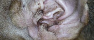
Inflammation of the eyelids
Inflammation of the eyelids is divided into two categories: simple and phlegmous .
Symptoms
The symptoms of the two types of eyelid inflammation are slightly different depending on the cause. With simple inflammation, the cat begins to be bothered by itching, and she tries to scratch the area. The edges of the eyelids take on a reddish tint and become thicker. If the inflammation is phlegmous , then the eyelid will be swollen and its skin will become tense. Purulent mucus begins to secrete from the reddened conjunctival sac.
Causes of inflammation
Superficial inflammation of the eye can occur due to ordinary vitamin deficiency or metabolic disorders. This type of inflammation can occur due to eczema. And the cause of phlegmous inflammation can be a deep wound, frequent scratching of the eyelid by the cat itself, or purulent inflammation of the meibomian glands.
Giving help
To treat simple inflammation, it is enough to rub an ointment containing broad-spectrum antibiotics, such as prednisolone and Oxycort, into the skin of the eyelid. It is much more difficult to cure phlegmatic inflammation. If an abscess forms, you should contact your veterinarian and open the abscess immediately. If it doesn't come to this, then it is best to wash your eyes with a 3% solution of boric acid or chamomile infusion. Additionally, the veterinarian prescribes an injection block of the anterior cervical nerve ganglion. To do this, use a 0.25% solution of novocaine with an antibiotic.