Hepatopathy in cats is a complex of liver diseases that includes almost all liver lesions: from cirrhosis to liver cancer and hepatargia.
Often hepatopathy acts as a primary or secondary disease: for example, as a consequence of pancreatitis or intestinal damage.
liver disease
Important! The danger of hepatopathy lies in its late detection. The liver has a high ability to regenerate, so clinical symptoms become noticeable at a late stage, when more than 70% of liver cells are affected.
Main signs of liver diseases and their types
Liver diseases in cats very often remain invisible to their owners, since signs of the disease can be confused with food allergies and bad mood. In this case, it is important to be very attentive to your pet.
Most often, cats suffer from the following liver diseases:
- Cirrhosis of the liver;
- Cholecystitis;
- Cholelithiasis;
- Hepatitis;
- Lipidosis;
- Cholangitis;
- Liver failure.
In some cases, malignant and benign tumors, amyloidosis, peliosis and other liver diseases are diagnosed.
All liver diseases can be divided into 2 types: primary and secondary.
- In primary diseases, the lesion is localized within the liver.
- Secondary liver diseases are caused by problems in the body that have nothing to do with this organ.
As a rule, liver diseases in cats have certain signs. The owner’s task is to detect the disease in a timely manner and provide quality treatment. Such actions will prevent the development of extreme stages of the disease that threaten the pet’s life.
Common signs of liver disease in cats:
- stomach disorders: vomiting and diarrhea;
- lack of appetite;
- weight loss;
- apathetic state;
- yellowing of the mucous membranes;
- change in color of stool and urine;
- severe itching;
- enlarged abdomen as a result of fluid accumulation in it;
- problems with blood clotting;
- protrusion of the liver;
- hemorrhages in the skin;
- When pressing on the area in the liver area, the cat shows severe anxiety.
The presence of one or more of the above symptoms is grounds for urgent contact with a veterinarian.
Diagnosis, treatment
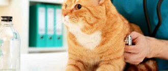
When the first symptoms of liver disease appear in your pet, you should contact a veterinary clinic. The specialist first conducts an external and physical examination. At this stage, the presence of ascitic fluid, hepatomegaly, yellowness of the oral mucosa, eyes, and skin can be detected. Next, the veterinarian prescribes additional research methods.
- A clinical blood test in the presence of an inflammatory reaction in the body is characterized by leukocytosis and an increase in ESR.
- A biochemical blood test is aimed at determining the concentration of pigments, alkaline phosphatase and other criteria that may indicate the presence of liver disease.
- X-ray of the abdominal organs can reveal an increase in the size of the liver, changes in its contours, the presence of abscesses and other pathologies.
- Ultrasound accurately determines structural changes in the liver; in case of cirrhosis and other pathologies, it is possible to detect areas with increased echogenicity. In addition, the study allows you to estimate the size of the organ.
Treatment of the disease depends on the diagnosis. Determination of timing, dosage and choice of medication is carried out by a veterinarian.
For a bacterial infection, the veterinarian will prescribe a course of antibiotic therapy. The choice of drug, its dosage and duration of use is based on the type of pathogen.
If liver changes are detected due to autoimmune processes, it is advisable to prescribe glucocorticosteroids.
Also read the article on how to feed a pill to a cat.
Cirrhosis does not respond to drug therapy; only relief of unpleasant symptoms is possible.
Therapy for helminth infestation involves taking special medications. In severe cases, surgery is possible.
An important component of treatment is diet, which helps reduce the load on the digestive organs and speed up the healing process.
Cirrhosis of the liver
Changes in the structure of the liver and overgrowth of connective tissue in it are called liver cirrhosis. This condition is characterized by loss of appetite, shortness of breath, and deterioration of heart function. The main sign of cirrhosis of the liver is an increase in its size and accumulation of fluid in the abdominal cavity of a cat. This liver disease can also be recognized by the appearance of jaundice and diarrhea, and redness of the conjunctiva.
Causes of liver cirrhosis:
- long-term intoxication of the body;
- hepatitis;
- infectious diseases caused by bacteria and viruses;
- insufficient amount of proteins and vitamins in the animal’s body.
Before starting treatment for liver cirrhosis, it is important to find out the cause of the disease. Based on these data, subsequent treatment methods are prescribed. As a rule, vitamins, diuretics, intravenous administration of salts, glucose and proteins are used to treat liver cirrhosis in cats.
To prevent this disease, it is recommended to undergo periodic veterinary examinations and feed your pet only high-quality food. The cat's heredity significantly influences the possibility of developing liver cirrhosis.
Diet and food for liver diseases
Gastritis in cats: symptoms and treatment at home
For any form of hepatopathy, the diet is prescribed and adjusted by a veterinarian. The brand of food, volume and number of servings, as well as the use of auxiliary products are determined based on analyzes and forecasts.
diet
For cats on specialized food, the following types of food are most often recommended:
- Purina Pro Plan Veterinary Diets;
- Farmina Vet Life Cat;
- Royal Canin Hepatic;
- Monge VetSolution Hepatic;
- Hill's Prescription Diet Feline.
Cholecystitis
Inflammation of the gallbladder (cholecystitis) occurs as a result of the formation of stones in it or the presence of giardiasis. Most often, this disease develops after a cat eats stale food contaminated with eggs and larvae of parasites, or poor quality food.
Symptoms of cholecystitis:
- elevated temperature;
- diarrhea;
- constipation;
- pain in the liver area.
To treat cholecystitis, special drugs are prescribed against microbes and parasites, as well as choleretic agents. The diet is reviewed and harmful foods are removed from the cat’s diet. The main preventive methods against the development of this disease are the use of anthelmintic drugs, timely vaccination, feeding the animal with high-quality feed and mandatory heat treatment of fish, which most often contain parasite eggs.
Anatomical and physiological data. The liver is a parenchymal organ, located mostly in the right hypochondrium and a smaller part in the left hypochondrium. The anterior convex surface of the liver is adjacent to the diaphragm, and the posterior concave surface faces the abdominal cavity. The liver is attached to the diaphragm by two ligaments, but they play a secondary role in fixing the liver. The position of the liver is mainly determined by intra-abdominal pressure. The inferior vena cava, into which the hepatic veins piercing the liver flow, is of known importance for fixing the liver.
On the outside, the liver is mostly covered with peritoneum. Under the peritoneal cover there is a fibrous membrane - Glisson's capsule. The suspensory ligament divides the liver into two lobes: the larger right lobe and the smaller left lobe. Between them on the posterior surface there are the gates of the liver, into which the hepatic artery and portal vein enter and the hepatic duct exits. All animals have a gallbladder, with the exception of the horse.
The liver consists of liver cells grouped into separate lobules, separated from each other by layers of connective tissue. The gaps between the liver cells are the primary bile ducts (capillaries). The bile capillaries unite into larger ducts and extend beyond the lobule. A vein runs through the center of the lobule, collecting blood from the lobule. The interlobular layers of connective tissue contain blood vessels, bile ducts and nerves. Kupffer cells are located in the endothelium of the capillaries of the portal vein. Thus, the liver contains specific hepatic epithelium and reticuloendothelial Kupffer cells with different functions.
Blood is delivered to the liver mainly by the portal vein, which collects blood from the stomach, intestines, and pancreas. glands and spleen, and to a lesser extent, the hepatic artery. The portal vein, formed from the confluence of the mesenteric and splenic veins, divides again in the liver, forming a dense network of capillaries. Several hepatic veins are formed from the capillaries, which flow into the inferior vena cava.
All the diverse activities of the liver are regulated by parasympathetic and sympathetic fibers of the autonomic nervous system. Through the vagus nerve impulses are transmitted that increase bile secretion and promote the accumulation of glycogen, and through sympathetic fibers impulses are transmitted that mobilize sugar from the liver, stimulate the formation of urea, etc.
The liver performs various and important functions for the body. It produces bile and releases it into the intestines. Bile plays an important role in the digestion of fats.
Increased dullness of the liver.
The liver takes part in metabolism, primarily in carbohydrate metabolism. Glucose entering the liver is converted into glycogen and stored there as a sugar reserve. As glucose is consumed in the body, liver glycogen is converted into glucose and enters the blood.
Therefore, the liver performs the function of maintaining blood sugar at the desired level. The liver plays a significant role in protein metabolism, as well as in water, mineral and vitamin metabolism.
The liver performs a barrier function, transferring protein breakdown products, alkaloids, heavy metals, etc. from a toxic to a non-toxic state. In the liver, microbes undergo phagocytosis, and the toxins of some of them are neutralized. The liver is a regulator of blood distribution in the body. When blood circulation slows down, a significant amount of blood can be retained in the liver and portal vein system.
Semiotics in liver disease. One of the most characteristic symptoms of liver and biliary tract diseases is jaundice—staining of visible mucous membranes and non-pigmented areas of the skin yellow. It depends on the increase in the amount of bilirubin in the blood. The intensity of jaundice does not always coincide with the degree of bilirubinemia. Jaundice coloring can be of varying intensity and with a variety of shades—golden, lemon yellow, yellow-green and dark brown.
The most intense coloring is obtained with obstructive jaundice, associated with an obstruction to the outflow of bile into the duodenum. In this case, bilirubin and other components of bile are absorbed into the blood and accumulate in large quantities. The cause of obstructive jaundice may be blockage of the hepatic duct by a stone, an ascaris that has crawled into it, or compression of the bile ducts by a tumor, scar, or enlarged lymph nodes. With obstructive jaundice, clayey, discolored feces are released.
In addition to obstructive jaundice, there may be parenchymal jaundice, which occurs due to damage to the liver parenchyma. Bilirubin is produced normally, but its release by hepatic epithelial cells into the bile ducts is disrupted, as a result of which bilirubin is absorbed into the blood. Parenchymal jaundice occurs in acute hepatitis and hypertrophic cirrhosis of the liver and, to a lesser extent, in phosphorus poisoning, sepsis, and lobar pneumonia.
It is necessary to take into account that, in addition to the two types of jaundice associated directly with liver disease, hemolytic jaundice also occurs. It occurs due to the breakdown of red blood cells due to the excessive formation of bile pigments. In this case, even a healthy liver cannot excrete all the bilirubin formed.
With mechanical and parenchymal jaundice, skin itching and bradycardia are observed as a result of irritation of the vagus nerve by bile acids.
In some liver diseases, abdominal dropsy—ascites—may develop. It occurs as a result of the exudation of blood plasma from the veins of the stomach, intestines and spleen into the abdominal cavity. The appearance of dropsy indicates difficulty in outflow through the portal vein.
A symptom of liver failure and a decrease in blood clotting in connection with this is hemorrhagic diathesis in the form of a tendency to bleeding and the appearance of hemorrhage on the mucous membranes. Hemorrhagic diathesis indicates a serious illness.
In severe forms of acute hepatitis, dyspeptic symptoms are observed, clinically expressed in decreased appetite. Severe forms of chronic liver diseases lead to exhaustion of the animal.
Other symptoms of liver disease and functional failure include symptoms of damage to the central nervous system in the form of depression, excitation, convulsions and drowsiness, as well as spasmodic pain that occurs in the form of attacks. The temperature in liver disease can be high with purulent cholecystitis and liver abscess and can be lower than normal.
Clinical study of the liver in animals presents significant difficulties. This is explained by the fact that it is deeply hidden in the abdominal cavity and the difference in anatomical relationships in individual animals. If palpation of the liver is possible in small animals through the abdominal walls, then in large animals this is possible only with a significant enlargement of the liver, and even then not in every animal. The most favorable location for research is the location of the liver in cattle, sheep and goats. In this species of animal, the posterior upper part of the liver is in contact with the costal wall for a long distance.
When examining the liver, first pay attention to the right hypochondrium, after which they begin to palpate the last four intercostal spaces. When palpating, pay attention to the palpable resistance to pressure and pain reaction. Large animals are examined standing, and small ones lying on the left side. In large animals, it is advisable to palpate the liver through the rectum. After this, percussion is performed. In ruminants, one can always detect normal hepatic dullness, which in cattle is located on the right in the upper part of the 10-12th intercostal spaces, protruding beyond the edge of the lung. In sheep and goats, hepatic dullness is determined on the right in the region of the 8th-12th intercostal space. Due to the filling of the rumen and intestinal loops with gases and feed masses, the sound during percussion from the liver changes in intensity and length. However, in all circumstances, the edge of the liver is several centimeters further than the posterior and inferior border of hepatic dullness.
An increase in hepatic dullness in ruminants is found in liver disease. The increase in liver volume occurs evenly in the caudal and ventral directions, and the anterior border of hepatic dullness remains unchanged. An increase in hepatic dullness in the caudal direction is possible with echinococcosis, abscess and neoplasms.
Significant enlargement of the liver in echinococcosis, chronic interstitial hepatitis and extensive tuberculosis causes protrusion of the subcostal and iliac region. In these cases, the anterior angle of the right hungry fossa is leveled, and upon palpation one gets the impression of tangible resistance. With palpation, you can feel the movement of a solid body, synchronous with breathing, and determine the condition of the surface, which can be smooth, rough and bumpy, for example with echinococcus, cancer and tuberculosis. In these cases, the liver can be easily palpated from the rectum.
A decrease in liver dullness is practically not detected, although in ruminants there are processes in which the liver decreases in volume (liver atrophy and cirrhosis).
In inflammatory processes, such as purulent hepatitis, sensitivity to pressure can be detected. Sensitivity to pressure is also found in the acute stage of hepatic helminthic disease in sheep. With all these processes, an increase in hepatic dullness is also detected.
In healthy horses, the liver is not detectable either by palpation or percussion. The horse's liver either does not extend beyond the pulmonary edge at all, or extends very slightly.
Inspection of both hypochondriums is carried out simultaneously with examination of the abdomen. Palpation of the liver is successful in those rare cases when the liver can be felt through the rectum. It is recommended to perform liver percussion on the right, in the area of the 10-17th intercostal space and on the left, in the area of the 7-10th intercostal space.
Minor liver enlargement in a horse is not clinically detectable, and only significant enlargement can be detected by percussion and palpation. The change in the abdomen, even with significant enlargement, is hardly noticeable. But with light palpation directly under the right costal arch, it is possible to palpate the liver in the form of a solid body, synchronously movable with respiratory movements. The appearance of absolute dullness of the liver is an indication that the liver is enlarged and very significantly. An increase is possible due to chronic hepatitis, purulent inflammation, cancer and some other diseases.
When examining the liver, it is necessary to pay attention to other clinical symptoms such as indigestion, intense jaundice, yawning, repeated fevers and sensitivity to pressure in the liver area (liver abscess).
Examination of the liver in carnivores is possible and valuable, since liver abnormalities in carnivores are quite common.
The study begins with examining both hypochondriums and comparing them with each other. Palpation is performed on a standing animal with both hands. Animals are covered on the right and left, and the fingers are brought under the costal arch. The pressure is increased gradually to avoid contraction of the abdominal muscles. Further research is carried out with the animal in a sitting position or in a position on its side and back. Palpation makes it possible to determine the boundaries of the liver, its thickness and consistency.
In healthy cats, the liver is detectable by palpation; on the contrary, the liver of healthy dogs is not accessible to palpation. In dogs, the right edge of the liver adjacent to the costal wall creates a band of bluntness from the 10th to the 13th rib. On the left, the dullness is smaller and reaches only the 12th rib. Physiological changes in hepatic dullness are relatively common. It is based on the animal’s fatness, body size and gas content in the gastrointestinal tract.
In pathological conditions, the liver increases in size and becomes denser. In these cases, the posterior border of the liver is located either in the region of the costal arch or beyond, especially on the right side, and becomes accessible to palpation. With congestive liver, amyloid, leukemia and pseudoleukemia, the upper surface of the liver appears smooth to the touch, and the liver itself feels like a solid body. With tuberculosis and purulent inflammation of the liver, its surface becomes lumpy and its edges uneven. With pneumothorax, exudative pleurisy and significant pulmonary emphysema, the liver is displaced back, due to which the area of hepatic dullness may be enlarged. On palpation, such a liver has a smooth surface, even edges and normal consistency. Limited enlargements in the liver area may be associated with liver abscesses
Enlarged periportal lymph nodes, echinococcosis and tumors.
Examination of the pig's liver can be done in the same way as in dogs, with which there are anatomical similarities, but the results of the examination are rarely positive due to the resistance and screaming of the animals and their significant obesity.
The study of the liver in rabbits and birds, although possible, has little practical significance.
One of the special methods for studying the liver is harpooning, which is especially recommended by Swedish scientists for diagnosing infectious anemia of horses.
Harpooning allows you to remove a piece of the liver for histological examination. The operation is performed with a special Wall knife. After preparing the surgical site in the area of the 14-15th intercostal space on the right, along the line of the macular or ischial tuberosity, the knife is inserted through the edge of the right lung and diaphragm into the abdominal cavity. As soon as the knife penetrates the liver, a feeling of density is created, which determines the depth of the knife insertion into the organ. Opening the knife and separating a small piece of the organ, remove it back. Harpooning does not pose a danger to the life of the animal. The extracted pieces are then examined for hemosiderosis, which is often decisive in making a diagnosis.
Hepatitis toxic and infectious
Hepatitis is a liver disease in which the basic functions of this organ, as well as the metabolic processes of the entire body, are disrupted as a result of the breakdown of liver cells.
Depending on the causes of hepatic hepatitis, it is divided into two types:
- Toxic - caused by poisoning from the ingestion of poisons, chemicals, drug overdose, and eating harmful plants;
- Infectious – caused by viral and parasitic diseases, can develop in parallel with other diseases.
Toxic hepatitis is characterized by the following symptoms:
- loss of appetite;
- temperature increase;
- apathetic state of the animal;
- weakening of the pulse;
- difficulty breathing;
- refusal of water;
- change in urine color.
When treating this liver problem, a diet and the use of special medications to maintain immunity, antibiotics, and vitamins are prescribed. To avoid a cat getting toxic hepatitis, it is enough to avoid poisoning the animal.
Infectious hepatitis has the following symptoms:
- yellowing of the mucous membranes;
- vomit;
- constipation;
- weight loss;
- temperature increase;
- increased feeling of thirst.
The treating veterinarian prescribes antibiotics, vitamins, glucose and antispasmodics to cats with this disease. At the same time, it is not allowed to give the animal dairy and meat products or broths at the beginning of treatment. You can feed them with porridge and drink decoctions and herbal infusions.
Preventative methods against infectious hepatitis include:
- timely vaccination;
- use of anthelmintics;
- availability of fresh air;
- minimizing contact between the domestic cat and stray animals;
- feeding thermally processed or frozen food;
- use of antiparasitic drugs in the proportions specified in the instructions.
Symptoms of the disease
The insidiousness of cirrhosis is that, unlike simple inflammatory processes in the liver, it has practically no signs in the early stages. Doctors explain this by the fact that the organ cells have high regenerative qualities. However, this positive aspect plays a bad role for the cat’s health, since it ends up on the operating table with an advanced pathology, which is almost impossible to cure.
However, owners, in any case, should carefully monitor symptoms of the disease such as:
- Lethargy, the cat’s loss of desire to walk and play with the owner. The animal prefers to sleep, and with physical activity, even minimal, it quickly gets tired.
- Appetite is significantly reduced, which leads to weight loss and further weakens the mustachioed friend.
- The cat often vomits, and bile impurities are clearly visible in the vomit.
- The process of defecation is disrupted, the cat alternates between diarrhea and constipation.
- The cat absorbs a lot of liquid.
- Uncontrolled urination, sometimes the pet does not have time to get to a place specially designated for this purpose.
- The cat's abdominal cavity swells as excess fluid accumulates there; this is called ascites in medicine (popularly called dropsy).
- In advanced forms, a clear sign of cirrhosis is yellowing of the mucous membranes of the animal.
- The coagulability of blood fluid decreases, and frequent bleeding is possible.
- Constant intoxication of the body causes encephalopathy, which provokes a loss of coordination of the cat's movements, increases the secretion of saliva and leads to a change in its behavior from calm to aggressive. A sick furry friend can meow loudly, thereby attracting the owner's attention to his problem.
- The color of the urine becomes significantly darker.
Unfortunately, when asked by owners whether it is possible to determine whether a cat has cirrhosis in the initial stages, doctors answer negatively. The disease develops secretly and, at the time of detection, it is often impossible to change anything.
Lipidosis
This disease occurs as a result of excessive accumulation of fat in the cells of the cat's body. Typically, lipidosis is caused by diseases such as diabetes mellitus or ulcerative colitis. Lipidosis is characterized by the accumulation of fat in the liver, causing the organ to become damaged and swollen. If this disease is not treated promptly, it can develop into kidney failure.
Symptoms of lipidosis:
- the appearance of excess weight;
- metabolic disease;
- loss of appetite.
Treatment is based on a special diet, as well as therapeutic methods prescribed by a veterinarian.
Effect of taking
The drug is prescribed to cats for pathologies of the liver and gall bladder. It has the following effects on the animal’s body:
- protects liver cells from harmful influences;
- prevents congestion in the liver;
- improves bile flow.
This is the most gentle drug for the treatment of cirrhosis in cats. It rarely causes side effects. This drug can be replaced with other human medications only after consultation with a specialist, since many drugs for humans have a negative effect on the animal’s body.
Liver failure
One of the most severe liver diseases is liver failure, which can occur in acute or chronic form. Among the main symptoms of this disease are yellowing of the mucous membranes, neuropsychiatric disorders, and hemorrhagic syndrome.
Acute liver failure is characterized by the following symptoms:
- bad breath;
- vomit;
- disorientation of consciousness;
- state of shock.
The causes of this disease can be severe poisoning and infections.
The course of the chronic form of liver failure occurs much more slowly than the acute form, so the initial symptoms may not be so pronounced. But over time, you may notice a loss of appetite, a decrease in temperature, an enlarged liver, vomiting and diarrhea. In some cases, you may notice blood in the stool.
As a rule, liver failure develops as a result of lack of treatment for certain diseases, such as diabetes mellitus or hepatosis. Older or overweight cats are at risk. The impetus for the development of the disease can be a stressful situation for the animal.
Prevention
It is easier to prevent any disease than to treat it. Therefore, you need to adhere to the preventive measures necessary for the health of your pet:
- It is better to purchase only high-quality food with a current expiration date. If you have doubts about the usefulness of the food, you can consult a veterinarian;
- It is also better to consult a specialist before taking vitamins or mineral supplements;
- it is necessary to comply with the schedule of mandatory vaccinations;
- the animal must not be allowed to come into contact with poisonous or toxic substances;
- It is worth remembering about antiparasitic treatment;
- It is better not to create situations that become a source of stress for the cat.
Important! If your pet is at risk (old age, past infectious diseases, weakened immune system), do not forget to visit a veterinarian in a timely manner to diagnose any problems at an early stage.
There are several types of hepatopathy caused by a complex of various factors. In all cases, the severity of the cat's liver problem and the severity of the symptoms must be assessed by a specialist who will make a diagnosis and prescribe effective drug therapy. Self-treatment with folk remedies will only worsen the situation, so if any alarming symptoms appear, you should immediately contact a veterinarian. Any liver damage without appropriate treatment can be fatal.
Cholangitis
Hepatic cholangitis is a disease characterized by severe pain in the side, accumulation of fluid in the abdominal cavity, and cutting sensations when urinating. At the moment, it has not been possible to find out the exact causes of this liver disease. There is an opinion that the impetus may be problems with the animal’s immunity.
With cholangitis, not only the liver, but also the stomach suffers. An increase in blood pressure is also often observed. Therefore, treatment of this disease requires mandatory consultation with a veterinarian.
Diagnosis of liver diseases in cats
Diagnosis of the disease involves a number of procedures, including drawing blood for analysis.
To determine the diagnosis, the following diagnostic measures are carried out:
- general and biochemical analysis of urine and blood;
- checking the concentration of bile acids and blood clotting;
- ultrasound examination of the liver (ultrasound);
- radiography;
- biopsy of affected tissue.
Good to know
- Cats can hide pain
- Serval cats
- Cat urine retention
- 8 Causes of Pain in Domestic Cats. An Approach to Recognizing and Providing Therapeutic Care to Your Pet
- Orofacial pain syndrome in cats (causes, pathogenesis, diagnosis and therapy)
- Catnip for cats (uses, similar plants). The world of smells in the animal world
- Basic Tips. How to Care for Your Aging Cat? Helping an Elderly Cat
- Bathing cats (practical and theoretical aspects). Methodology, tools. How to dry it properly? Optimal water temperature?
- 5 Tips: How to Choose a Kitten That's Right for You!
- 4 Facts About Feline Pneumonia: What You Need to Know!
- 7 Causes of Sneezing in Cats: What Pet Owners Need to Know?
The structure of the liver in cats
There are two surfaces on the liver: the diaphragmatic surface, which is convex, and the visceral concave surface, facing the intestines and stomach. On the left side of the dorsal obtuse edge of the liver is the notch of the esophagus, and on itself the dorsal obtuse edge of the liver bears the notch of the caudal vena cava. The ventral, right and left lateral edges of the liver have a sharp shape.
The right and left lobes of the liver are connected into a deep sagittal median notch. The round ligament is located along this notch in adult animals, and the umbilical vein in fetuses. The round ligament is continued by the falciform ligament to the diaphragm. To the right, the quadrate lobe of the liver separates from the round ligament. Just on the quadrate lobe lies the gallbladder, separating the larger right lobe to the right. The latter, in turn, is divided into the right lateral and medial lobes. The portal of the liver is located above the quadrate lobe, and the portal vein and hepatic artery with the same nerves enter, and the bile duct exits. The caudate lobe of the liver is located dorsal to the hilum. From the caudate lobe, the caudal process extends to the right lobe with a pronounced renal depression, to the left of which is the mastoid process, which is placed between the leaves of the lesser omentum.
There is also an indentation from the stomach in the liver; it is visualized in the left half of the liver. Below the gastric indentation there are indentations of the jejunum from the loops, and to the right there is an S-shaped indentation of the duodenum.
Liver veins in cats
The portal vein forms a blood network in the liver. Initially, in the tissues of the organ, it is divided into numerous smaller veins that pass through the stroma of the organ. Those, in turn, are divided into interlobular veins and approach the liver lobules.
Inside each hepatic lobule, interlobular veins form a capillary network. The capillaries of each lobe form the central vein of the lobule in the center of each lobule, then they form the sublobular veins. The sublobular veins, in turn, merge with the vessels of the same name and form the hepatic veins, which open into the caudal vena cava.