Based on materials from www.merckmanuals.com
Oral diseases in cats can be caused by infections, injuries, tumors or inflammatory diseases. Ideally, a full oral examination should be a regular part of routine periodic veterinary examinations, as oral diseases are more effectively treated in the early stages. Otherwise, many diseases can proceed covertly, gradually developing to serious conditions.
Oral inflammation and ulcerative diseases in cats.
Gum disease is the most common cause of oral problems. Inflammation in a cat's mouth can be caused by some viruses, including feline herpesvirus, feline calicivirus, feline leukemia virus, and feline immunodeficiency virus. Symptoms vary greatly depending on the severity and extent of the inflammation, but loss of appetite is common. With inflammation in the mouth and tongue, there is often bad breath, drooling, sometimes with traces of blood. Painful sensations can manifest themselves in the fact that the cat rubs its mouth with its paw and resists attempts to examine it. Lymph nodes may be enlarged.
Stomatitis in cats.
With stomatitis, inflammation in a cat progressively develops in the oral cavity, on the gums, and in the upper part of the throat. The reasons are not exactly known; stomatitis may be associated with an inadequate reaction to substances with which the tooth surface comes into contact. The most obvious symptom is severe pain when opening the mouth. The cat may scream and twitch when it yawns or opens its mouth to eat. There is bad breath, increased salivation, and difficulty swallowing. Cats often change their behavior - as hunger approaches, they will approach food and then run away hissing to avoid pain. With severe or prolonged stomatitis, noticeable weight loss is possible. With a diet of foods that are soft and palatable, stomatitis can become quite severe before symptoms become noticeable.
Because of the pain experienced, veterinarians often need to use anesthesia to examine a cat's mouth. Medical history, oral examination, identification of general diseases (eg, kidney failure) and viral infections are taken into account when making a diagnosis. In addition, tissue samples may be taken for a biopsy to rule out oral cancers and other specific diseases of the mouth.
Surgical removal of all premolars and molars, as well as the connective tissue that attaches the tooth to the jaw bone, is the only treatment that can achieve some improvement and long-term control of the disease. If the teeth are removed in time, before the disease has progressed too far, the condition, in most cases, improves significantly or the inflammation even stops completely. When bacterial infections are detected, antibiotics are used. Changing your cat's diet and using antiseptic mouthwashes can also help reduce symptoms. Cats that are unable or unwilling to eat or drink may be given intravenous fluids or tube feeding to prevent dehydration and malnutrition. Small feedings using tasty liquids and soft foods encourage the cat to eat.
Fungal stomatitis.
Fungal stomatitis
causes excessive proliferation of the fungus Candida albicans. This disease is rarely the cause of mouth inflammation in cats. The main symptom is the appearance of creamy white patches (plaques) on the tongue or lining of the cat's mouth. Fungal stomatitis is thought to be associated with other oral diseases, long-term use of antibiotics, or a suppressed immune system. Ideally, treatment of both the underlying disease and the fungal infection is necessary. A balanced diet is prescribed. If the underlying disease cannot be cured, the prognosis is quite unfavorable.
Inflammation of the tongue in cats.
Glossitis
is an inflammation of the tongue caused by infection, irritation, injury, disease, or other causes such as electrical burns or insect bites. Fibers, threads, and other foreign objects can get stuck under the tongue. The cat may drool and refuse to eat, but the reason for this becomes clear only after a thorough examination of the mouth. In such cases, the treatment of glossitis requires the removal of foreign objects and all remnants of broken or diseased teeth.
If glossitis is caused by an infection, antibiotics are prescribed. In some cases, washing wounds and antiseptic rinses have a good effect. Your cat may need to be put on a wet food diet or receive intravenous fluids. If the cat cannot eat for a long time, tube feeding is used.
Short-term glossitis can be caused by insect bites, and sometimes emergency treatment is required. In cases where glossitis is a consequence of other diseases, treatment of the underlying disease is necessary. The tongue tissue usually heals quickly once the irritation is relieved and the infection is cleared.
Oral soft tissue injuries in cats.
Mouth injuries can cause severe inflammation but are usually treatable.
Cheek bite.
A wound on the inside of the cheek can be caused by the cat itself while chewing food. To prevent the injury from worsening, “extra” cheek tissue is surgically removed.
Mouth burn in cats.
There are thermal, chemical and electrical burns of the mouth. In the case of a burn, the cat must be examined for damage to other organs of the body, which in some cases can be life-threatening. Cats with burns in the mouth exhibit “hesitation” when trying to eat or drink, may drool, and resist examination of the mouth. Inflammations and wounds may appear in the mouth, which can easily become infected. If the burn occurred in front of your eyes, tell your doctor all the details. If the burn causes only redness, without tissue damage, treatment will consist of a diet of soft or liquid food until the condition returns to normal. If the soft tissue in the cat's mouth is significantly damaged, the veterinarian can wash it with an antiseptic and remove any dead parts. Antibiotics may be prescribed to reduce the risk of infection.
Tumors in a cat's mouth.
Tumors in the mouth and throat are less common in cats than in dogs. Unfortunately, tumors that do arise are often malignant.
Benign tumors.
Gum fibroma
(gingival fibroma) a benign (non-growing) growth that usually occurs near the gum line. The growth is relatively insensitive and hard, having either the color of normal gums or a slightly paler color. The amount can be large enough to completely cover the surface of several teeth. The usual treatment is surgical removal of the fibroid. After the operation, daily rinsing is prescribed until the cat recovers completely.
Epulis
(supragingival, Epulides) is another type of benign tumor-like formation that occurs on the gums. In practice this is rare. This type of tumor usually affects only the area around one tooth. A biopsy of tissue samples may be performed for proper diagnosis and treatment.
Malignant tumors.
Squamous cell carcinoma is the most common malignant tumor in the oral cavity of cats. Usually occurs on the gums and tongue, then quickly spreads throughout the mouth.
Symptoms depend on the location and size of the tumor. Typically, symptoms include bad breath, refusal to eat, and excessive drooling. If the swelling affects the back of the mouth or throat, swallowing may be difficult. The tumor often has ulcers and bleeds. Your cat's face may become swollen as the tumor enlarges and invades surrounding tissue. Nearby lymphatics often enlarge before the tumor itself becomes noticeable. For diagnosis, a biopsy of tissue samples is usually performed.
Treatment and prognosis depend on the type of tumor and its stage. Malignant melanomas are highly invasive and grow rapidly, so the prognosis is poor. Surgical removal improves the chances of survival and may even eliminate the tumor, but recurrences are common. Squamous cell carcinoma has a poor prognosis and survival is only possible with early diagnosis and treatment. Removing a tumor often requires removal of the lower jaw.
Salivation disorders in cats.
Saliva moistens the mouth, helping to begin digestion. A cat's salivary glands, like any part of the body, can experience medical problems. Disorders include salivary gland cysts (mucoceles), excessive salivation, and salivary gland tumors.
Excessive drooling.
Excessive drooling
(Hypersalivation, ptyalism or sialosis) can develop for two main reasons - if too much saliva is produced (a condition called ptyalism or sialosis) or if the cat is unable to effectively swallow the resulting saliva. In any case, there is drooling. A more serious cause of drooling is rabies, so this is the option veterinarians check first. Treatment requires identifying the underlying cause. If the skin is not kept as dry as possible, irritation of the lips and face may begin in a short time. In such cases, the veterinarian recommends cleaning with suitable antiseptic agents.
Salivary gland cyst in a cat.
Salivary gland mucocele
(sialocele, salivary gland cyst) is an accumulation of saliva surrounded by granulation tissues, developing due to a violation of the integrity of either the salivary gland itself or its duct.
In a mucocele
, saliva accumulates (cysts form) under the skin after damage to the salivary ducts or glands. Although any of the salivary glands can be affected, those under the tongue and in the jaw are most often affected. The cause usually remains unclear. Signs of the disease depend on the location where saliva accumulates.
The first sign of the disease may be painless, slowly enlarging formations, most often in the neck area. A mucocele under a cat's tongue may not be noticeable until it breaks down and starts bleeding. Pharyngeal (throat) mucoceles can block the air passages, causing difficulty breathing. If the mucocele becomes infected, pain or fever may occur. To distinguish mucoceles from abscesses, tumors, and other types of cysts, veterinarians take samples of fluid from the cyst using a special needle.
For treatment, surgery is often recommended to remove the affected salivary glands and ducts. For mucoceles in the neck or under the tongue, if surgery is not possible, periodic drainage may be prescribed. For mucoceles in the throat, complete removal of the glands and ducts is often recommended to prevent the possibility of life-threatening air obstruction.
Tumors of the salivary glands.
Tumors of the salivary glands
occur rarely in cats (but are approximately twice as common as in dogs). As a rule, cats over 10 years of age are affected. Malignant neoplasms in the oral cavity make up the majority of the tumors that form - these are, most often, various types of carcinoma and adenocarcinoma. Typically, tumors spread to nearby lymph nodes and lungs. Tumors removed by surgery tend to recur, so radiation therapy (in addition to surgery or alone) is usually prescribed.
Dry mouth (xerostomia).
Dry mouth
develops due to decreased saliva production. One sign is behavior in which the cat is clearly interested in food, but refuses to eat as if the food is bad. Another sign is lip smacking and excessive tongue movement when eating. The gums and mucous membranes of the mouth become dry, and a thick layer of plaque usually forms on the teeth. The risk of disease is higher for older cats with kidney disease. Treatment consists of supportive care - artificial saliva substitutes to moisten food. The disease rarely goes away on its own.
Sialadenitis
Sialadenitis in dogs is usually called inflammation of the sublingual or submandibular gland. The cause of inflammation can be injuries, blockage of the salivary ducts by foreign bodies, past illnesses, etc. It manifests itself in the form of severe swelling of the affected area. If the sublingual area is damaged, the dog has difficulty moving its tongue. If the submandibular gland is damaged, the tumor can be felt in the intermaxillary space. Treatment will depend on the stage and complexity of the disease. If there is a purulent formation in the gland, the affected area must be opened and cleaned. In other cases, you can do without surgery.
Are you here
Description and reasons
Mucocele of the salivary glands (sialocele, salivary gland cyst) is an accumulation of saliva in the tissues leading to the formation of a cavity. The disease develops when the salivary glands or their ducts are damaged, which leads to the leakage of saliva into the surrounding tissues, in response to this the body tries to delimit the lesion with granulation (scar) tissues, resulting in the formation of a cavity.
Dogs and cats have four pairs of salivary glands - parotid, mandibular, sublingual and zygomatic. Damage to any of these glands (or their ducts) leads to the formation of a mucocele. Probable causes of damage may include factors such as trauma (impacts, chain tension, etc.), penetrating wounds (eg bites), foreign bodies, salivary gland stones (sialoliths) and neoplasms. In most cases, the exact cause of mucocele formation cannot be determined.
Depending on which particular salivary gland the integrity is damaged, several forms of the disease are distinguished. An accumulation of saliva between the branches of the lower jaw closer to the neck is called a cervical mucocele. The formation of a cavity in the sublingual area is called a ranula or sublingual mucocele. Pharyngeal (pharyngeal) mucocele is noted when a cavity forms in the tissues adjacent to the pharynx. A zygomatic mucocele develops when a cavity filled with saliva forms below the eyeball. In rare cases, the formation of several cavities is noted and this form of the disease is referred to as a compound or complex mucocele.
Clinical signs
In dogs, a predisposition to the disease has been noted in breeds such as the poodle, German shepherd, dachshund and Australian silky terrier. In cats, compared to dogs, mucocele is much less common; they most often have a sublingual form of mucocele (ranula).
Signs largely depend on the specific form of the disease. In case of cervical localization, swelling is detected in the area of the connection of the branches of the lower jaw with the neck; this swelling, as a rule, does not cause concern in the animal. When a mucocele is localized under the tongue (ranula), a cavity (pouch) is identified under the tongue, disturbances in food intake and the appearance of blood due to injury are likely. In the zygomatic form, the cavity with saliva can put pressure on the eye, which is likely to cause retraction of the eyeball and squint. The most dangerous form of mucocele is pharyngeal, which interferes with food intake and can block the airway, causing the animal to suffocate. Without treatment, the disease progresses and the cavities tend to increase in size.
Diagnostics
Diagnosis in most cases is not difficult; a presumptive diagnosis of high probability is established on the basis of characteristic clinical manifestations as well as examination of the contents of the mucocele under a microscope. Definitive diagnosis may require histopathological examination, where a piece of excised tissue is examined under a microscope by an experienced veterinary pathologist, but this type of diagnosis is extremely rare.
Treatment and prognosis
Only in isolated cases does a mucocele go away after a single puncture and drainage of saliva, so the basis of treatment for mucocele is complete surgical removal of the affected salivary gland along with the duct. In emergency cases, with a significant volume of the cavity and compression of vital organs (example: pharyngeal form of mucocele with breathing problems), the fluid is pumped out with a syringe. Fluid suction is used only as a temporary measure; with prolonged attempts at conservative treatment, this can lead to the formation of abscesses and excess scar tissue, which reduces the likelihood of success of surgical treatment.
The prognosis after proper surgical correction is favorable. Due to the fact that the animal has four pairs of salivary glands, the removal of one of them does not in any way affect the digestive function as a whole.
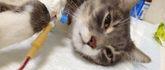
Photo 1 . Appearance of lesions in a young cat with a mucocele at a veterinary clinic appointment.
Photo 2 . View of the pumped out fluid from a cat with 1 photo.
Veterinary clinic of Dr. Shubin, Balakovo
Inflammation of the salivary gland in cats (sialadenitis, sialadenitis) is an acute or chronic disease, predominantly of a viral nature, in which the inflammatory process can affect any salivary glands and their ducts.
What is this?
Saliva production is an important part of digestion. Enzymes that are released during salivation simplify the swallowing process. A healthy dog produces about 1 liter of saliva per day, depending on weight and lifestyle.
Main major salivary glands:
- Parotid. They are located in the area of the ears, and their main feature is their small size;
- Submandibular. They are located in the back of the maxillofacial apparatus between several veins;
- Sublingual. They are located under the tongue and secrete saliva, which contains a protein secretion and is rich in mucin.
The secretion secreted by the salivary glands differs in its composition and functions. Inflammation of even one pair leads to painful and unpleasant sensations, as well as problems with the digestive system.
Types of salivary glands and their functions in the body
Salivary glands in cats are secretory structures of the anterior digestive tract. Located in the oral cavity and beyond. The main function is the production of saliva - a transparent, colorless liquid, which not only contributes to the process of eating food, but also protects the oral mucosa from drying out, mechanical, thermal, and chemical damage.
Types of salivary glands in cats:
- submandibular;
- sublingual;
- indigenous;
- infraorbital;
- zygomatic (orbital);
- parotid.
Important! The main major and minor salivary glands in cats are paired.
The main functions of the salivary glands in the animal body:
- Secretory. Production of a secretion that moisturizes the oral cavity. This is necessary for the two-way transport of chemicals between saliva and mucosa.
- Excretory, transport. The salivary glands take part in the transport of substances from the blood plasma into the secretion. Together with saliva, hormones (insulin, thyroxine, steroids) enter the oral cavity.
- Regulatory. The salivary glands have endocrine functions, stimulate protein synthesis, and promote the healing of mucosal damage.
Cat salivary glands:
- The parotid gland is the largest, paired, located at the base of each ear behind the temporal bone.
- The submandibular gland is a steam gland, located under the root of the tongue.
- The sublingual gland, the ducts of which exit under the tongue of the animal.
- Molar gland - in the front part of the lower jaw with ducts emerging in the area of the front teeth.
- The infraorbital gland, or zygomatic gland, is located in the bones of the upper jaw, very close to the lower orbital circle.
Reasons for the development of inflammatory processes
An infection caused by pathogenic microorganisms penetrates the salivary glands of animals through the blood, lymph, and ducts.
Reasons for the development of inflammatory processes in the salivary gland:
- mechanical damage, trauma to the glands and their ducts;
- concretions, stones in ducts;
- benign, malignant tumors in the gland;
- viral, parasitic, bacterial infections, diseases;
- dental diseases, inflammatory processes in the oral cavity;
- foreign objects in the ducts of the salivary glands, blockage of the ducts;
- acute, chronic intoxication of the body (poisoning with salts of heavy metals, toxins);
- colds, respiratory diseases, ear diseases (sinusitis, rhinotracheitis, otitis);
- failure to maintain pet oral hygiene;
- thermal, chemical burns of the mucous membrane;
- allergic reactions in the body;
- autoimmune disorders, diseases;
- stress, hypothermia, and other negative factors that weaken the cat’s body;
- hypersecretion of the salivary glands;
- mycoses.
Important! Sialadenitis can also be transmitted by contact if the main cause of the disease is infection.
Inflammation of the salivary glands is noted against the background of leukemia, plague, chlamydia, intestinal, streptostaphylococcal, adenovirus, herpesvirus infections. The cause of sialadenitis in cats can also be a surgical operation performed by an unqualified veterinarian in violation of the rules of asepsis.
Surgery:
- Drainage of salivary gland ducts.
- Removal of the salivary gland.
- Restoration of ducts.
- Doping - ligation of the ducts.
- Opening and removal of the cyst (mainly for sublingual and pharyngeal cysts).
The definitive treatment, which will completely eliminate the problem, is performed by surgical drainage of the cyst and subsequent removal of the affected salivary glands along with the ducts (Fig. 3, Fig. 4).
Ranulas and pharyngeal mucoceles are treated with marsupialization. This is one of the surgical methods for treating various cysts, not only in the area of the salivary glands. It is based on the transformation of a closed cyst cavity into an open pocket, that is, the opening of one of the walls of the cystic cavity (Fig. 5). In this case, a permanent large hole is created leading from the pathological focus of the cyst into the oral cavity. This is achieved by placing sutures on the walls of the cyst. As a result, the accumulated saliva flows directly into the oral cavity without creating swelling.
Types of inflammation of the salivary glands
Depending on the cause and mechanism of development, sialadenitis is classified into:
- traumatic;
- viral (infectious);
- contact;
- hematogenous;
- lymphogenous;
- post-operative.
Based on localization, the following types of inflammation are distinguished:
- Parenchymatous. Affects the parenchyma of the organ.
- Sialodohit. Affects the excretory ducts.
- Interstitial. The parenchyma of the salivary gland is affected.
Veterinarians also note the most common types of inflammation of the salivary glands in animals. Depending on which gland is affected by inflammation in cats, the nature of the inflammation is diagnosed: sialadenitis, mumps, sialocele (mucocele), sialodenosis.
Sialadenitis
Sialadenitis is an infection of the salivary glands of non-infectious etiology, mainly of a nonspecific nature. Primary or secondary pathology occurs due to blockage, obstruction of the salivary ducts with stones, conglomerates, foreign bodies, as well as due to hypersecretion of the gland.
Most often, one or two parallel salivary glands are affected. Multiple lesions of any glands are also possible. The disease occurs in acute or chronic form, often accompanied by suppuration.
The animal is lethargic, experiences pain and discomfort when eating or drinking water. Swallowing is difficult. Cloudy saliva constantly flows from the mouth. Anorexia (complete refusal to eat) depletes the pet's body. Due to tissue swelling, the shape of the cat’s skull and head changes. Palpation of swollen areas causes severe pain.
Mumps
Mumps in a cat is an inflammation of the parotid salivary gland. This pathology most often occurs against the background of concomitant viral, bacterial, parasitic, and respiratory ailments. The infection penetrates hematogenously, lymphogenously from foci of chronic inflammation.
Severe swelling of the tissues in the area of the parotid salivary gland, pain, enlargement of the gland, refusal of food, water, lethargy, high temperature, fever are the main signs of mumps in cats. Swallowing is difficult. When opening the mouth and palpating the swollen areas in the gland area, the cat experiences severe acute pain.
Diagnostic methods
If you suspect mumps, your pet should be seen by a veterinarian. After a clinical examination, a biopsy will be taken from the animal and a general clinical blood test will be performed. Based on the results of the biopsy, the veterinarian will be able to exclude a neoplastic cause of the pathology and determine the causative agent if the disease is infectious.
An informative diagnostic method is an x-ray examination of the dog’s skull in two projections. X-ray will help detect a neoplasm, blockage of the lumen of the gland with mineral formations, an anomaly in the development of the organ.
Acute and chronic forms of inflammation
Acute and chronic forms of inflammation of the salivary glands have different intensity of manifestation of their characteristic symptoms, depending on the location of the lesion.
- At the beginning of the inflammatory process, the gland is swollen and very edematous.
- The tissues at the site of inflammation are hard.
- The temperature rises sharply by 1-2 degrees from the physiological norm.
- Flakes, particles of pus, and blood are noticeable in the saliva.
The pet's behavior changes. Attacks of depression are replaced by anxiety. The appetite is reduced or the cat completely refuses to eat.
Important! In the acute form of the disease, symptoms appear intensely and suddenly, but can also disappear quickly, which does not mean that the pet is healthy. The pathology can become chronic.
If treatment is not started, the inflammatory process will affect nearby tissues. Necrosis and sepsis may develop.
Sialadenitis mainly affects kittens and young cats. In older animals, blockage of the salivary ducts, disturbances in salivary excretion due to age-related changes, neoplasms, and conglomerates in the glands are noted.
Forms of the disease
A severe form of the disease is the plasmacytic-lymphocytic form . It is caused by various viruses - rhinotracheitis, panleukopenia, hyperreaction of the immune system. This form has complications. The gums begin to bleed and swell. Without treatment, complications develop.
The dental form of the disease is accompanied by sore gums, bleeding and swelling. Diseased tissues become bluish in color. The submandibular lymph nodes begin to swell. Unlike the plasmacytic form, the disease is caused not by a virus, but by accumulated plaque. The dental form is treated faster and rarely causes complications.
Juvenile gingivitis is a form of the disease characteristic of cats under the age of 1.5 years, also called juvenile gingivitis. Inflammation of the gums and pharynx occurs immediately after the eruption of permanent teeth. If left untreated, periodontitis may develop.
Symptoms and first signs of inflammation
The clinical picture and intensity of manifestation of signs of the disease depend on the stage, inflammatory process, age, conditions of detention, individual physiological characteristics of the body, and the state of the pet’s immune system.
Symptoms and first signs of inflammation in cats:
- decreased appetite, refusal to eat (dysphagia), increased thirst;
- weight loss;
- nasal and eye discharge;
- fever, fever, heat;
- salivation (salivation);
- changes in heart rate, pulse;
- nausea, gagging, vomiting;
- lacrimation;
- bloody, purulent discharge from the oral cavity;
- swelling of the muzzle on the side of the affected gland;
- pain when swallowing, opening the mouth;
- unpleasant odor from the mouth;
- viscous muddy saliva.
The oral mucosa is hyperemic and edematous. On the surface you can notice small ulcers and bleeding lesions. Breathing is difficult, frequent (dyspnea). The animal behaves restlessly.
The cat meows pitifully, attracting the owner's attention. The animal rubs its muzzle with its paw and experiences discomfort. Opening the mouth and palpating the swelling causes acute pain.
Symptoms
Some diseases may not appear. For example, epulis, when small in size, does not cause much discomfort or pain in the pet.
Signs of the appearance of growths and compactions:
- bad odor, often putrid, from the mouth;
- salivation (intense secretion of saliva) mixed with blood;
- tooth decay with subsequent bleeding;
- distortion of the muzzle due to an increase in tumor volume;
- itching in the mouth area, so the cat is constantly scratching;
- sneezing caused by irritation of the mucous membrane;
- refusal to feed, inactivity, lethargy;
- emaciation.
With the subsequent development of the process, the animal stops eating, the coat becomes coarser and dull, and signs of dehydration appear, since drinking water causes pain.
Symptoms of the severe stage are blackening of the stool due to swallowing a large amount of blood from the affected gums; chamfering of teeth.
Methods for treating inflammation of the salivary glands in cats
Before treatment is prescribed, a comprehensive examination is carried out. The treatment regimen is selected individually and depends on the degree of the disease.
Treatments for salivary gland inflammation in cats include:
- drug, imptomatic therapy;
- physiotherapy (electrocution, UHF therapy, gland massage, solux);
- surgical intervention;
- alternative medicine.
Important! Before prescribing medications, it is necessary to establish the root cause, determine the type, nature, and nature of infectious agents.
To relieve intoxication, animals are injected with physiological solutions intravenously and detoxification therapy is prescribed.
Surgical treatment involves:
- resection, removal of the affected salivary gland in severe cases, for example, with neoplasms, multiple stones, advanced sialadenitis;
- surgical removal of stones and conglomerates to restore the functions of the ducts;
- opening the abscess cavity, draining abscesses, salivary gland ducts;
- doping (ligation) of ducts;
- removal of tumors.
When purulent fluctuating foci appear, they are opened and the exudate is subsequently removed. The wound cavity is washed with antiseptic and antibacterial solutions.
The prognosis after surgical treatment is generally favorable if the owner follows all the veterinarian’s recommendations during the recovery period.
Etiology of damage to the salivary glands:
- Trauma – blunt or penetrating wounds. Iatrogenic injuries (received during the performance of any diagnostic procedures) are possible.
- Foreign bodies (accidentally eating sharp and hard blades of grass and leaves can lead to blockage of the ducts of the parotid salivary gland).
- Bacterial and viral infections (eg leukemia).
- Formation of calculi (stones) in the ducts of the salivary glands.
- Immune-mediated diseases.
- Neoplasia (tumors).
- Idiopathic (unknown) etiology.
- Breed predisposition in cats is uncharacteristic.
- Age predisposition: mostly young animals are affected; older animals are more likely to have tumors.
The main danger of the disease lies in the neglect of the process, which leads to indigestion and perverted appetite, but the most terrible consequence may be a cancerous tumor, which often develops in the tissues surrounding the affected salivary gland as a result of the constant irritating effect of saliva.
Cat feeding diet
The veterinarian will select the optimal nutrition and diet for a cat with inflammation of the salivary glands. Food should be easily digestible, nutritious, and have a liquid consistency so that the animal can easily swallow it.
With a natural diet, increase the amount of fermented milk products. Cats are given low-fat cottage cheese, skim milk, kefir, and natural yogurt. It is advisable to give boiled meat in the form of minced minced meat. Cereals are well boiled in low-fat meat broth. You can feed your cat only fresh food at room temperature. There was always fresh drinking water in the bowl.
With a ready-made diet, cats are prescribed medicinal ready-made food in the form of mousses and pates. The drying is soaked in kefir, yogurt, broth, and water.
Prevention measures
Proper care, a balanced diet, and good living conditions will help to avoid the development of the inflammatory process.
- Monitor your pet's immunity. Do not skip scheduled vaccinations or revaccinations.
- Do not give your cat fish with bones or tubular bones.
- Pay attention to your cat's oral hygiene.
- Treat dental, other systemic, viral, and concomitant diseases in a timely manner.
- Do not allow your pet cat to come into contact with stray animals.
- Protect your pet from stress, hypothermia, and any negative factors that weaken the body’s immunity.
- Take your cat to the clinic two to three times a year for preventive examinations.