Indications for use
Esophagoscopy in dogs and cats
is a very useful tool for diagnosing and treating esophageal diseases. It is a highly reliable diagnostic method for assessing esophageal disorders that affect the mucous membrane or alter the lumen of the organ.
Esophagoscopy of cats and dogs
allows you to obtain samples for cytology and histology. The most common disorders of the esophageal mucosa and lumen diagnosed by esophagoscopy are foreign bodies, esophagitis and strictures. Esophageal ulcers, fistula, and neoplasia are more common in dogs and cats. Megaesophagus, diverticula, vascular ring abnormalities, and digestive disorders are best studied with contrast radiography, but endoscopy can provide more accurate and supportive information for these pathological conditions. Indications for esophagoscopy include clinical signs related to esophageal disease, including regurgitation, dysphagia, unophagia, and unexplained salivation.
Esophagoscopy in cats and dogs also has a useful therapeutic role. The main therapeutic indications for esophagoscopy are removal of foreign bodies and dilation of esophageal strictures under direct visualization. A series of grasping forceps assists the endoscopist in grasping and carefully removing the foreign body. Accurate assessment of the esophagus after removal is important to assess mucosal damage and rule out perforation. Conservative treatment of benign esophageal strictures is currently the most reliable approach to these pathologies in animals and humans. Mechanical dilatation of strictures is achieved by dilation of a balloon catheter. The author proposed a technique using endoscopic electrocautery incisions of the stricture before balloon expansion.
Which cats are at risk?
The impetus for the development of esophageal diseases is overeating or, conversely, starvation.
Diseases of the esophagus can be congenital and are diagnosed in kittens and young animals, or acquired, which occur in old cats. Risk factors:
- neurological problems;
- incorrectly selected food;
- fasting or overeating;
- inflammation of the mediastinum;
- brain pathologies;
- inflammatory process in muscles.
Patient preparation
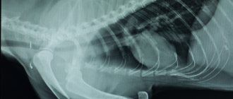
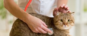
For patients undergoing esophagoscopy, a 12-hour fast is required. Contrastography studies should be avoided before esophagoscopy because barium may interfere with the examination. If necessary, water-soluble, non-ionic iodinated agents are preferred, although mucous viscous suspensions of barium (for example, kefir) are better for contrast studies of the esophagus. Salt rinsing or aspiration can be used to remove residual contrast medium. General anesthesia is required; The patient should be placed in the left lateral position, with an endotracheal tube and oral dilator. Thoracic radiography should be carefully checked to exclude esophageal perforation, as esophagoscopy should not be performed in this case.
Colon diseases
Anatomy of the large intestine
- Macroscopic anatomy
- Microscopic anatomy
- Physiology of the large intestine
- Concepts of colon diseases
Acute colitis
- general information
- Clinical diagnosis
- Treatment
Idiopathic colitis
- Etiology
- Clinical diagnosis
- Treatment
Parasitic colitis
- Etiology and pathogenesis
- Treatment
Histiocytic colitis
- general information
- Clinical diagnosis
- Treatment
Granulomatous colitis
- general information
- Clinical diagnosis
- Treatment
Eosinophilic colitis
- general information
- Clinical diagnosis
- Treatment
Irritable bowel syndrome
- Etiology
- Clinical diagnosis
- Treatment
Constipation
- Etiology
- Clinical diagnosis
- Treatment
Pseudocoprostasis
- general information
- Clinical diagnosis
- Treatment
Megacolon
- Etiology
- Clinical diagnosis
- Treatment
Colon tumors
- Etiology
- Clinical diagnosis
- Treatment
Rectal stricture
- Etiology
- Clinical diagnosis
- Treatment
Rectal prolapse
- Etiology
- Clinical diagnosis
- Treatment
Anal sinus diseases
- Etiology
- Clinical diagnosis
- Treatment
Anal tumors
- Etiology
- Clinical diagnosis
- Treatment
Esophagoscopy procedure and normal results
Due to its simple tubular structure, the esophagus is usually easily examined with an endoscope. With the patient's head and neck extended, the endoscope follows the dorsal aspect of the endotracheal tube in the mouth until the upper esophageal sphincter reaches dorsal to the larynx.
The entrance to the cervical esophagus is close, but provides low resistance to minimal endoscope tip pressure. If any resistance to this maneuver is felt, the endoscope should be pulled back and redirected to a central and dorsal position.
Before moving into the esophagus, air is pumped until the lumen is clearly visualized. The cervical esophagus has flexible longitudinal mucous folds that disappear after air insufflation.
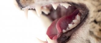
The normal mucous membrane of the dog's esophagus is pale pink or grayish, the surface is smooth and shiny. Pigmented mucosal patterns can be seen in pigmented dog breeds such as Chow Chow, Shar Pei, etc. Some of the peredophageal structures leave an imprint on the flaccid wall of the esophagus. The outline of the trachea can be observed by making a curved impression against the ventral wall of the cervical esophagus.
In the middle third, when the esophagus approaches the base of the heart, the aortic arch is visible pulsating towards the wall of the organ. By moving the tip of the endoscope caudally, the imprint of the left main bronchus is clearly visible. Sometimes the imprint and pulsation of the left subclavian artery (proximal to and on the same side of the aortic arch), the left atrium (just distal to the left bronchus) and the veins of the azygos (in the distal third of the esophagus) are visible.
By slowly moving the endoscope along the esophagus, the gastroesophageal sphincter is easily accessible. At the gastroesophageal junction, the color of the mucous membrane changes sharply from pale pink to red relative to the gastric mucosa. The landmark between two different epithelia (esophageal lining and gastric cuboidal epithelium) is usually characterized by an irregular edge, sometimes protruding several millimeters into the lumen of the esophagus. Various aspects of this area may be observed in the average patient and should not be considered pathological. The lower esophageal sphincter may appear as a mucous rosette, but varying appearances may be normal. The normal lower esophageal sphincter is usually closed, although this function is largely related to the anesthetic protocol used. Minimal or no resistance is usually encountered when advancing the endoscope through the sphincter, but sometimes left deviation of the abdominal esophagus requires slight deflection of the endoscope tip to complete this maneuver. Examination of the lower esophageal sphincter is completed only after visualization of its gastric side by endoscopic retroviration (“J” maneuver).
The cat esophagus differs from the dog esophagus in the presence of clearly visible submucosal vessels and circular rings formed by circular folds of the mucous membrane, which gives a typical image of the distal tract. It is generally difficult to obtain esophageal mucosa in dogs and cats. Obtaining a mucosal sample may be necessary if a mass lesion is present or if esophagitis is suspected. While proliferative lesions are easily biopsied with traditional biopsy forceps, the esophageal mucosa is tough and usually cannot be cut with these forceps. The tubular anatomy of the esophagus complicates the procedure. A valid alternative is a forceps with a central spike attached to the esophageal mucosa or capsule for suction biopsy. A cytological sample obtained by brushing may be useful in cases of esophageal neoplasia.
Structure and functions
A cat's esophagus is a long, hollow tube that connects the pharynx and stomach. The organ is designed to move the food bolus from the oral cavity further into the gastrointestinal tract. Located in the neck, passes near the diaphragm and heart, connecting to the stomach. Thanks to muscle tissue, the esophagus can push food in both directions - from the mouth to the stomach and vice versa. This helps the animal get rid of the fur accumulated inside. In the absence of food, the walls of the organ fit tightly to each other. The structure of a cat's esophagus is shown in the table:
| Organ structure | Characteristic |
| Shells | Mucous |
| Submucosa | |
| Muscular | |
| Adventitial serous | |
| Musculature | Ellipsoidal, spiral and longitudinal direction of muscle fibers |
| Blood supply | Carotid, bronchoesophageal artery |
| Jugular vein | |
| Innervation | Branches of the vagus nerve |
| Sympathetic trunk | |
| Lymphatic drainage | Cervical and mediastinal lymph nodes |
Good to know
- Histopathological changes in small animal hepatobiliary diseases
- Clinical characteristics of hepatocellular carcinoma
- Clostridium perfringens and Clostridium difficile in dogs and cats
- Giardia duodenalis and Cryptosporidium spp. humans from domestic dogs
- Platynosomum fastosum infection in outdoor cats
- Complex cholangitis/chonlangiohepatitis in cats against the background of Platynosomum fastosum infestation
- Histopathological features, immunophenotyping, cloning and eubacterial fluorescence in situ hybridization in cats with lymphocytic cholangitis/cholangiohepatitis
- Differentiating benign and malignant causes of lymphocytosis in the bone marrow
- Prognostic markers of hepatic lipidosis in cats: a retrospective study of cats
- Pancreatitis and diabetes mellitus in cats
Causes and symptoms: what ailments are there?
Dilatation of the esophagus
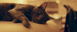
Megaesophagus in cats is characterized by stretching of the organ, which results in a protrusion of the wall. Stretched muscles cannot contract normally and move food, causing the food eaten to form a lump. The animal vomits after drinking liquid. The cause is hereditary diseases, disruption of the autonomic nervous system, endocrine diseases, heavy metal poisoning, and chronic inflammation of the esophagus. Symptoms:
- frequent vomiting of foam, undigested food or water;
- dehydration;
- weight loss;
- fragility and dullness of wool;
- purulent runny nose and cough;
- pneumonia due to inhalation of vomit.
Diverticulum
A diverticulum occurs when foreign bodies enter the body and become stuck in the organ.
The disease is characterized by a blind protrusion of the organ wall. It develops against the background of pathology of the vascular ring, in which unnaturally located arteries - the aorta and the Batal duct - compress the esophagus. It is also caused by foreign objects stuck in the organ and preventing the movement of the food bolus. Esophageal diverticulum manifests itself as follows:
- pain after eating;
- hyperthermia;
- the cat cannot eat, even if it wants to eat;
- regurgitation immediately after eating.
Inflammation of the esophagus
Veterinarians note that cats have very delicate digestive organs, so esophagitis can develop if an irritating substance affects the structures of the esophagus for 20-25 minutes.
Esophagitis in cats is characterized by an inflammatory process that occurs due to accidental ingestion of alkaline or acidic solutions. Another cause is the cat’s careless consumption of hot liquids or food. The inflammatory process occurs when food is thrown back from the stomach (reflux gastritis), in which gastric juice irritates the walls of the esophagus. Symptoms:
Inflammation of the esophagus in an animal is accompanied by characteristic symptoms such as difficulty swallowing and regurgitation.
- pain while eating;
- difficulty swallowing (dysphagia);
- regurgitation;
- salivation;
- aerophagia;
- temperature increase;
- weight loss;
- loss of appetite.
Stricture
It is characterized by the proliferation of fibrous tissue and narrowing of the organ. It develops due to the entry of foreign objects into the cavity of the esophagus, as a result of tumors, traumatic injuries, incorrectly performed surgical intervention, uncontrolled use of medications to the pet by the owner, inhalation or ingestion of toxic substances. Manifestation:
- pain when eating;
- vomit;
- swallowing disorder;
- increased salivation;
- weight loss
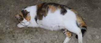
Foreign body jam
The pathological process in the esophagus develops when accidentally swallowing small hard sharp objects - fish bones, fragments of plastic toys. The condition is dangerous due to organ blockage, internal bleeding and secondary infection. Doctors identify the following signs:
Excessive drooling in a pet may be a symptom of the presence of sharp objects stuck in the organ.
- difficulty or inability to swallow;
- refusal of food;
- lethargy, reluctance to play;
- pain in the esophagus;
- constant reflex swallowing;
- the cat shakes his head;
- anxiety and restlessness;
- profuse salivation, sometimes mixed with blood.
Laboratory techniques and biopsy collection
Fecal analysis
- Established order
- Trypsin test
- Microscopic examination of feces
- Test for worm eggs
- Bacteriological research
Dynamic tests
- Xylose Absorption Test
- Quantitative Fat Absorption Test
- N-benzoyl-L-tyrosyl-p-aminobenzoic acid test
- Ammonia tolerance test
- Bromosulfthalein retention test
Biopsy collection
- Fiberendoscopy
- Esophageal/gastric examination
- Colon examination
- Small intestine biopsy
- Liver biopsy
- Abdominal paracentesis
Hematology and clinical biochemistry
- Routine hematology
- Whey proteins
- Sodium to Potassium Ratio
- Trypsin-like immunoreactivity (TLI) test
- Whey Folate and Vitamin B12
Study for liver diseases
- Introduction
- Serum alanine aminotransferase
- Serum aspartate aminotransferase
- Gamma glutamine transferase
- Serum alkaline phosphatase
- Serum bilirubin
- Serum bile acids
- Plasma ammonia
Diseases of the digestive system of cats
Common diseases
In cats, the most common diseases of the digestive system are: stomatitis, pharyngitis, acute and chronic catarrhal gastritis, gastric ulcer, enteritis, peritonitis, ascites, acute parenchymal hepatitis, inflammation of the biliary tract and gallbladder.
Prevention of digestive diseases
To prevent diseases of the digestive system of cats, it is necessary that the animal’s diet is balanced in calories, vitamins and minerals, and also contains a sufficient range of high-quality feed. It is necessary to ensure that toxic or poisonous substances do not enter the animal’s food. In addition, it is necessary to control the purity of drinking water.
If there are diseases of other body systems, they should be treated promptly. In addition, it is necessary to protect cats from local and general hypothermia and prevent feeding them cold and hot food.
Therapy
In mild cases, medication and symptomatic treatment may be prescribed:
- Parenteral nutrition, when nutritional solutions are administered intravenously. The veterinarian may also resort to injecting saturated broths directly into the stomach, which is done using a catheter.
- If inflammation is present, broad-spectrum antibiotics are prescribed.
- The esophagus is washed with sucralfate solution.
- Affected and ulcerated tissues are cut away during endoscopy.
- Treat secondary diseases using recommended therapy.
But in difficult situations, all this gives either a weak effect or does not lead to improvements at all. Then only surgery will help. During surgery, the “swollen” part of the esophagus is cut out, suturing the edges with a purse-string suture. The regeneration time is about two weeks, during which the cat is fed through a tube or resorted to parenteral feeding. After this period, the animal is gradually transferred to normal food, first giving preference to saturated broths and vegetables, boiled and mashed through a sieve.
Treatment of infectious diseases of the digestive system of cats.
Bacterial and parasitic diseases are usually treated with drugs that kill the infection. There are no specific drugs for the treatment of viral diseases yet. Antibiotics (effective against bacteria) are usually given orally to the cat for several days until recovery is evident, although their effectiveness in treating digestive diseases is not recognized by all doctors. For septicemia (blood poisoning), antibiotics are administered by injection. The veterinarian prescribes antibacterial treatment, taking into account the cause of the disease, the results of previous treatment and the cost of medications.
Progress in the study of the life cycle of parasites and the improvement of antiparasitic drugs have made it possible to treat cats for parasites. Response to treatment is usually rapid, and a single course of treatment is often sufficient if the infection is recent and the damage caused by the parasites is not severe.
Preventing cat infestation with parasites largely comes down to compliance with sanitary requirements. This is achieved primarily by providing adequate space for the cat, regularly cleaning the living area and cleaning the litter box. In addition, proper nutrition and living conditions will minimize stress on your cat, helping to keep it healthy.
Inflammation of the esophagus as a cause of its expansion
A fairly common cause of dilation of the esophagus is its own inflammation. When organ tissues are affected by some negative environmental factors, including the action of irritating and caustic substances, they lose elasticity and firmness, which leads to the formation of a “pocket”. Because of this, esophagitis, which is not taken seriously by many specialists, is actually quite a serious disease that needs to be treated immediately.
How can you tell if your cat has an inflamed esophagus? The clinical picture of the pathology is not very characteristic, the symptoms are few. So, the cat may vomit, he is reluctant and eats little (it hurts), he tries to drink more or prefers semi-liquid, soft food.
Predisposing factors
"Megaesophagus" may be the only manifestation of a systemic disease (such as myasthenia gravis). Megaesophagus is also one of the signs of dilatation (expansion) of the stomach and a general disorder of motility of the gastrointestinal tract. The disease can be congenital or acquired, primary or secondary. In the first case, it develops as an independent disease, in the second, it is a consequence of pathologies already existing in the animal. Megaesophagus of the primary type is often considered an idiopathic disease, since no objective reasons for its occurrence can be identified.
Some researchers believe that the likelihood of this disease occurring is closely related to the feeding characteristics of the animal. So, when a cat eats exclusively dry food, the risk increases. This is due to the fact that cats, due to the specific, “predatory” structure of their jaws, do not particularly chew food. Accordingly, it is weakly wetted by saliva. If a cat eats irregularly (that is, he has not developed a feeding regimen), then he overeats every time, trying to eat as much food as possible at one time. Over time, this can lead to chronic enlargement of the esophagus. Accordingly, to prevent this from happening, it is necessary to give preference to canned or “natural” food.
Treatment
Therapy includes elevated feeding and/or gastrostomy tube placement and nutritious, high-calorie diets. Surgical treatment remains controversial.
Pharmacological management using drugs that relax the gastroesophageal sphincter or enhance the strength of esophageal reduction has been reported to be disappointing, although sildenafil, a phosphodiesterase-5 inhibitor, has been reported to have a pronounced effect on esophageal contractility in cats, altering the propagation and amplitude of esophageal contractions. Nifedipine, a calcium channel blocker, resulted in transient clinical improvement (2 months) in an adult German shepherd dog with megaesophagus.
Cat digestive system and diseases
Teeth play an important role in the digestion process in cats. They are necessary for biting and crushing food. A cat has 30 teeth, and they are arranged as follows:
- lower jaw - 6 incisors, on each side a canine and three molars;
- upper jaw - 6 incisors, on each side a canine and four molars.
The main signs of diseases of the digestive system in cats.
Symptoms of diseases of the digestive system
may include excessive drooling, diarrhea, constipation, vomiting or regurgitation, loss of appetite, bleeding, abdominal pain and bloating, and dehydration. A cat may react to stomach pain by meowing loudly, howling, or adopting unusual postures (such as squatting and arching its back).
The location and nature of the disease can often be determined by external signs. For example, unusual biting, chewing and swallowing often indicate diseases of the mouth, teeth, jaws or esophagus. Nausea is usually associated with inflammation of the lining of the stomach or intestines (gastroenteritis) caused by infection or irritation. However, non-digestive problems such as kidney disease can also cause nausea.
Diarrhea can be a symptom of diseases of the digestive system, but there are many reasons for its occurrence. It is important to prevent prolonged diarrhea in your cat, as dehydration and salt (electrolyte) imbalance are possible. If there is a large loss of fluid, the cat may go into shock.
Changes in stool color, consistency, or frequency may also be signs of problems with your cat's digestive system. Black, tarry stool may be a symptom of bleeding in the stomach or small intestine. Straining during bowel movements is usually associated with inflammation of the rectum and anus. Bloating (flatulence) can occur as a result of a buildup of gas, fluid, or food you've eaten—usually due to reduced activity of the muscles that move food through the digestive system. Bloating can also be caused by a physical obstruction, such as a foreign object or intussusception (the penetration of one part of the intestine into another), or simply by overeating.
Diseases of the digestive system in cats.
Based on materials from www.merckmanuals.com
Digestive system of a cat
includes all the organs used in the process of receiving and assimilating food - mouth, esophagus, stomach, liver, pancreas, intestines, rectum and anus.
The digestion process begins from the moment food enters the mouth and the cat begins to chew it. Enzymes found in saliva begin the chemical breakdown of food. Then the process continues - after swallowing, additional breakdown of food occurs in the stomach, absorption of nutrients in the intestines, and removal of waste from the body. Digestion is critical not only for obtaining nutrients, but also for maintaining the proper balance of fluids and electrolytes (salts) in a cat's body.
The functions of a cat's digestive system can be divided into four categories: digestion of food
,
nutrient absorption
,
peristalsis
(movement through the digestive tract), and
excretion of feces
.
When treating digestive disorders, the doctor's first goal is to identify the part of the digestive system where the problem is present, then determine the causes and select appropriate treatments.
Anthelmintic and Gamavit for cats
When carrying out deworming, you need to remember that modern drugs for helminths are selectively toxic to parasites and practically safe for mammals. This is due to the huge differences between the organisms of parasites and warm-blooded animals. However, there have been cases where, after deworming, animals experience undesirable consequences (vomiting, diarrhea, etc.).
The reason is the absorption of helminth decay products by the intestines, if cases of overdose are excluded. Modern drugs for helminths, when used correctly (strictly according to the instructions), kill parasites directly in the intestines (or other place where they live), and do not simply expel them from the body. Due to the decomposition of worms, their toxins are absorbed by the intestines and enter the bloodstream, causing intoxication of the body.
Thus, it turns out that the more effectively the drug helps get rid of helminths, the stronger the adverse consequences for the animal’s body (more helminths die, more toxins enter the body).