3974Pavel
In most cats and cats, bilateral inflammation develops, which is not localized, but covers the entire thoracic region. Lack of timely monitoring can lead to death. With pleurisy, the pulmonary region and heart are at risk, and these are vital systems of the pet’s body.
In case of illness, the pleural surface is filled with moisture. In a normal healthy state, the pleura in the chest also releases moisture, but this amount is enough for “basic” processes to occur. When the fluid volume is exceeded, the pressure on the organs increases. If treatment is not organized, pathologies with the lungs and heart cannot be avoided.
© shutterstock
How to suspect pleural effusion?
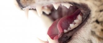
Signs of the presence of pleural effusion are inspiratory dyspnea - rapid breathing with difficulty exhaling and rapid heartbeat - tachycardia. There may also be signs of cardiac and respiratory failure - cyanosis of the mucous membranes of the oral cavity and tongue. If these symptoms are present, it is imperative to examine the animal to confirm the presence or absence of pleural effusion and to determine the reasons that provoked it.
Since the accumulation of fluid in the chest cavity causes compression of vital organs such as the heart and lungs, it is necessary to remove this fluid as quickly as possible. The amount and nature of the fluid may vary, which determines the severity of symptoms.
In our clinic, there were cases when it was necessary to remove 150-180 ml of fluid from the right and left halves of the chest cavity, respectively, and in total it reached 350 ml. In such conditions, due to strong compression, the work of the animal’s heart and lungs is significantly hampered, creating a life-threatening situation.
Diagnosis of pathology
A veterinarian can make a preliminary diagnosis based on a visual examination of a sick cat. When percussing the chest, the physician will detect dullness in the lower part of the lung. The upper limit of dullness is located in the horizontal direction, even if the position of the animal’s body changes.
An effective diagnostic method for suspected hydrothorax is an X-ray examination of the chest area. The accumulated serous fluid is detected in the form of a continuous darkening of the lower half of the pulmonary field. The horizontal upper limit of darkening fluctuates during the animal's breathing period.
X-ray of the chest of a cat with hydrothorax
A diagnostic method such as transudate examination is informative. A fluid sample is obtained by puncture. A distinctive feature of transudate with thoracic hydrops is its protein content below 3%.
First of all, it is necessary to exclude pleurisy in the animal, which usually occurs with an increase in temperature. If a higher than 3% protein concentration is detected in the test fluid taken during puncture of the chest cavity, the doctor may suspect pleural inflammation in the pet.
In order to carry out a differential diagnosis, the animal is prescribed a clinical and biochemical analysis of blood, urine, coprological examination, electrocardiography of the heart, ultrasound examination of the myocardium, liver, kidneys and other organs. The prognosis is often cautious or unfavorable.
To see what hydrothorax looks like in a cat during an ultrasound examination, watch this video:
Types of pleural effusion
Depending on the underlying disease that caused this symptom, pleural effusion may differ in cellular composition and physicochemical characteristics. In some cases, the fluid may show signs of different types of effusions, since there may be a combination of several reasons that lead to the accumulation of fluid in the chest cavity.
Transudate is a relatively clear liquid, contains few cells, has a low relative density, and low protein content. The causes of transudate are diseases in which there is a low concentration of albumin in the blood - liver failure, enteropathy and protein-losing nephropathy, prolonged fasting, loss of plasma due to extensive skin damage (for example, burns).
Exudate is most often a turbid, viscous liquid containing a large number of cells with a predominance of neutrophils and has a high concentration of protein. There are septic and aseptic exudates. Septic exudate is characterized by the presence of microorganisms.
The cause of exudative pleural effusion can be an inflammatory process of the chest cavity, fungal or bacterial granulomatous processes. Exudative effusion often develops with feline infectious peritonitis.
Aseptic form of exudate, i.e. without the participation of microorganisms, most often develops when neoplastic or cystic formations rupture.
Hemorrhagic effusion - characterized by a significant content of red blood cells and a high concentration of protein in the liquid contents. As a rule, it develops as a result of an injury.
Chylous effusion is a white liquid, contains a significant amount of protein and triglycerides, and has a high density.
The causes of the development of chylous effusion are lymphangiectasia (expansion) of the thoracic lymphatic duct, lymphoproliferative diseases of the mediastinal lymph nodes and their vessels, compression of the lymphatic vessels of the thoracic cavity by a tumor, torsion of the lung lobe, cardiovascular pathologies, dirofilariasis, diaphragmatic hernia. Less common is chylous effusion into the pleural cavity as a result of rupture of the thoracic lymphatic duct.
Causes of the disease
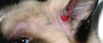
The appearance of a focus of inflammation on the pleura can be caused by many different reasons, the most common of which include:
- various pathogens of diseases in cats, namely worms, harmful bacteria, viruses, fungi and protozoa;
- consequences of inflammation occurring in the immediate vicinity of the serous membrane. This is especially true for lung diseases such as pneumonia or abscess;
- oncological pathologies, for example, lymphosarcoma or mesothelioma. They often lead to such a terrible disease as metastatic pleurisy, which is extremely difficult to treat, due to the fact that the pet’s body is already weakened by a malignant tumor;
- hypoalbuminemia, that is, a low amount of protein in the blood fluid, which is caused by problems with the liver and gastrointestinal tract;
- mechanical damage to the chest resulting from trauma;
- when drugs such as Furadonin or its closest analogues are used to treat a cat, without the supervision of a specialist;
- with a hernia of the diaphragm;
- the presence of a blood clot in the cat’s lungs, which prevents normal blood supply to this organ;
- blood (hemothorax) or lymph (chylothorax) entering the pet's chest;
- for pancreatitis and renal failure.
Owners should also pay attention to the fact that the cause of cat pleurisy may well be prolonged stress, vitamin deficiency, poor diet, overwork, and hypothermia.
Symptoms of hydrothorax: how to recognize the disease in your pet?
The first thing you need to pay attention to is your pet’s behavior. With hydrothorax, shortness of breath gradually develops, the body temperature remains normal, but due to internal changes, the mucous membranes become bluish (cyanosis). When palpating the chest area, there is no pain, but there is swelling.
One of the features of hydrothorax is its stages. Periods of exacerbation may be followed by temporary relief. However, don’t mistake this for recovery! Remember that hydrothorax in cats requires professional veterinary treatment.
Signs of hydrothorax (dropsy) in cats:
- general weakness,
- fast fatiguability,
- increasing shortness of breath,
- appetite disorders,
- cyanosis of the mucous membranes (pressure restores the natural color),
- swelling in the perineum, chest, eye area and paws.
A cat suffering from dropsy loses the desire to play. She spends more time in solitude, has virtually no contact with people, and does not go into arms. With hydrothorax, animals lie down with their paws widely spread forward and their heads stretched upward. Breathing becomes heavier (long inhalation and short exhalation).
Hello student
DIVISION OF THE LUNGS INTO LOBE AND BRANCHING OF THE BRONCHIAL TREE
The division and designation of the lobes of both lungs is carried out in accordance with the branching of the bronchial tree, arbor bronchalis. The transition from the trachea to the lungs is formed by the main bronchi, bronchi principales, which are further divided into lobar bronchi, bronchi lobares. The structure of the main and lobar bronchi is similar to the structure of the trachea. The right main bronchus has a noticeably larger diameter compared to the left. At the site of the division of the trachea into the main bronchi there are bifurcations, bifurcatio tracheae, the latter form an angle from 63° to 68° in a dog, and from 30° to 40° in a cat (Bitter, 1974; Schaake, 1974). In this case, the left main bronchus deviates more strongly from the axis of the trachea, forming an angle from 45° to 50° with it in the dog. Both main bronchi run first caudolaterally, then caudally and pass into the caudal lobar bronchi.
Directly behind the bifurcation from the right main bronchus, bronchus principalis dexter, the right cranial lobar bronchus, bronchus lobaris cranialis dexter, extends almost at a right angle. Soon after this, the middle lobar bronchus, bronchus lobaris medius, departs ventro-laterally. The next branch is the ventro-medially directed accessory lobar bronchus, bronchus lobaris accessorius. The distances between the branches vary depending on the size of the dog. On average, the distance is equal to the diameter of the branching lobar bronchus. In a cat, the distances are 5 - 8 mm. From the left main bronchus, bronchus principalis sinister, the left cranial lobar bronchus, bronchus lobaris cranialis sinister, extends cranially. In a cat, the branch site is 7-8 mm away from the bifurcation. After the branch of this bronchus, the left main bronchus continues as the left caudal lobar bronchus, bronchus lobaris caudalis sinister.
Segmental bronchi, bronchi segmentales, of various sizes depart from the lobar bronchi. They are divided depending on their direction into dorsal and ventral segmental bronchi (Adrian, 1959; Felder, 1962).
The lungs of dogs and cats are distinguished by very deep interlobar fissures, fissurae interlobares. These slits extend through the dorsal margin almost to the base of the lung. Sometimes the left interlobar fissure is not so deep.
The left lung, pulmo sinister, is divided into two approximately equal lobes, cranial and caudal, lobus cranialis and lobus caudalis, the cranial lobe is further divided into cranial and caudal parts, pars cranialis and pars caudalis. Due to the limited space between the heart and the lateral chest wall, this part of the lung can expand only to a small extent. The caudal part slightly overlaps the cranial part posteriorly. In the region of the obtuse (dorsal) edge, margo obtusus, there is no separation between the parts. The caudal lobe is compact, pyramidal in shape with its base on the diaphragm.
Rice. 5 and 6. Lobes, bronchial tree and lymph nodes of the lungs of dogs and cats, diagram, dorsal view (after Mtiller, 1938) a lobus cranialis; b lobus medius; with lobus caudalis; d lobus accessorius (located in depth and indicated by a dashed line)
1 incisura cardiaca pulmonis dextri or sinistri; 2 tracheas; 3 bronchus principalis; 5 - 8 bronchi lobares: 5 bronchus lobaris cranialis, 6 bronchus lobaris medius, 7 bronchus lobaris accessorius, 5 bronchus lobaris caudalis; 9 I or III ventral bronchus segmentalis of the caudal lobe, 10 I dorsal bronchus segmentalis of the caudal lobe; 11 other bronchi segmentales; 13 In. tracheobronchalis sinister; 14 In. tracheobronchalis dexter; 75 In. tracheobronchalis medius; 16 Inn. pulmonales
The right lung, pulmo dexter, in dogs and cats is divided into the cranial lobe, lobus rranialis, and the middle lobe, lobus meatus. and the caudal lobe, lobus caudalis, with an accessory lobe, lobus accessories. The cranial lobe is almost completely separated from the remaining lobes by the cranial interlobar fissure, fissura interlobaris cranialis. The cranial contour of this lobe is more rounded in a dog, while in a cat it is pointed. The middle lobe is located between the cranial and caudal lobes, not reaching the dorsal edge of the lung. A pointed end protrudes from it ventrally. Directed cranial sharp edge, margo acutus. The middle lobe forms, together with the caudally directed sharp edge of the anterior lobe, the cardiac notch, incisura cardiaca. At this point the pericardium contacts the right chest wall. The caudal lobe of the right lung is similar in shape and position to the caudal lobe of the left lung. On its surface, bordering the diaphragm, medially there are uneven depressions caused by the adjacent accessory lobe. The accessory lobe in dogs and cats differs most noticeably from the other lobes in form. Its middle part is relatively thick and has three branches. directed dorsally, ventrally and laterally. The accessory lobe is fused with the medial surface of the caudal lobe. It juts into the mediastinal recess and literally “sits astride” the posterior vena cava. Thus, its caudal surface is adjacent to the pericardium, the medial surface is adjacent to the caudal mediastinum, the caudal surface is adjacent to the diaphragm, and part of the lateral surface is on the medial side to the mesentery of the posterior vena cava.
Rice. 7 and 8. Left lung of a dog and cat, fixed in situ, view from the medial side (after Nickel Wilkens, 1987)
a lobus cranialis, partes cranialis et caudalis; with lobus caudalis
1 fissura intralobaris; 2 fissura interlobaris; 3 incisura cardiaca pulmonis sinistri; 4, 7 facies mcdialis: 4 pars vertebralis, 7 pars mediastinal is; 5 margo acutus with margines ventralis and basal is (6); 8 facies diaphragmatica; 9 hilus pulmonis; 10 aortic depression, 12 depression from a. subdavia sinistra, 13 impression from a. thoracica interna (dog), 15 esophageal depression; 18 ligamentum pulmonale; 19 bronihus principalis; 20 a.m. pulmonalis; 21vv. pulmonales
Rice. 9 and 10. Right lung of a dog and cat, fixed in situ, view from the medial side (after Nickel Wilkens, 1987)
a lobus cranialis; b lobus medius; with lobus caudalis; d lobus accessorius
1 fissura interlobaris cranialis, 2 fissura interlobaris caudalis; 3 incisura cardiaca pulmonis dextri; 4, 7 facies medialis: 4 pars vertebralis, 7 pars mediastinalis; 5 margo acutus with margines ventralis et basalis (6); 8 facies diaphragmatica; 9 hilus pulmonis; 10 aortic depression, 12 depression from a. and v. thoracica interna (dog), 13 impression from v. cava caudalis, 14 impression from v. cava cranialis, 15th impression from v. azygos dextra (cat), 16 depression from v, costocervicalis (dog), 21 esophageal depression, 22 tracheal depression (cat), 24 depression from the ribs; 25 ligamentum pulmonale; 26 bronchus principalis; 28 a. pulmonalis; 29vv. pulmonales
Treatment
Most often, removal of the damaged lobe of the lung is indicated. The animal is placed in a hospital. There the patient receives the necessary infusion therapy, oxygen therapy, and painkillers. The attending physician monitors all vital signs: heart rate, breathing, blood pressure. The animal is preparing for surgery.
After the operation, the patient remains in the hospital until all physical parameters are completely stabilized. The outflow of fluid from the chest, as well as the sanitation of the chest, is carried out through installed drainages.
The prognosis for spontaneous torsion of the pulmonary lobe and timely surgical intervention is good. In the case of concomitant pathologies, it depends on the cause that caused the problem.
Treatment of thoracic hydrops in cats
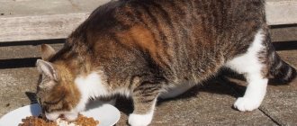
Treatment of hydrothorax in cats is based on an integrated approach. It is necessary to eliminate the cause of the disease, as well as its symptoms. The cat needs to be provided with complete peace and comfortable conditions. It is necessary to completely exclude liquid food from the diet and reduce the consumption of water, milk and other liquids.
You need to feed your pet often, but in small portions. The diet should be balanced; if necessary, the veterinarian will give dietary recommendations.
If chest dropsy is caused by an infectious disease, then treatment is aimed at combating its causative agent. If a parasitic infection is detected, deworming is carried out in the clinic against parasites. If the disease was provoked by cachexia (depletion of the body), then a therapeutic diet, vitamin intake and supportive therapy are provided.
At the same time, symptomatic treatment is prescribed. The first thing the veterinarian does is remove the accumulated edematous fluid (transudate) using a puncture of the pleural cavity. Then, to restore the function of the heart muscle and reduce the load on it, heart medications are prescribed.
At the discretion of the doctor, maintenance therapy may include not only diet and vitamin therapy, but also infusion therapy. The dropper contains glucose and a calcium chloride solution. They ensure rapid recovery of the body of an animal exhausted due to a serious illness. Additionally, diuretics are prescribed.
What symptoms indicate illness?
The symptoms of atelectasis are almost identical to pneumonia. A pet owner can suspect the occurrence of a disease based on the following signs:
One of the signs of this condition in a fluffy is a dry cough.
- Cough. Exudate accumulates in the lungs, which irritates the nerve fibers and causes coughing. At the initial stage of development, the cough is dry, but as the pathology progresses it becomes wet.
- Breathing problems. Deterioration in the functioning of the organ causes difficulty breathing. The cat begins to breathe with difficulty due to the accumulation of mucus and disruption of gas exchange.
- Wheezing. They can be heard not only by the veterinarian while listening to the lungs, but also by the owner by placing his ear to the sternum of the pet.
- Nasal discharge. Not always observed, but quite often. The discharge is mucous in nature, sometimes admixtures of pus are noticeable.
- Increased body temperature.
In addition, the cat may experience heart rhythm disturbances, a change in the pink tint of the mucous membranes to blue, weakness, lethargy and loss of appetite.
Prevention of hydrothorax
Prevention of thoracic hydrops in cats is aimed at preventing factors that can lead to this disease (mechanical injuries, exhaustion of the body, infectious and parasitic diseases). Pay attention to several useful recommendations that, in some cases, will help avoid the development of hydrothorax in cats:
- Vaccinate your pets against infectious diseases in a timely manner at the veterinary clinic. It is important to consider which infections often affect animals in the region where you live.
- Carry out antiparasitic treatment (deworming) 1-2 times a year. This applies not only to those cats that have the opportunity to go outside, but also to pets.
- Always consult a veterinarian for diseases of the cardiovascular system, kidneys and liver. This will avoid the possibility of developing thoracic hydrops in cats as a concomitant condition with the disease.
- Pay attention to your pet's diet . It must be healthy and balanced. To provide additional prevention of hydrothorax, try to fortify it with vitamins C and K.
Remember that the main thing with chest dropsy is to promptly seek help from a veterinarian!