Fibrosarcoma in cats is a malignant neoplasm that develops in the skin, as well as in the subcutaneous tissue and soft tissues; there are cases when fibrosarcoma can grow into the fascia, bones, ligaments, great vessels and nerves. Fibrosarcoma develops from skin fibroblasts.
Fibrosarcoma is a malignant neoplasm, but among sarcomas it is not the most aggressive, but is prone to metastasis. The mortality rate from fibrosarcomas among cats is low.
Of the total number of cats with oncology who come to the clinic, animals with fibrosarcoma make up a very small percentage.
In some cases, fibrosarcoma has clear boundaries and is localized as a round or oval formation; in other cases, its boundaries cannot be determined due to strong infiltration into the soft tissue.
Also, this tumor can be classified according to the degree of malignancy into highly and poorly differentiated.
If the tumor is differentiated, then it has a cellular-fibrous structure, and in it the cellular component always predominates over the fibrous one.
A poorly differentiated tumor mainly consists of immature polymorphic cells with a large number of mitoses, because of this it has a more pronounced malignancy and is more likely to give metastases.
One of these formations is post-vaccination feline sarcoma. This is a malignant formation of mesenchymal nature, which appears after subcutaneous injections of vaccines or drugs. This formation has rapid growth but low metastatic effect. This formation does not appear immediately after the injection, but quite a long time may pass from the injection to the first signs of development of the formation. They most often occur after the administration of rabies vaccines.
Sarcoma (cancer) – what is it?
Sarcoma (cancer) in cats is an insidious disease. Characterized by the formation of malignant tumors. They consist mainly of connective tissue.
The peculiarity of sarcoma is aggressiveness. With this form, metastases affect neighboring organs in a short period of time. Unfortunately, it is almost impossible to determine the development of the disease in the early stages, since it occurs without visible symptoms.
Soft tissue sarcoma in cats and other types of disease most often lead to death. The main reason for such serious consequences is late diagnosis. As a rule, surgical intervention no longer brings positive results.
Currently, many types of sarcoma are known. They are studied by scientists. In their opinion, the most dangerous are the following:
- fibrosarcoma;
- liposarcoma;
- myxosarcoma.
Modified cells originate in synovial tissue. Due to their rapid growth, the connective tissue is damaged in the shortest possible time. Malignant tumors can affect both tissue and bones of the animal. As a rule, they arise suddenly, localizing in any place without exception.
Causes
Modern science has not found out the exact cause of the appearance of malignant cells. At the moment, there are only a number of factors and assumptions that can affect the development of a tumor or predisposition to it.
One of the most likely causes of the development of osteosarcoma is frequent bone injuries; the risk especially increases with constant damage to the same section. There are also cases of the development of the disease at the site of attachment of implants during the reposition of fractures.
The most predisposed to the formation of bone tumors are cats of short-haired breeds, as well as individuals older than 7 years.
Despite the statistics on the formation of the disease after orthopedic intervention or fractures, the majority of cases of bone tumors occur for no apparent reason against the background of complete well-being.
Classification
Sarcoma in cats is divided into two groups according to location, affecting either soft tissue or hard tissue.
This disease can also be classified according to other parameters. Depending on them, the following types are distinguished:
- Post-injection – a tumor forms in the withers area.
- Rhabdomyosarcoma - affects striated muscle tissue.
- Liposarcoma is a cancer of the fat layer that often causes lumps to form on a cat's abdomen.
- Fibrosarcoma is a lesion of fibrous tissue.
- Osteosarcoma is a malignant process that occurs in the bones.
In veterinary practice, the latter type is found in 80% of cases, so it is considered the most common. As metastases grow, any organs and lymph nodes can be affected.
Stages
Like any cancer, sarcoma in cats has four stages of development. They directly influence the choice of treatment and prognosis:
- First stage. The disease occurs without symptoms. It is already possible to palpate the formations, but they are quite small in size - up to 5 cm. The tumors have clear boundaries. Metastases have not yet formed. When the disease is diagnosed at this stage, most animals have a good chance of recovery. Veterinarians believe that the tumor will respond well to treatment.
- Second stage. Tumors (bumps) on a cat’s stomach or anywhere else reach sizes of more than five centimeters. They lose clarity of boundaries. There is a tendency to rapidly increase, but metastases have not yet formed.
- Third stage. It differs from the first two in that adjacent lymph nodes are affected by metastases.
- The fourth stage is the last and most dangerous. With it, metastases have already spread to all organs. Therapy is selected in such a way as to simply alleviate the cat’s condition. The prognosis is unfavorable. If the disease is diagnosed at this stage, it is recommended to euthanize the animal, since the chances of recovery are zero.
Treatment of bone tumors in dogs and cats. Interview with the chief physician
May 6, 2014
Oncological diseases of domestic animals are, unfortunately, not a rare phenomenon. The chief physician of the Biocontrol veterinary clinic, Candidate of Biological Sciences Evgeniy Aleksandrovich Kornyushenkov talks about what types of bone tumors there are, about the experience of the Biocontrol clinic in their treatment and what the owner should pay attention to first.
What is a bone tumor? A bone tumor is a pathological process in bone tissue that leads to its destruction.
What types of bone tumors occur in animals? Of course, tumors are different. Chondrosarcoma, synovial sarcoma, fibrosarcoma are not the most common types of tumor; most often, in 90% of cases, osteosarcoma occurs in animals.
Who is more likely to have bone tumors? Bone tumors are a problem most often in large dogs in the second half of life, after 6-7 years of age.
What are the first symptoms of a bone tumor? The leading symptom is lameness. Often, unfortunately, such patients take anti-inflammatory drugs for weeks. This is precisely the reason why the patient comes to us not at the initial, but at a later stage of the tumor process. From what? For lameness, a general veterinarian prescribes anti-inflammatory drugs purely empirically (without much analysis). Yes, there is a positive reaction to them, lameness decreases, but the process of bone destruction, nevertheless, continues, hiding from us the first symptom - lameness.
The next sign is a pathological fracture, after which it becomes clear that the lameness was something more . As symptoms develop, the following sequence can be characterized: lameness, then swelling, then increased bone contours, tenderness, and finally a pathological fracture.
What stages of tumor development do oncologists distinguish? Unfortunately, bone tumors, in particular osteosarcoma, as the most common tumor, do not behave openly. Owners do not notice the first manifestations of diseases. At the appointment, the doctor notes lameness and arthrosis in the anamnesis, and on x-rays the initial stage of the development of osteosarcoma may be missed. Unfortunately, this means that the first stage is almost elusive and the patient comes to oncologists already from the second stage of osteosarcoma.
The classification itself is a complex question, but to understand what is happening there is a simplified version of the stages of development:
- I – there is swelling, but no lameness, no visible manifestations
- II – the tumor becomes visible to the naked eye (swelling, edema), there is pain
- III – pathological fracture
- IV - metastasis to distant organs, swollen and disintegrated limb, cachexia, lack of appetite, shortness of breath.
What examination is performed when an animal is admitted with a suspected bone tumor? First, a medical history is taken (have there been any previous operations due to cancer, fractures), then a clinical examination, X-ray examination (we look for signs of osteodestruction (destruction of bone tissue), especially in the metaphysis areas - this is a common location for osteosarcoma). If all the data is available, a preliminary diagnosis is made. Only trephine biopsy will finally confirm our suspicion. If changes are visible in the image, that is, classic manifestations are already present, then distant metastasis must be excluded. According to statistics, animals that come to us in 10-20 percent of cases already have metastases to vital organs, in particular to the lungs.
A cytological and histological report is mandatory, but in general, the first stage of treatment can begin after radiography. Of course, done in a specialized clinic, and not in a general practice clinic. Here the experience of the person who watches the animal plays a big role. When this is an oncologist specializing in the treatment of osteosarcomas, the likelihood of error will be very small.
Blood tests are also taken - we are interested in the change (increase) in alkaline phosphatase, it increases in the blood during the breakdown of bone tissue. But again, this is a very specific method that is necessary for an established diagnosis and treatment, and not at the differential stage.
Treatment depending on the stage. Is it possible to cure this type of cancer? First of all, the owner must understand that the treatment of bone tumors must be considered not from the position of a surgeon, but from the position of an oncologist. Sometimes I often hear: “we have a good surgeon, we will turn to him.” However, when treating osteosarcoma, surgery alone is not enough. This tumor has a high potential for metastasis, and therefore cannot be treated without chemotherapy or immunotherapy. The main task is not only to perform the operation, it is important to stop the oncological process, to prevent metastases, so that after the operation the animal lives not 1-2 months, but several years, which we still manage to do to this day.
If we are talking about the stages of treatment, then the first stage in stages I and II is chemotherapy, in some cases it is combined with radiation therapy (in the presence of swelling, a soft tissue component). For bone tumors alone, radiation therapy is not a specific therapy, but in combination with chemotherapy, it helps the animal improve the quality of life and cope with pain. This serves as a preparatory stage for surgery and prevention of metastasis.
The second stage is the use of our unique technique, namely allogeneic bone marrow transfusion. Bone marrow is taken from the donor dog, divided into cells and transfused into the recipient (our patient). This procedure is performed a couple of days before organ-conserving surgery or amputation. What does it give? Delay of metastasis by several months compared with those groups that received chemotherapy alone.
The third stage is a radical operation using a method that has long been developed and used in our clinic - implantation of irradiated bone.
Irradiated bone implantation, how does it happen? We perform a wide segmental resection of the bone along with healthy tissue, place it in liquid nitrogen and irradiate it at our source. Then in the operating room we return the irradiated bone without cancer cells to its place.
The second method of replacing defects during safe operations is transplantation of donor deimmunized bone. Within a week, the recipient's stem cells are injected into it so that they are distributed there and the fusion stage is easier to complete.
Dog Nyura, two weeks after placement of the bioimplant
How long does this operation take? This operation lasts 4-6 hours, and can last up to 8. During bone irradiation, the animal lies under anesthesia in the operating room. The time depends on the complexity of the metal structure and skin grafting. The duration of the operation is not a big problem; we have a fairly powerful anesthesiology service that allows us to carry out long operations.
What happens after surgery? We must achieve fusion of the bone segments. In our experiment we already had 10 animals in which we used similar methods for treating osteosarcoma. Rehabilitation begins immediately if we do not see complications. The animal walks for the first two weeks in a protective splint, then the stitches are removed, and from the first day without the splint the animal is already trying to lean on its limb. After surgery, in cases of osteosarcoma, chemotherapy courses are also carried out.
Does osteosarcoma occur in cats? In cats, osteosarcoma behaves less aggressively, and its degree of malignancy is lower. Today we treat them without the use of chemotherapy. If it is chondrosarcoma, there is always surgery, no chemotherapy.
Patient: cat Tas. Placement of tibial bioimplant
Bioimplant of the tibia placed in the cat Tas
Cat Tas, 45 days after transplantation
Is home treatment carried out as prescribed by the doctor? Specific home treatment for osteosarcoma, in addition to what is carried out in the clinic, is not required. We categorically disagree with colleagues who claim that it is necessary to discharge the animal from the hospital as quickly as possible and it will recover faster at home. Thus, we lose control over the patient. After all, complications may arise; the owner may not notice the problems that have arisen in time and will not promptly contact the clinic. And time will be lost.
Plus, in my opinion, the treatment of the animal should fall on the shoulders of the doctor, and not the owner, who actually becomes a nurse, performing injections and dressings, and not going about his daily life.
How often should I see a doctor after the postoperative rehabilitation period is over? Immediately after the operation, we conduct 2-3 courses of chemotherapy, which means you need to prepare for two to three months of frequent visits to the clinic. It is best, both for the animal, and for the doctor and owner, to leave the animal in the clinic during the procedures and pick it up when there are breaks between courses. In the future, we let them go for a month or three, and they come only for medical examinations and control X-ray examinations. Here everything is decided individually for each patient.
How long do operated animals live? From our experience, the maximum period is that the animal lived for 5 years and died from the onset of another cancer. The average life expectancy of an animal with osteosarcoma with complex treatment according to the literature is 1 year, however, we have many examples of long-livers, 2 - 3 - 4 years. Later, since these were most often older animals, they died, but not from osteosarcoma.
Is amputation always indicated for osteosarcoma? Amputation is not a death sentence, but rather discomfort and, of course, it does not lead to “disability” of the animal. However, for some animals, amputation may be an exit procedure. We perform amputations only in stage 3 cases, when the tumor size is very large or there is a pathological fracture.
Are there cases when surgery should not be performed? Direct contraindications for generalization of the process (metastasis to vital organs), over 12-13 years of age, and in the presence of concomitant diseases. In cases of synovial sarcoma, and we currently have two such patients, there is a very good result with chemoradiotherapy. The observation period has already been 6 months - the animals were at risk of amputation, but for now we have suspended the process. One animal even underwent surgery for a mammary tumor.
Were there any cases when the implant did not take root? Since we are discussing a new treatment method, we cannot yet talk about reliable statistics - we only had 10 animals. When it reaches 20, then we can talk about any reliable statistics. At the moment there were none. During bone irradiation, the issue of survival depended on the presence of infectious complications, the occurrence of the process at the implantation site itself, but we are also trying to solve this problem by installing an intravenous port.
Is an intravenous port also an intraclinical development? A permanent intravenous port improves the quality of life of animals, because with numerous infusions, doctors do not have to injure the veins each time and “open the gates” of infection. The solution we found, taken from our medical colleagues, gives good results in the prevention of septic problems.
What should a general practitioner veterinarian know in order not to harm the patient ? As I said at the very beginning, the doctor should be alert to lameness of the animal, especially if it occurs over the age of 6 years. He must definitely order an X-ray examination and, if this doctor does not have experience in treating bone tumors, then refer such a patient to a specialized clinic where there is a team of oncologists, surgeons, anesthesiologists, chemotherapists and rehabilitation specialists with experience in working with this pathology.
Shrek the dog. Bioimplant placement
Shrek the dog - 30 days after surgery
Shrek the dog, one year after transplant
Elena (06/22/2015 at 07:37 pm): I don’t live in Russia and I don’t trust local veterinarians. What should I do, how can I help? my dog was diagnosed with sarcoma of the bone of the hind limb. Local conditions do not allow the operation. I would be grateful for any advice.
Reply | to write a message
- Biocontrol (06/23/2015 at 09:01): The only answer we can give in your situation is to come and bring the dog. Let's see what can be done.
Reply | to write a message
Tatyana (06.10.2015 at 14:44): Good afternoon. I have a 2.5-year-old Labrador retriever, a girl. A hard lump appeared on the side of my hand on the front paw, we went to the vet, took an x-ray in two projections, the surgeon stretched out the lump with a syringe, Having ruled out the accumulation of pus, she said that it was a sarcoma and was prescribed Narocarp 100, a course of a week. The lump itself does not bother the girl at all, does not limp, does not hurt on palpation, excellent appetite, as fast as an electric broom. Maybe this is an erroneous diagnosis. We live in Novomoskovsk, Tula. Please tell me what this could be and what should we do?
Reply | to write a message
Tatyana (06.10.2015 at 14:51): Good afternoon. I have a 2.5-year-old Labrador retriever, a girl. A hard lump appeared on the side of the hand on the front paw, we went to the vet, took an x-ray in two projections, the surgeon pierced the lump with a syringe, Having ruled out the accumulation of pus, she said that it was a sarcoma and was prescribed Narocarp 100, a course of a week. The lump itself does not bother the girl at all, does not limp, does not hurt on palpation, excellent appetite, as fast as an electric broom. Maybe this is an erroneous diagnosis. We live in Novomoskovsk, Tula. Please tell me what this could be and what should we do?
Reply | to write a message
- Biocontrol (06.10.2015 at 15:33): Tatyana, good afternoon! In order to confirm or refute the diagnosis of sarcoma, it is necessary to do a histological analysis. We recommend that you contact us for an appointment with an oncologist or a surgeon. If you have any additional questions, ask them on our forum, in the “Questions to an oncologist” section, here:
Reply | to write a message
Natalya (12/07/2015 at 00:20): Hello. Please tell me which pain reliever is best for dogs. With the lowest risk of stomach and intestinal bleeding. Is it possible to give a dog analgin, baralgin? Some doctors recommend Rimadyl, others recommend Movalis. A 10-year-old dog has had a tumor on his front leg on his elbow for two years now. Big and hard. Now I just started to get sick. Appetite preserved. She began to limp heavily. Thank you.
Reply | to write a message
- Biocontrol (12/07/2015 at 12:04): Natalya, good afternoon! Ask your question directly to our doctors on the forum:
Reply | to write a message
Yulia (06/18/2016 at 12:25): good afternoon, how much does the implantation surgery and all the necessary further treatment cost, setter, 8 years old, you took histology, the diagnosis was not confirmed, but yesterday they took an x-ray, there is a sarcoma of the radius, right leg . the doctor suggests removing it, we are located in the city of Khabarovsk, write at least approximately the cost to guide us, thanks in advance
Reply | to write a message
- Biocontrol (06/20/2016 at 09:23): Julia, good afternoon! Patients with osteosarcomas are supervised by our chief physician, Evgeniy Aleksandrovich. You can communicate with him directly on our forum, in the “Questions to the Chief Doctor” section. Follow the link:
Reply | to write a message
Yulia (03.10.2016 at 20:24): German shepherd, 14 years old, boy. He has heart ischemia. But he is in good physical shape. My left paw hurts, they took an x-ray. One doctor says joint destruction, second arthrosis, bursitis, third bone osteosarcoma. We don’t know what to do, could you look at the pictures and tell us what to do next. I beg you to take a look, the dog is suffering .
Reply | to write a message
- Biocontrol (04.10.2016 at 09:37): Julia, hello. You can communicate directly with Evgeniy Aleksandrovich and show pictures in our forum, in the “Questions to the Chief Doctor” section, here:
Reply | to write a message
Mikhail (10/18/2016 at 10:29): My dog is a 12-year-old Rotfeller, spayed female. My legs gave out. Tumor on the elbow joint. We went to the vet. They took a primitive x-ray and used it to diagnose osteosarcoma. The photographs do not show any metastasis and the lungs are clean. Appetite is good. Weakness in legs when rising, preventing walking. The dog was preserved very well. There are no signs on the body or skin other than swelling on the joint. The vet said that nothing could be done. I’m just shocked and I love my dog very much and I don’t know how to help her and who to trust! I need your advice. Is there anything I can do to help? Looking forward to your reply. Michael
Reply | to write a message
Biocontrol (10/18/2016 at 12:36 pm): Mikhail, it is better for you to consult directly with our chief physician at our Bioforum. He cares for patients diagnosed with osteosarcoma. Ask your question in the “Questions to the Chief Doctor” section here:
Describe your medical history in detail and attach pictures and test results.
Reply | to write a message
Irina (03/19/2017 at 20:35): Good afternoon. My cat is 11 years old. He has osteonic sarcoma of the metacarpal bones of the right forelimb, diagnosed by X-ray. How to help him. The cat is weakened, the paw is increasing in size very quickly.
Reply | to write a message
- Biocontrol (03/28/2017 at 17:19): Irina, good afternoon! An X-ray does not make a definitive diagnosis. We recommend that you contact a veterinary oncologist to clarify the diagnosis. Once it is established, it will be possible to talk about treatment options.
Reply | to write a message
Natalya (23.05.2017 at 20:16): Good day to you! Rottweiler dog, eight years old, right front leg is swollen and cannot walk. I’ve lost weight and have constant pain. I have an X-ray but I don’t know how to attach it to the letter.
Reply | to write a message
- Biocontrol (05/24/2017 at 09:27): Natalya, good afternoon! It is better for you to discuss the case with your dog with our chief doctor directly, on the “Questions to the Chief Doctor” forum, here: You can attach an x-ray to the message in the forum.
Reply | to write a message
Grigory Evgenievich (06/13/2017 at 10:53): Good afternoon, your colleagues from the Krasnodar veterinary clinic Doverie are bothering you. A 2-year-old Great Dane came to me for treatment with a neoplasm in the forearm area, the size of a chicken egg; on x-ray, the bone tissue was discharged with degenerative changes in the periosteum, with growths along the periphery, regional lymph nodes are not enlarged, chest x-ray and ultrasound of the abdominal cavity did not reveal distant metastases. The neoplasm was removed surgically without bone removal. According to histology, the result was systemic histiocytoma. Is chemotherapy effective for this tumor, and if so, what protocol do you use?
Reply | to write a message
- Biocontrol (06/13/2017 at 13:07): Grigory Evgenievich, good afternoon! You can communicate directly with our oncologists on the “Questions for an Oncologist” forum, here:
Reply | to write a message
Olga (01/19/2018 at 07:50 pm): Good afternoon! We do not live in Moscow and we were diagnosed with osteosarcoma, this was confirmed by a biopsy. The dog is 1.5 years old. Initially, we were diagnosed with osteomyelitis and we took and are still taking anti-inflammatory drugs. Please tell me how much on average it costs to treat our disease in your clinic, provided that the dog stays in your hospital. Thank you in advance.
Reply | to write a message
- Biocontrol (01/20/2018 at 20:21): Olga, hello! It is better to discuss questions regarding osteosarcoma with the chief physician, Evgeniy Aleksandrovich, directly. This can be done on the Bioforum (link in the top menu on the right), in the “Questions to the Chief Doctor” section.
Reply | to write a message
YULIA (07/30/2018 at 13:55): Good afternoon. Caucasian Shepherd dog is 6 years old. The X-ray shows that the elbow joint is swollen and is recognized as magpie. Tell me, if the leg is removed without chemotherapy, is there a chance that the dog will still live? And how many? I wouldn't want to torture an animal. I read that if you remove it, it will be easier for her... I don’t know what to do, please advise.
Reply | to write a message
- Biocontrol (06.08.2018 at 14:39): Hello, Yulia! Our chief physician, Evgeniy Aleksandrovich, deals with osteosarcoma issues. Write to him directly on the Bioforum, there you can discuss the current situation with him:
Reply | to write a message
Olga (12/04/2018 at 10:43): Good afternoon, my dog has a sarcoma of the frontal part, a very large lump, they did an X-ray, but the blood showed nothing, what should we do, is there a chance of recovery?
Reply | to write a message
- Biocontrol (12/05/2018 at 12:15): Olga, hello! It is better for you to consult our oncologists directly on this issue. This can be done on the forum:
Reply | to write a message
Olga (01/03/2019 at 19:39): Good afternoon! I really need advice. We visited many clinics and ended up doing a myelogram on the cat. Doctors said he had a tumor in the T13 vertebra. It's like it destroys the bone. Is there a chance that this is not a tumor? Neither x-rays nor tests showed anything. The cat is only one and a half years old.
Reply | to write a message
- Biocontrol (01/04/2019 at 18:43): Olga, hello! For a one and a half year old cat, an oncological diagnosis is less likely than for an older cat. Moreover, the x-ray showed nothing. You better consult our surgeons. This can be done on the forum, in the “Questions to the surgeon” section. Describe the situation in detail and attach pictures with test results. Forum link:
Reply | to write a message
Elena (02/11/2019 at 23:38): !As urgent as possible!
Help me make a diagnosis. Dog, Rottweiler, 10 months old, swelling appeared 2 months ago. It is painful when pressed. The skin is clean, there are no discharges. The injury, if any, was not noticed.
https://imgur.com/MihTAAo https://imgur.com/E0VLtSo
Reply | to write a message
- Biocontrol (02/12/2019 at 09:42): Elena, hello! You can consult with Evgeniy Aleksandrovich on our forum, in the “Questions to the Chief Doctor” section, here:
Reply | to write a message
Alla (02.16.2019 at 21:18): How much does it cost?
Reply | to write a message
- Biocontrol (02/18/2019 at 11:57): Hello Alla! Each case is individual, so it is impossible to give an average cost of treatment.
Reply | to write a message
Evgenia (08/19/2019 at 16:21): Hello! We took an x-ray of the dog's paw, and the doctor advised me to send the picture to you! How can I do that? We live in Voronezh
Reply | to write a message
- Biocontrol (08/20/2019 at 09:07): Evgenia, hello! You can write to our chief doctor, Evgeniy Aleksandrovich, on the forum - he directly deals with cases of bone tumors. The forum has a function for attaching an image:
Reply | to write a message
Sophia (07/16/2020 at 18:19): Good afternoon! A 15-year-old cat was diagnosed with osteosarcoma of the jaw - within 2 days a bone compaction appeared. The cat does not meow, eats with appetite, and generally does not seem to be in pain - the back teeth were removed to reduce potential pain from the seals. However, the diagnosis was made in a general clinic without any tests or x-rays (they decided not to do it, since the cat was old - unnecessary radiation), the doctor said that the cat had a few months left and prescribed antibiotics. The cat is in another city, we want to understand whether it makes sense to take the cat to the clinic - given its age? What do you recommend doing? Maybe collect some tests for the doctor so that he can understand the picture and let him know whether it makes sense to come to you? I will be very grateful for the answer!
Reply | to write a message
- Sophia (16.07.2020 at 18:22): one of the unpleasant manifestations is general lethargy for several months, however, the appetite is good, + more than six months ago a hernia was removed on the abdomen
Reply | to write a message
Sophia (16.07.2020 at 18:28): the voice has become quieter, the cat has begun to ask to go outside less (it used to actively walk), saliva leaks involuntarily
Reply | to write a message
Biocontrol (07/17/2020 at 09:09): Hello, Sophia! It is best for you to discuss this issue with our oncologists directly. This can be done on the Bioforum, in the “Questions to the Oncologist” section, where you can also attach photographs, x-rays and test results. Follow the link and register on the forum:
Reply | to write a message
Sophia (07/16/2020 at 06:39 pm): there is a noticeable asymmetry on the muzzle, no soft seals can be felt - only hard ones along the entire underside; Interspectin injections were prescribed for 6 days; the cat has a hard time with moving, but we are ready to bring it if there is a prospect of treatment
Reply | to write a message
Click to cancel reply.
Clinical manifestations
It is useful for all owners to know how sarcoma manifests itself in cats. This will help detect the disease at an early stage. The main thing, even at the slightest suspicion, is to immediately contact a veterinary clinic for an examination.
So, let's look at the signs of sarcoma:
- Problems with movement, often lameness.
- Decreased activity.
- The appearance of tumors, after a certain time they increase.
- Fracture of limbs.
- Decreased appetite or complete refusal to eat, resulting in anorexia.
- Severe painful spasms, due to which the animal’s behavior radically changes. It is imperative to take painkillers, as the pet may die from pain shock.
Post-vaccination complications
Post-vaccination sarcoma in cats is a type of cancer in which a malignant tumor forms in the places where vaccinations were given. Localization area: withers. Why is this happening? Doctors still cannot give a clear answer to this question. There is a version that modified cells begin to grow due to inflammation of the injection site. It manifests itself in the formation of a rather large lump. It will have an irregular shape. The tumor grows into neighboring tissues. It will feel quite hard to the touch and may expand to large sizes. This type of sarcoma affects neighboring tissues in just a few weeks. The animal suffers greatly during this time and quickly dies.
Diagnostics
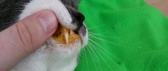
Only the owner will be able to notice the first symptoms and changes in the pet’s behavior. But diagnosis and treatment should be made by a qualified specialist. When you contact the clinic, a blood test is prescribed. The animal is also examined and the formations are palpated. Their nature can be determined after a biopsy. To do this, cells are taken from the tumor for research. You can determine the extent of organ damage by taking an x-ray of the cat.
Based on the results obtained, the doctor prescribes treatment. What therapy will be depends on the stage of the sarcoma.
Illustrations
Osteogenic sarcoma of the spine
Osteosarcoma of the hind limb
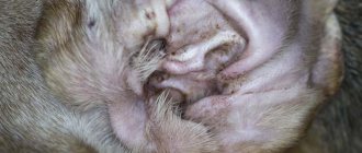
Osteosarcoma of the mandible
Osteosarcoma of the lower jaw, x-ray
Treatment
Most cancers are difficult to treat. The fact is that tumors are affected by strong chemicals, which significantly worsen the general condition of the animal. In some cases (with stage four sarcoma), doctors frankly tell the owners that the treatment will not bring the desired effect, so it is recommended to euthanize the animal. With this development, this is the only humane way that will free your pet from suffering.
Sarcoma in cats can also be treated surgically. However, this method is only possible if the formation has not had time to metastasize.
It is worth noting that in the initial stages, properly selected drug therapy and surgical intervention give a positive result.
If a single type formation is detected, it is recommended to remove it. All affected areas are also excised. If the tumor appears on the paw, the limb is amputated.
For post-vaccination sarcoma, not only surgical removal of the tumor is recommended, but also mandatory drug treatment. It is taken before and after surgery, even if x-rays have not shown the presence of metastases.
A doctor may also diagnose an unresectable sarcoma in a cat. In this case, the animal is prescribed a course of chemotherapy.
It is worth noting that surgical intervention will significantly worsen the animal’s quality of life, but will save it. This is the only way to completely defeat cancer in the early stages. As for chemotherapy, everything will depend on the overall health of the pet. Young individuals, although it is difficult, still tolerate such treatment. But cats that are more than 10 years old rarely survive.
Adjuvant chemotherapy protocol
• Doxorubicin - 30 mg/m2 intravenously once every 3 weeks, 3-5 courses.
• Doxorubicin – 30 mg/m2
• Cyclophosphamide - 300 mg/m2 - once every 3 weeks - 3-5 courses.
We begin chemotherapy on the 10th to 14th day after surgery. It should be remembered that doxorubicin is a fairly toxic chemotherapy drug. It causes various anaphylactic reactions, myelosuppression, cardiotoxicity in dogs at cumulative doses greater than 180 mg/m2, and nephrotoxicity in cats. All this must be taken into account when conducting a course of chemotherapy. As an additional drug treatment after surgery, metronomic chemotherapy can be used, which is aimed at slowing angiogenesis in the tumor and suppressing regulatory T cells that are necessary for tumor growth. In this protocol, chemotherapy drugs are given in reduced doses on a daily basis for an extended period of time. We use a combination of piroxicam at a dose of 0.3 mg/kg and cyclophosphamide at a dose of 15 mg/m2 daily. It is still premature to draw conclusions about the effectiveness, however, there are positive reviews in the specialized foreign literature.
In the complex treatment of soft tissue sarcomas, rhabdomyosarcoma should be especially emphasized. This tumor is one of the most aggressive among soft tissue neoplasms. However, it responds better to radiation and chemotherapy than other sarcomas. In animals, it is most often localized on the limbs, but can also appear in other parts of the body (breast, lower jaw). For the treatment of rhabdomyosarcoma, we always use adjuvant radiation therapy, regardless of the grade of the tumor and the condition of the resection margins. Rhabdomyosarcoma actively metastasizes, so adjuvant chemotherapy should be part of complex treatment.
Preventive measures
There are no specific preventive measures. The only thing that can be recommended to owners is to limit exposure to carcinogenic substances as much as possible. Also, do not forget about strengthening your immune system. Currently, stores sell a variety of vitamins and complex supplements that will help your pet’s diet become complete. You should not refuse vaccination, because injections can be given intramuscularly.
It is important to understand that sarcoma cannot be treated at home. Moreover, there are no effective folk methods to combat it. The owner will only lose precious time, but will not save the life of his pet.