What you need to know about ICD in cats
Urolithiasis can also occur in cats. It most often occurs in cats due to the structure of the urethra (narrow and curved in the shape of the letter “S”).
At risk:
- overweight animals;
- castrated cats;
- long-haired breeds;
- unsterilized cats.
Sterilized cats get sick less often than unsterilized cats and those whose estrus does not end with pregnancy. In males, on the contrary, urolithiasis is more common in castrated cats than in non-castrated cats. It is believed that early castration changes hormonal levels, and this serves as a provoking factor for changes in metabolism and leads to a narrowing of the urethra.
The average age of onset of ICD is 6-7 years.
The disease is diagnosed when stones have already formed in the cat’s kidneys or bladder. In cats, the disease begins to manifest itself as red urine (from blood) and then problems with urination. The diagnosis is confirmed by ultrasound.
There is no single cause for the disease. The main factor in the development of KSD is a high concentration of salts in the urine. The release of salts, in turn, is affected by feeding and the course of metabolic processes in the body.
Causes
It is impossible to identify the main source of the pathology. Experts have identified many factors that result in urolithiasis in cats. The causes of this pathology are mainly related to poor nutrition, lifestyle, care, and heredity.
Most often the disease is caused by:
- genetic predisposition;
- congenital abnormalities in the development of the genitourinary system and anatomical features (too thin or long, as well as curved urethra);
- drinking water of poor quality containing many minerals, for example from a tap;
- diet with low fluid intake;
- disorders leading to decreased metabolism;
- dysfunctions in the digestive tract;
- mixing or alternating natural and industrial dishes (dry snacks, canned food) during the day or in one feeding most often provokes the appearance of metabolic disorders, as a result of which urolithiasis occurs in cats;
- excessive amounts of minerals in the diet, for example, with a large amount of fish or fatty foods;
- low-quality cheap feed;
- overfeeding leading to obesity;
- inactivity;
- infections (streptococcus, staphylococcus), injuries to the pelvic bones, inflammatory processes and neoplasms in the genitourinary organs.
What types of stones do cats have with urolithiasis?
There are mainly two types of urinary stones in cats:
- oxalates;
- struvites.
It is these two species that play the main role in the development of ICD in cats. Now their prevalence is the same.
It has been noted that urolithiasis in cats is more often of the struvite type, while oxalates are detected in 70% of cats.
Increasingly, cases where, with neutral acidity of urine, both struvite and oxalates are formed simultaneously. It is much more difficult to cure a mixed type of disease.
There are other types of uroliths:
- xanthines;
- cystines;
- apatites;
- urates;
- potassium and magnesium pyrophosphates.
Diagnosis and treatment of these types are the same as for struvite and oxalate.
Feeding cats with mixed type of urolithiasis
In some cases, cats develop mixed types of uroliths. Quite often, this problem occurs after surgical removal of stones and the transition to a new diet - with insufficient medical supervision, an excessive pH shift and the formation of a new type of stones are possible. Remember that only a veterinarian can prescribe a therapeutic diet for urolithiasis! For cats, the risk of developing the struvite-oxalate type of urolithiasis is quite high; measures for treating these stones are diametrically opposed - acidification of urine in the struvite-oxalate type of urolithiasis and the use of treats for cats leads to the formation of oxalates. For prevention, the most suitable pH level is 6.8 – 6.9. This type of stone requires particularly careful monitoring of treatment, regular urine tests and ultrasound.
In our center you can order a calculation of a home diet or a selection of a therapeutic commercial diet from a veterinary nutritionist.
Causes of struvite in cats
Phosphorus salts participate in the formation of struvite or phosphate. Struvite stones are usually shaped like a circle, oval or tetrahedron. Mainly localized in the bladder.
The main predisposing factors for the formation of stones:
- Feeding. A diet that is dominated by foods high in magnesium and low in protein and fat promotes the formation of uroliths. Supplements that alkalinize urine, low calorie feed and low moisture content increase the risk of disease.
- Infectious diseases . Struvite caused by infection is rare and is more common in kittens (under 1 year of age) and older animals (over 10 years of age).
- Age, gender, breed. Cat breeds with long hair such as Persian, Himalayan, Oriental, Ragdoll are predisposed to urolithiasis. And vice versa, in Russian Blue, Siamese, Abyssinian, Burmese cats, struvite-type urolithiasis is much less common.
- Lifestyle. At risk are animals with extra pounds, castrati, leading a sedentary lifestyle, and consuming little fluid. It has been noted that cats with constant access to the street are less likely to suffer from ICD.
According to average indicators, struvite in the urine of a cat is formed at the age of 2-6 years.
Conditions of occurrence
For struvite to form, three conditions must be met:
- The main reason is the high level of concentration in the urine of the components that make up the stones (magnesium, phosphate, ammonium). The higher the level, the faster the formation of urolith.
- Struvite in urine is formed if there is a base (core) around which sand and crystals accumulate. According to a number of observations, desquamated epithelial cells, bacteria, and suture threads (in the case of a previous operation) can serve as such a basis.
- Acidity of urine. Struvite often forms in an alkaline environment (pH level greater than 7). The risk of stone formation is reduced at a pH less than 6.4. This acidity promotes the dissolution of existing stones. However, crystals or stones are also found in a neutral environment. Therefore, bringing urine to a neutral or acidic level cannot be the only remedy that can cure urolithiasis.
The process of formation of uroliths in ICD, if it begins, proceeds very quickly. Otherwise, the crystals would be washed away by urine and would not have time to turn into stones.
Features of the disease
KSD is a chronic pathology: the animal develops salt deposits in the form of stones or sand in the kidneys, urinary ducts, and bladder. Every fourth individual is at risk of experiencing these symptoms. Animals from two to six years old, cats with impressive weight, breeds with long hair, castrated males and unsterilized females are susceptible to the disease.
Manifestations
Males experience the pain of passing stones or sand more often, since their urethra is much narrower than that of females. Periods of exacerbation include the beginning of autumn, as well as the time from January to April.
At first, the disease is not noticed because it does not manifest itself. However, further formations will only increase in size, and one day a moment will come when the formation will move away from the wall of the bladder or kidney and begin to move with the flow of secretions.
Even the smallest formation causes extreme pain to the cat, causing mechanical damage to the delicate surface of the urethra. Impressive-sized salt formations can clog the urinary duct - this is called obstruction. The fluid stagnates, burning pain and intoxication appear. In this case, prompt emergency assistance must be provided, otherwise the animal will simply die.
Types of formations
ICD manifests itself in two types of formations: struvite and oxalate. For the first, an alkaline environment is needed - if there is an excess of phosphorus and magnesium in the body, then the urine becomes alkaline. Struvite has a harder structure.
Oxalates are formed if the urine pH is very acidic. It is caused by high amounts of calcium. Oxalates are special in that they have sharp edges and a loose structure.
Signs of urolithiasis in cats
If urolithiasis is suspected in cats, symptoms and treatment depend on a number of factors:
- location of the stone;
- how long does it stay in the organ;
- stone type;
- presence of concomitant diseases.
Symptoms of kidney stones
Kidney stones are located in the kidneys and/or ureters (nephrolithiasis). In cats, this type of disease occurs in 5% of cases, is asymptomatic or manifests itself with the following signs:
- pain when palpated in the kidney area;
- fever;
- blood in urine;
- drooping state;
- loss of appetite;
- The cat may lick excessively in the kidney area.
However, these signs are not characteristic only of ICD. The animal may experience frequent and difficult urination, unquenchable thirst, weight loss, vomiting, and drooling. Therefore, to make a diagnosis, you need to do an x-ray of the kidneys.
If there is a blockage of the ureter of one kidney, then the second kidney is also affected in cats - renal failure develops.
Symptoms of bladder stones
If stones in a cat’s bladder do not block the urethra and do not have sharp edges, then they do not make themselves felt for a long time. Very often, formations are discovered by chance during the diagnosis of another disease. This is especially true for struvite, which has a smooth surface. So a smooth urolith with a diameter of 2 cm may not manifest itself in any way and not bother the pet. Oxalates have an uneven surface and rub against the mucous membrane, which leads to irritation and inflammation.
If symptoms of urolithiasis do appear, the owner often does not notice them because cats prefer to go to the litter box in privacy.
The first signs that you can use to understand that your cat has problems:
- visits to the toilet became more frequent;
- began to urinate in the wrong place;
- urine is colored due to blood;
- discomfort is noticeable in the animal during urination;
- cats begin to lick their genitals more actively to make going to the toilet easier.
Signs of urolithiasis in cats may appear and disappear periodically. However, without treatment they will not disappear completely.
Reasons to contact a veterinarian:
- The frequency of trips to the toilet increases, and the portion of urine is small, only drop by drop can be released. The cat practically does not leave the toilet. Takes unnatural poses, hunches over.
- The urge to urinate causes pain.
- Blood in the urine is clearly visible.
- Intense, unnatural thirst.
- Dramatic weight loss.
- Depressed state.
In severe forms of urolithiasis, the urethra (urethra) is completely blocked. A calculus or plug consisting of many crystals blocks the canal, and the cat cannot get out of the way.
Complete urethral obstruction does not occur in cats, only in cats.
Due to stagnation of urine and damage to the bladder mucosa by stones, inflammation and spread of infection to the kidneys can occur. Vomiting begins, thick, viscous saliva appears in the corners of the mouth, kidney failure, dehydration, poisoning of the body develop, convulsions and coma may develop.
If measures are not taken within the first 2 days, blockage of the urethra in 80% of cases leads to the death of the cat.
How does pathology manifest itself?
Finding out that a pet has urolithiasis at the beginning of its development will not work: he cannot complain of discomfort or problems with urination, so owners learn about the presence of a dangerous pathology when it has gone too far. You need to run to the clinic if the following symptoms of ICD appear:
Sign of urolithiasis based on the cat's posture
- the cat goes to the toilet not in its usual place, but anywhere;
- little urine is excreted; grains of sand and blood may be visible in it;
- the urge to urinate, on the contrary, becomes frequent;
- pain and irritation of the urinary tract by sand cause the cat to lick the urethra.
Gradually, the pet’s body temperature rises (up to 40˚C), he refuses food, and moves little. When urine cannot pass through the channels, the cat becomes very anxious, meows, and takes a characteristic pose to facilitate the outflow.
It is especially important to see a veterinarian in time if the cat is in critical condition, which is characterized by the following symptoms of urolithiasis:
- the stomach thickens, its volume becomes noticeably larger;
- since urine can no longer pass out, it stagnates in the bladder, causing severe tissue intoxication;
- the cat hardly moves;
- foamy saliva is released from the mouth;
- the animal’s temperature drops, the pet is trembling;
- vomiting is possible.
In the absence of timely assistance, the animal dies.
Important: intoxication occurs within a day after urination stops!
Diagnostics
It is important for the doctor to determine at what stage of development the cat’s urolithiasis is in order to choose the right treatment.
Diagnosis of kidney stones
For stones localized in the kidneys, X-rays show good results. Determining stones using ultrasound is less accurate, but ultrasound has the advantage of detecting other pathologies in the development of the kidneys, for example, enlargement of the pelvis or dilation of the ureter. Stones in the ureter or pelvis lead to blockage and expansion. The accuracy of diagnosis increases if both x-rays and ultrasound are performed.
If these methods do not allow recognizing uroliths, they resort to MRI (magnetic resonance imaging) or PAP (percutaneous antegrade pyelography), when a contrast agent is injected into the area of the pelvis. It is important for the doctor to exclude diseases such as hydronephrosis (enlargement of the pelvis or hydrocele of the kidney) and chronic kidney pathology.
Cultures, urine analysis for the presence of crystals, and x-rays can suggest the type of stones.
Determination of uroliths in the bladder
The most accurate way to determine the presence of stones with a diameter of more than 3 mm in the bladder is by x-ray. Small uroliths are detected using cystography - the introduction of a special dye into the organ through a catheter. Research is necessary to exclude another disease - idiopathic cystitis. This disease usually manifests itself at a younger age - 1-2 years. Therefore, if there are problems with bowel movements in an adult animal, the doctor is more likely to suspect stones rather than cystitis.
If the cat’s condition is serious, the doctor can use his fingers to feel for the enlargement of the organ and the presence of a stone in it. Even the owner can determine this by palpating the cat's abdomen. With multiple small stones, a characteristic crunching sound may appear on palpation.
A urine test is also performed. But it does not provide complete information. The test can show that urine contains crystals and determine its acidity. However, sand in the bladder does not mean there are stones. And more often than not, the sand found does not match the type of stone. To determine what type of stones they need to be examined in the laboratory, but this is possible after extraction.
In addition, collecting a urine sample can be difficult and the material must be examined immediately after collection. Cooled biomaterial may show a false result.
Conservative treatment
Therapeutic procedures are prescribed to restore urinary outflow and relieve the inflammatory process that causes urolithiasis in cats. Treatment should not only eliminate pain, but also be aimed at prevention, eliminating relapses and complications.
Obstruction most often occurs due to muscle spasm, which is caused by irritation and mechanical damage to the mucous membrane of the urinary ducts. The animal is prescribed a course of medication that eliminates stagnation of urine and restores the patency of the ureters. For this purpose, sedative medications and antispasmodics (baralgin, spasmolitin, atropine and others), as well as antibiotics and homeopathy (magnesia, cantharis, apis and others) are used. This stops an attack of urolithiasis in a cat and improves the patient’s condition. In combination with medications, lumbar novocaine blockade and heat are used.
Treatment of urolithiasis
Urolithiasis in cats is difficult to treat. Both medical and surgical treatment of urolithiasis has many complications. It is rare for an animal to recover completely.
Treatment for kidney stones
Kidney stones are treated only in the following cases:
- provoke problems with the outflow of urine;
- cause pain;
- have reached a large size and deform the organ.
For struvite in the kidneys, it is recommended to dissolve them with a special diet and medication. This is a long process. Oxalate stones do not dissolve. An attempt to dissolve them is always unsuccessful.
The consistency of stones in the ureter cannot be changed. To do this, they need to be pushed into the bladder so that they are surrounded by urine. Therefore, in case of blockage of the ureter, its lumen is increased using a stent. The second method is removal with an endoscope.
Treatment of urolithiasis in cats begins with the prescription of diuretics - drugs that increase the rate of urine formation. This is necessary to move the stone from the kidneys into the bladder. However, after such therapy, 75% of animals require surgery.
Surgical treatment is carried out in different ways:
- Removal of part of the ureter.
- Dissection of the wall of the ureter.
In cats, surgery often ends in complications. The most common is urine leakage, which can lead to partial or complete disruption of its outflow.
In the most extreme case, doctors decide to remove the kidney. In this case, an important condition is that the second kidney must work normally.
Stone crushing is not carried out in cats. Oxalates are difficult to treat, and with an increase in energy flow, the animal's kidneys and ureters are damaged.
After treatment, to avoid relapses, the stones are examined and recommendations are given depending on their type. The main preventative measure is to maintain urine acidity levels in the range of 6-6.5 pH. However, in a third of animals, urolithiasis occurs again.
Treatment for urinary stones
In some cases, urolithiasis may not manifest itself. And if the stones were discovered by chance, then regular monitoring of them using x-rays is required.
If you notice the first symptoms of urolithiasis, you should consult a doctor. If tests suggest struvite in a cat's urine, an acidifying diet and drugs that destroy stones are possible.
Oxalates and other stones in a cat’s bladder that contain calcium do not dissolve either with therapeutic feeding or with medication. The only way to treat urolithiasis in cats of the oxalate type is surgical.
If you suspect a blockage of the urethra, especially if the cat cannot go to the toilet for 6-8 hours, or is experiencing pain, emergency help is needed. The first thing you can do to help is to eliminate the spasm and give a painkiller tablet or give an injection (No-shpa, Papaverine). Then the cat should be treated only in the clinic.
Urethral stones can be treated in several ways:
- In case of acute urinary retention, the primary task is to empty the bladder of its contents. This can only be done in a clinical setting using catheters. During catheterization, local or general anesthesia is performed. In some cases, the canal may need to be flushed to push the crystals back into the bladder. If the outflow has returned to normal, then in the future it all comes down to preventing relapse. Drugs are prescribed that relax the urethra to improve the flow of urine without a catheter (Cornam drug). Catheterization allows the urolith to be advanced or broken, but is considered dangerous. The catheter often injures the mucous membrane and causes infection.
- Pushing the stone from the channel into the bubble. It is performed under general anesthesia or strong sedatives. A puncture is made in the bladder through the abdomen to allow urine to pass through a tube. Using a catheter, syringe and saline solution, the stone formation from the urethra is pushed back into the bladder. Not used in cats.
- In the case of small uroliths in females, the bladder is washed.
- Destruction, crushing of stone. Laser is most often used. The procedure is performed primarily on females.
- Massage – indicated for the formation of sand plugs.
After urination has been restored, the cat needs droppers for the first few days to restore the condition, increase the outflow of urine and eliminate intoxication. Anti-inflammatory drugs and antibiotics are prescribed for 2 weeks if the temperature is elevated and there are signs of infection. Injections for a cat with urolithiasis are given if there is blood after urination has been restored.
When stones cannot be dissolved, there are a lot of them or they are large, removal of stones from the bladder is carried out by opening it. In severe conditions, when the stones are large and they do not pass back into the bladder, the catheter is left for a while or a cystostomy is performed (a strip operation in which the bladder is cut). If the problem occurs again, an artificial urethra (cystostomy) is made.
After recovery, lifelong dietary therapy with medicinal food and regular monitoring in a veterinary clinic are prescribed.
Operation
Surgical removal of stones is the leading method of treatment. Disturbances in the outflow of urinary fluid and kidney function, leading to hydronephrotic transformation and attacks of pyelonephritis in the acute stage, hematuria and severe pain - such complications are caused by urolithiasis in cats. In most of these cases, surgery is simply necessary.
Depending on the type of formation, the veterinarian chooses urethrostomy or cystotomy. In the first case, an outlet channel is artificially created that reaches the area of obstruction. Cystotomy is considered a more complex abdominal operation. It is used when the size of large urolithic formations exceeds the diameter of the urethra.
After surgery, the outflow of urine is restored, but the animal requires an additional course of antibacterial and anti-inflammatory therapy.
Therapeutic nutrition for urolithiasis
The only treatment method that does not threaten the life of the animal is the dissolution of uroliths using a therapeutic diet. This method is applicable only for struvite, urate and cystine and can be done at home.
In addition, the use of medicated feed to a certain extent reduces the risk of the appearance of both struvite and oxalates.
Depending on the urine test, a diet is prescribed. There are 2 types of food in the form of canned food and dry granules for struvite and oxalate type of disease. The therapeutic diet is available from such brands as Hill's (SD), Royal Canin's (SO), Purina (UR).
Medicinal feeds have the following effects:
- acidify the urine (to pH 6.0), which causes the dissolution of struvite;
- increase the secretion of urine, which promotes the removal of salts.
The proportion of magnesium, phosphorus and calcium in medicinal feeds is reduced. But they may contain salt to make you thirsty. Therefore, if salt is contraindicated for your pet (heart failure, kidney failure, heart disease), such a diet is not acceptable.
If the diet is followed correctly, struvite dissolves in an average of 1 month. During the entire period of therapeutic nutrition, monitoring is carried out using x-rays. Therefore, when uroliths are detected, but there is no information about their type, a trial of specialized food is prescribed. In this case, any other products, nutritional supplements, or treats are excluded. If the condition is not complicated by infection, then the diet leads to improvement already on the 5th day.
The animal has a chance to fully recover if within 3-4 weeks the uroliths have become smaller or dissolved.
After effective completion of treatment, the owner must conduct a urine test every six months, ideally once every 3 months.
It is not recommended to feed young cats (under 1 year) with medicated food. If a kitten gets sick, then surgery is considered the best option.
Oxalates cannot be dissolved by diet. Medicinal foods, for example, Hill's (XD), Eucanuba Oxalat Urinary Formula can only act as prevention.
When eating natural foods, you should follow these recommendations:
- food should be as hydrated as possible;
- avoid overfeeding;
- provide soft water for drinking;
- The menu should include a balance of calcium and phosphorus.
If a cat is on homemade food and cannot or does not want to eat commercial medicinal food, there are special additives that acidify urine.
The goal of any diet is to make the urine acidic (pH 6.0). Only in this case conditions are created that are unfavorable for the activity of bacteria and the formation of stones.
What to feed a cat with urolithiasis
Only with proper nutrition can a cat diagnosed with ICD live painlessly for several more years. Since some pets prefer exclusively dry food, while others prefer homemade food, approaches to diet will vary.
Dry food for a cat with ICD: which one to choose
Most dry food is completely unsuitable for feeding a cat with urolithiasis - they contain too many mineral salts. But there are also special mixtures that can be selected depending on the type of urinary stones, for example:
- Oxalates – Royal Cannine Urinary S/O LP34, Hill's PD Feline K/D;
- Struvite – Purina Pro Plan Veterinary Diets UR, Hill's Prescription Diet C/D.
You only need to buy food that belongs to the premium and super-premium class.
How to feed your cat homemade food
Home feeding of a cat with urolithiasis also depends on the type of stones. Since the high acidity of urine is caused by calcium, you need to limit your pet to eggs and milk (and their derivatives). Vegetables rich in this element should also be excluded from the cat's diet. In addition, with oxalates, it is extremely undesirable to give your pet offal, since they contain a large amount of oxalic acid.
Monotony in food should be avoided. The cat's menu should be based on meat dishes, while adding industrial food of any kind to the food is prohibited.
It is important to provide the animal with free access to water. Since cats drink little, you need to try to teach your pet to regularly visit the watering hole. The water bowl should not be located next to the food bowl so that the cat does not turn his attention to the food.
Forecasts
After surgical treatment of the kidneys, the prognosis of how quickly the organs will recover depends on the duration of the disease and the qualifications of the veterinarian.
The prognosis for urinary stones is favorable because urethral blockage is rare. And the problem can be solved either by diet or removal. In case of struvite, you can prevent their re-formation with specialized food.
The risk of relapse increases with oxalates. In this case, the cat needs to be fed with special industrial food and taught to consume more water.
Prevention
Once the pet’s well-being has stabilized, it needs lifelong preventive measures. KSD is not completely curable and cats are at risk of relapse. It is better to take some time to care for your furry friend than to treat the consequences of this serious problem. Prevention of urolithiasis in cats includes the following measures:
- proper nutrition: medicinal food developed for pets with urolithiasis, or a natural diet selected together with a specialist;
- weight control of a cat: body weight should be within 3.5-4.5 kg;
- herbal medicine with diuretic preparations;
- avoid the animal's thirst, use only fresh and filtered water for drinking;
- eliminate passivity in your pet’s life, since activity is one of the best preventative measures;
- every 6-12 months, control ultrasound of the bladder and kidneys and once every 4-5 months, urine analysis to assess its composition;
- mandatory medical examination in case of exacerbation;
- periodic consultation with a veterinary specialist.
By adhering to these simple rules, your furry pet will have the opportunity to live many more years of a fulfilling life.
Struvite and oxalates in the life of dogs and cats
Urine
is the end product of kidney function. It is the result of ultrafiltration of blood in the renal glomeruli, transport processes in the renal parenchyma, processes of concentration and dilution of urine, synthesis of biologically active substances by the kidney and its metabolic function.
During the day, different species of animals produce a certain, characteristic volume of urine. The main component of urine is water, in which the end products of metabolism and cellular elements that join the urine during its formation and movement through the urinary tract are dissolved. Thus, it distinguishes between an organized and an unorganized cage.
In this article we will look in more detail at unorganized urine sediment, namely crystalline salts consisting of magnesium ammonium phosphate (struvite) and calcium oxalate (oxalates).
Crystalline salts can be detected in clinically healthy animals during routine urine analysis, in particular sediment microscopy. In other cases, their presence in the urinary tract can lead to the manifestation of the clinical picture of the disease. In this case, crystalluria is the initial stage of urolithiasis
, which can develop into the formation of sediment (“sand”) and urate stones (uroliths).
Uroliths
are complexes of crystals and matrix that are present in one or more parts of the urinary tract, with hypersaturation of urine with substances leading to crystallization. Each urolith is multilayered, it consists of a core, a central substance that makes up most of it, followed by a peripheral substance - a layer of precipitates, which is covered by a crystalline shell. According to research results, urate stones predominate, consisting of one mineral substance, but there are also mixed representatives that have a core of one mineral composition, while the outer layers may consist of others.
From the above, it becomes clear that the crystals are formed from mineral metabolic products that precipitate from the urine solution. If these crystallized minerals are retained in the urinary system, they can grow and aggregate to form urolith stones.
Various physical and chemical factors lead to the formation of urinary stones in patients. They vary depending on the type of crystal. The main and common factor for their occurrence is the concentration of urine. The lower the density of urine, the less saturated it is with electrolytes, which are responsible for the formation of crystals, which means this reduces the likelihood of stone formation. The pH of urine also plays an important role in this process. Knowing whether the urine environment is acidic or alkaline, one can predict the mineral composition of crystals and uroliths.
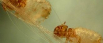
Struvite
composed of magnesium ammonium phosphate, they are the result of the crystallization of ammonium, phosphate and magnesium ions. It is a colorless substance that often forms crystals of various sizes. They have the shape of a prism with a number of faces from three to eight, needles or flat crystals with beveled edges. These crystals most often form in alkaline urine, where the pH is greater than or equal to 7.0. In dogs, the formation of these crystals often occurs in conjunction with a bacterial infection with urea-producing bacteria such as Staphylococcus spp. or Proteus spp. However, in cats, they can appear without accompanying infectious agents, which, according to recent studies, is associated with the excretion of ammonium ions by the renal tubules. In dogs, struvite is more often diagnosed in females than in males. This is because bitches are at increased risk of developing a urinary tract infection. Approximately 95% of struvite stones in dogs are found in the lower urinary tract, with the remaining 5% located in the upper urinary tract (pelvis and ureter). According to statistics, about one third of all detected uroliths in the upper urinary tract in dogs were of the struvite type.
Unlike dogs, in cats, in 95% of studied cases, struvite stones are formed without the participation of an infectious agent. The main role in their formation is played by changes in the physical and chemical parameters of urine, such as pH, density, mineral saturation with magnesium and phosphorus. It was also noted that increased fiber consumption plays a role in the formation of struvite. In cats, during clinical observations, no gender predisposition to the appearance of certain stones was identified, but a number of breeds were noted that are at risk ( Ragdoll
, Himalayan, domestic shorthair). The age range of the disease ranges from 4 to 7 years, and the average age of affected cats is 5.7 years. Quite rare cases of struvite formation associated with urinary tract infections can occur in cats aged 1 year or older than 10 years.
Calcium oxalates (oxalates) come in two forms such as dihydrates and monohydrates. Crystals of calcium oxalate dihydrate occur much more often and are colorless octahedra of various sizes; these octahedrons resemble a small envelope or a Maltese cross. They can be found primarily in urine, which has an acidic pH. Crystals of calcium oxalate monohydrate are colorless, they have a flat shape with pointed ends, similar to picket boards, and they can also take the shape of a spindle or dumbbell.
If a urine sample taken for analysis is not examined in the near future or the urine is oversaturated with bacterial flora, then calcium oxalate dihydrate crystals may form in vitro in large quantities, which will distort the analysis data. In clinically healthy animals, this type of element may be a common component of urine sediment. Diseases such as hyperadrenocorticism, hypercalcemia, hypercortisolemia, hyperoxaluria and a number of others can contribute to the formation of calcium oxalates in dogs. In a number of studies, it has been shown that acidosis and aciduria may contribute to the formation of this type of stones due to increased urinary calcium excretion and decreased excretion of citrate, which is a competitive inhibitor of calcium oxalate crystal formation. Unlike struvite, the solubility of the described crystals does not depend on the pH of the urine. According to statistics, the prevalence of uroliths consisting of calcium oxalate is similar to that for struvite stones and ranges from 40 to 50% of all stones studied in dogs and cats. According to sexual distribution, males are at risk in relation to females, in dogs the ratio is 2:1, and in cats it is 1.5:1. Breed predisposition also plays a role; dog breeds most susceptible to the formation of oxalate stones are: Mittel and Miniature Schnauzers, Lhasa Apso
, Yorkshire Terrier, German Spitz, Shih Tzu, Chihuahua, Dachshund and a number of others, Ragdoll cats, British Shorthair, Scottish Fold, Havana, Persian. Urinary stones of calcium oxalate composition are found predominantly in the lower urinary tract in both dogs and cats, and 2-3% are localized in the renal pelvis or ureter. When examining uroliths from the kidneys and ureters, oxalt origin was revealed in one and a third of cases in dogs and in almost all cases in cats. Like struvite stones, calcium oxalate stones are radiopaque, allowing them to be detected using x-rays.
Clinical manifestations of the presence of crystaluria and uroliasis may vary, depending on the progression of the disease. At the initial stage, they may not be noticeable at all for both the pet and the owner, but then the disease can progress and lead to acute or chronic cystitis, the formation of polyps, changes in the wall of the bladder and urinary tract, up to acute obstruction of the urethra or ureter .
Ultrasound
full bladder. All stones are high-density structures that do not conduct echo rays; therefore, on the device’s monitor screen they are visualized as white formations with dark acoustic shadow tracks behind. Stones are distinguished by their mobility; unlike other pathologies, they are not attached to the walls of the bladder and can easily change their location. You can also use this study to detect the presence of sediment in the bladder, which will look like flakes. It changes its location when pressing or braking the organ under study. Particles in the sediment have increased echogenicity; they can form clusters, and when the process is advanced, form hypo- and hyperechoic structures. The echo suspension can be attached to the walls of the bladder; during examination, it looks white because it does not transmit rays further; if the bladder is well filled, particles as small as 1 mm can already be distinguished.
X-ray examination is also used for complex diagnostics.
. Struvite and oxalate stones, for the most part, are contrasting and are clearly visible in photographs. Sometimes false negative results are obtained, which are due to the fact that stones less than 3 cm in diameter may go unnoticed when taking x-rays without contrast. Such a study helps to identify the presence of pathological inclusions of uroliths, both in the bladder and in the renal pelvis, ureters and throughout the urethra.
If doubts remain, after manipulations about the presence of stones or sand in the urinary sediment, they resort to additional research, such as double-contrast cystography or cystoscopy, if the anatomical structure of the urethra allows.
After verification of the diagnosis, treatment and further prevention of the described pathology begin.
If uroliths and crystalluria do not threaten the patient’s life, then the dissolution process begins.
The procedure for dissolving struvite stones differs between dogs and cats. Due to the fact that the formation of stones in dogs is associated with the presence of an infectious process, the use of competent antibiotic therapy is an important part of the dissolution process. When stones dissolve, bacteria can be released from them, therefore, it is important to perform bacteriological culture of urine to avoid antibiotic resistance. For dogs and cats, it is necessary to use specialized diets that are aimed at dissolving struvite. These foods help lower urine pH, reduce the intake of minerals that are found in stones, reduce the concentration of urine, and reduce the formation of urea, which is used as a substrate by bacterial urease (which is only present in dogs). The key to successful treatment is to achieve an acidic urine reaction, which increases the solubility of struvite crystals. Feeds designed to dissolve struvite stones contain a low concentration of magnesium, since it has been scientifically proven that an excess of this element in food is one of the predisposing factors for the formation of struvite in cats. They also have a high sodium content, which stimulates the animal to increase water consumption, which entails increased diuresis and a decrease in the concentration of urine, which means it will be less saturated with stone-forming minerals. On average, such dietary therapy is necessary for dogs for 3 months, and for cats for 20 days, but it can reach up to 55; all these indicators directly depend on the initial extent of the disease. During the procedure for dissolving stones, it is necessary to strictly monitor the dynamics of the patient’s condition to assess its effectiveness. After successful dissolution of struvite, it is necessary to use special food for feeding pets, designed to prevent relapses. An important point for the patient’s further well-being is careful attention to signs of recurrent urinary tract infection, since timely failure to provide assistance can lead to the re-formation of struvite stones, especially in animals predisposed to this pathology.
Calcium oxalate uroliths do not undergo dissolution and therefore their treatment is possible only through surgical intervention. Studies have revealed that after removal of oxalate urinary stones, the rate of relapse of the disease is 50% of cases. Based on this, important preventive measures are measures to prevent the occurrence and progression of this type of urolithiasis. A pronounced positive effect in dogs was found from consuming food rich in phosphorus, potassium, magnesium and reducing carbohydrates in the diet. The key to preventing the formation of calcium oxalate urinary stones is to increase your water intake to reduce urine concentration. The optimal specific gravity of urine, to reduce the risk of recurrence, will be 1.035 - 1.040 in cats and up to 1.020 in dogs.
If the stones are large or can threaten the patency of the urinary system in the form of its complete or partial obstruction, they are removed surgically
from the place where they are located. If the physiological characteristics of the animal allow and the location of the stone is optimal, then a non-invasive method such as laser lithotripsy is used. Its essence lies in the fact that under the influence of a laser through a fiberglass endoscope, the element is fragmented into such component parts that can independently come out with the patient’s urine current or be captured using an endoscopic clamp - a manipulator in the form of a loop. In humane medicine, uroliths located in the kidneys and ureters are treated with extracorporeal shock wave lithotripsy, which is not yet widespread in veterinary medicine.
Mineral types of stones have characteristic visual features, such as shape, color and surface character, and they can have completely different crystalline compositions or consist of several types. In this regard, the macroscopic picture of the sample is incomplete and uninformative for determining the exact composition of the resulting material. Therefore, after removal of the urolith, it is necessary to conduct a thorough laboratory analysis. As mentioned above, some stones may consist of several layers of crystals of different compositions. Their alternation of different substances and layering may reflect different periods of growth during which different types of minerals were deposited or alternations of different proportions of stone occurred, so that a urolith consisting of less than 70% of one type of mineral is mixed. A detailed analysis of the stone allows you to more accurately prescribe treatment and the necessary further preventive measures.
After treatment or a procedure for dissolving stones, prevention of recurrence of urolithiasis is necessary, since there is a high probability of repeated formations. Prevention strategies are aimed at eliminating or controlling the causes that underlie the formation of different types of uroliths. If identifying the causes is difficult, then the main element of prevention is minimizing the factors associated with the formation of stones, namely, optimal diet therapy. It must correspond to the identified type of stone or sediment and be species specific. It is also necessary to regularly carry out medical examinations for animals, such as urine tests, ultrasound of the abdominal cavity and the urinary system in particular, since timely detection of the disease is the main key to its successful treatment and the well-being of the pet.
Feeding with feed
When using industrial products, it is better to feed your furry friend with special food. They have a special content of minerals, for example, phosphorus (not higher than 0.8%), magnesium (less than 0.1%). These minerals in large quantities provoke the appearance of tripelphosphate stones, which are most often found in ICD. Cheap economy class food is prohibited. If the animal drinks little, then it is better to soak dry snacks or abandon them in favor of special canned food for cats with urolithiasis.
Prevention of urolithiasis (UCD) in castrated cats, their “walking” brothers and cats
photo from website: tinydog.ru
The goal of prevention is to prevent relapse. Doctors recommend:
- Make sure there is water in your pet's bowl. The cat must drink a lot. This is justified by the need to increase diuresis. You can give your pet not only water, but also chamomile decoction, which reduces inflammation.
- Reduce calorie intake (for cats prone to obesity).
- Monitor the functioning of the gastrointestinal tract. If constipation occurs, it is necessary to give the animal a mild laxative that does not provoke an imbalance of electrolytes.
- Avoid hypothermia - the pet must be kept warm for 24 hours. So winter gatherings near a window open for ventilation are strictly prohibited.
- Stick to a diet - it is chosen depending on the type of stones and their chemical composition.
We previously wrote about how to choose dry food for a cat. We remind you that the wrong choice of diet can lead to a greater exacerbation or a quick relapse - especially if you prefer to save on the animal and feed it with budget options, which have as little benefit as meat. Next, we will talk about what recommendations to follow when caring for a pet diagnosed with urolithiasis.
Natural nutrition
An incorrectly selected diet is one of the common causes of the appearance or recurrence of urolithiasis. The ideal option is to develop a diet for cats with urolithiasis with a specialist in this field.
When eating natural foods, vitamins A and B are additionally prescribed. For oxalate urolithiasis, carrots, boiled eggs, and white beets are recommended for pets, and for struvite urolithiasis, cheese, cottage cheese, boiled meat and rice. Food must be freshly prepared.
You should exclude pork, chicken, fish, sausages, canned food and caviar from your furry friend’s diet. Dishes should be dietary, that is, non-acidic, low-fat, non-spicy and unsweetened. They should not contain excessive amounts of protein.
ICD - statistics and background
Urolithiasis is a disease that occurs under the influence of many interrelated phenomena, rather than any single cause. That is why during treatment you have to work on several “fronts” at once. Two types of factors influence the development of the disease:
- Endogenous - those that act from the inside.
- Exogenous – causes from outside.
photo from the site: arhcat.ru
ICD is most often diagnosed between the ages of one and six years. More often, cats are brought to see a veterinarian - the peculiarities of the anatomical structure of the urinary system organs in males make them the No. 1 target for a dangerous disease. Cats suffer less from it, but this does not eliminate the risk of developing the disease in females. Exacerbations occur in autumn and spring. Recently, visits to veterinary clinics with KSD have become more frequent. Experts attribute this to a number of factors:
- Changes in the diet of domestic cats and kittens (the predominance of dry food in the diet, often of low quality).
- The sedentary lifestyle of pets, whose pastime is reduced to alternating wakefulness in a static position and sleep.
- The emergence of new breeds that are characterized by a genetic predisposition to the disease. Also, many of them do not have time to adapt to unfavorable climatic conditions.
- Ecological deterioration - not only people, but also their pets suffer from environmental pollution.
- The presence of chronic urinary tract infections.
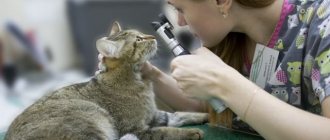
How is urolithiasis diagnosed?
With such symptoms, the animal must undergo an ultrasound of the urinary organs.
To determine an accurate diagnosis, it is necessary to urgently take your pet to a veterinary clinic. If nephrolithiasis or urolithiasis is suspected, the animal is sent for urine tests, which will show which salt predominates in the composition of the stones. The genitourinary system is examined using ultrasound. If KSD develops in non-castrated cats, a differential diagnosis must be prescribed, which can exclude pathologies similar in course:
- nephritis;
- cystitis;
- urethritis.
It is strictly forbidden to give diuretics to an animal during an acute attack of urolithiasis, because due to a violation of the outflow of urine, the drug will not help, but will only worsen the situation and the animal will die.